Abstract
Many independent lines of evidence indicate that the lactose permease of Escherichia coli (LacY) is highly dynamic and that sugar binding causes closing of a large inward-facing cavity with opening of a wide outward-facing hydrophilic cavity. Therefore, lactose/H+ symport catalyzed by LacY very likely involves a global conformational change that allows alternating access of single sugar- and H+-binding sites to either side of the membrane (the alternating access model). The x-ray crystal structures of LacY, as well as the majority of spectroscopic studies, use purified protein in detergent micelles. By using site-directed alkylation, we now demonstrate that sugar binding induces virtually the same global conformational change in LacY whether the protein is in the native bacterial membrane or is solubilized and purified in detergent. The results also indicate that the x-ray crystal structure reflects the structure of wild-type LacY in the native membrane in the absence of sugar.
Keywords: lactose, permease, symport, transport, membrane proteins
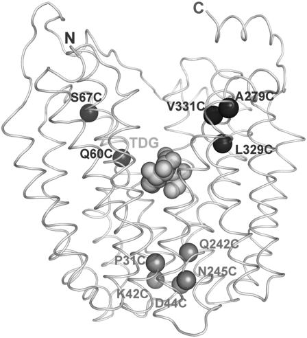
The lactose permease of Escherichia coli (LacY), a paradigm for the Major Facilitator Superfamily of membrane transport proteins, has been solubilized, purified, and reconstituted into proteoliopsomes in a fully functional state (1). X-ray crystal structures of a conformationally restricted mutant (2–7) have been solved in an inward-facing conformation (8, 9), and the crystal structure of wild-type LacY exhibits the same conformation (10, 11). Both structures have 12 transmembrane α-helices, most of which are shaped irregularly, organized into two pseudosymmetrical six-helix bundles surrounding a large interior hydrophilic cavity open only to the cytoplasm (Fig. 1). The sugar-binding site and the residues involved in H+ translocation are at the approximate middle of the molecule and are distributed so that the side chains important for sugar recognition are predominantly in the N-terminal helix bundle, and the side chains that form an H+-binding site are mainly in the C-terminal bundle (12). The periplasmic side of LacY is tightly packed, and the sugar-binding site is inaccessible from that side of the molecule.
Fig. 1.
Single-Cys LacY mutants. Positions of Cys replacements are shown on the backbone of LacY (Protein Data Bank ID code 1PV7; www.pdb.org) viewed perpendicular to the membrane with the N-terminal helix bundle on the left and the C-terminal bundle on the right. Black spheres indicate single-Cys replacements on the cytoplasmic side of the sugar-binding site: 60, 67 (helix II), 279 (helix VIII), 329, and 331 (helix X); grey spheres indicate single-Cys replacements on the periplasmic side of the sugar-binding site: 31 (helix I), 42, 44, (helix II), 242, and 245 (helix VII). The light grey spheres at the apex of the inward-facing cavity represent TDG.
Wild-type LacY is highly dynamic. Hydrogen/deuterium exchange of backbone amide protons in wild-type LacY occurs at rapid rate (13–15), and sugar binding is entropic (5), inducing widespread conformational changes (reviewed in refs. 16–18). More specifically, site-directed alkylation (reviewed in refs. 19–21), single-molecule fluorescence resonance energy transfer (6), double electron–electron resonance (7), site-directed cross-linking (22), and, most recently, tryptophan-quenching studies (23) each provide independent evidence that sugar binding increases the open probability of a sizeable hydrophilic cleft on the periplasmic side of LacY and closing of the cytoplasmic cavity. Furthermore, the periplasmic cleft must close, as well as open, for translocation of sugar across the membrane to occur (22, 24, 25). Notably, in the conformationally restricted mutant C154G LacY, the periplasmic cleft is paralyzed in an open conformation (6, 7, 21). However, all x-ray structures of LacY (C154G, as well as wild-type LacY) exhibit the same inward-facing conformation. Therefore, it is likely that the crystallization process selects a single conformer of LacY that is in the lowest free-energy state.
In studies described here, site-directed alkylation with the hydrophobic, membrane-permeant thiol reagent tetramethylrhodamine-5-maleimide (TMRM) (20, 21, 26) was used to examine the reactivity/accessibility of 10 strategically placed single-Cys LacY mutants (five on the periplasmic side of the sugar-binding site and five on the cytoplasmic side) (Fig. 1). The studies were carried out with right-side-out (RSO) membrane vesicles or with LacY solubilized and purified in n-dodecyl β-D-maltopyranoside (DDM). As shown previously with RSO membrane vesicles (reviewed in refs. 19–21), ligand binding increases the rate of TMRM labeling of the five single-Cys replacements on the periplasmic side and decreases the rate of labeling of the five mutants on the cytoplasmic side. Of great importance, as shown here, purified LacY in DDM exhibits essentially the same behavior. The results indicate strongly that purified wild-type LacY in DDM micelles has much the same inward-facing conformation as in the native bacterial membrane, as well as in the crystal structure. Moreover, sugar binding induces a similar change to an outward-open conformation both with purified LacY in DDM and with LacY embedded in the native bacterial membrane.
Results
N245C Single-Cys LacY.
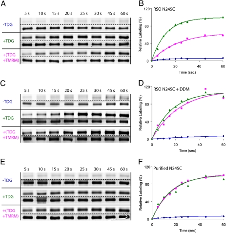
As shown (20, 27), the reactivity/accessibility of single-Cys N245C, located at the periplasmic gate of LacY (Fig. 1), is very low in the absence of β-D-galactopyranosyl 1-thio-β-D-galactopyranoside (TDG) (Fig. 2 A and B). However, preincubation with TDG increases TMRM labeling by ~15-fold (Fig. 2 A and B and Table 1). The rate increases linearly for ~15–20 s and then slackens. When TDG and TMRM are added simultaneously, the rate of labeling also increases, but only by ~7-fold (Fig. 2 A and B), and the effect of preincubation with TDG is unaltered by the presence of valinomycin and nigericin. Addition of 2.0% DDM to the membrane vesicles has little or no effect on TMRM labeling, but the effect of preincubation with TDG is abolished (Fig. 2 C and D). Moreover, when single-Cys N245C LacY is purified and subjected to the same labeling protocol, the observations are indistinguishable from those obtained with membrane vesicles in the presence of DDM (Fig. 2 E and F). That is, TMRM labeling is hardly detectable in the absence of TDG, and addition of TDG with or without preincubation increases the labeling rate by ~15-fold (Table 1).
Fig. 2.
TMRM labeling of periplasmic N245C single-Cys LacY. (A and B) TMRM labeling in RSO membrane vesicles. (C and D), TMRM labeling in RSO membrane vesicles after addition of 2.0% DDM. (E and F), TMRM labeling of purified protein in DDM. Samples were incubated with 40 μM TMRM (A–D) or 4 μM TMRM (E and F), for the times indicated at 0 °C in the absence of sugar (−TDG; blue ◆), were preincubated with TDG (+TDG) for 10 min before addition of TMRM (green ▲), or TMRM and TDG were added simultaneously [+(TDG+TMRM); pink ■]. Samples then were subjected to minipurification with monovalent avidin, and the TMRM-labeled proteins were subjected to SDS/PAGE and assayed for fluorescence (lanes above dotted lines) and protein (lanes below dotted lines). The data then were treated semiquantitatively as described in Experimental Procedures by setting the 60-s point in the presence of TDG to 100%. Although not shown in A or B, when valinomycin (10 μM, final concentration) and nigericin (0.1 μM, final concentration) were added, no significant deviation from the pink curve (pink ■) shown in A was observed.
Table 1.
TDG changes the rate of TMRM labeling of single-Cys mutants
| LacY mutant | Helix | Fold change in TMRM labeling in RSO vesicles | Fold change in TMRM labeling in DDM |
| Periplasmic | |||
| P31C | I | 13.6 | 8.9 |
| K42C | II | 8.5 | 5.9 |
| D44C | II | 8.2 | 6.2 |
| Q242C | II | 5.2 | 3.2 |
| N245C | VII | 14.9 | 6.9 |
| Cytoplasmic | |||
| Q60C | II | −7.2 | −3.8 |
| S67C | II | −14.7 | −8.3 |
| A279C | IX | −13.1 | −3.9 |
| L329C | X | −2.6 | −3.0 |
| V331C | X | −5.1 | −4.3 |
Rates of TMRM labeling were obtained from the time courses shown in Figs. 3–5 as described in Experimental Procedures. For each mutant, the ratio of the estimated initial rate of TMRM labeling in the presence of TDG relative to that observed in the absence of TDG was calculated. Positive numbers indicate an increase in the relative labeling rate caused by the addition of TDG (periplasmic) , and negative numbers indicate a decrease in the relative labeling rate caused by the addition of TDG (cytoplasmic).
S67C Single-Cys LacY.
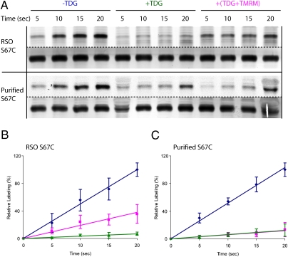
In contrast to N245C LacY, TMRM labeling of single-Cys S67C, located on the cytoplasmic side of the sugar-binding site, is rapid both in RSO membrane vesicles and with the purified protein in the absence of sugar (Fig. 3 A–C). Moreover, TDG now inhibits the rate of TMRM labeling by ~15-fold in RSO membrane vesicles and ~8-fold with the purified protein (Fig. 3 A–C and Table 1). When either the intact vesicles or purified S67C LacY are preincubated with TDG before addition of TMRM, labeling is abolished almost completely. However, when the sugar and the alkylating reagent are added simultaneously, the labeling rate of the mutant in RSO vesicles is inhibited only partially (Fig. 3 A and B). With purified protein in DDM, the same low rate of labeling is observed with or without preincubation with TDG (Fig. 3 A and C). Thus, TDG is readily accessible to the sugar-binding site with purified LacY in DDM but is significantly less accessible with intact RSO membrane vesicles.
Fig. 3.
TMRM labeling of cytoplasmic S67C single-Cys LacY. (A) SDS/PAGE gels showing TMRM labeling in RSO membrane vesicles or with purified protein (fluorescence above dotted line; protein below). (B) Time course of TMRM labeling in RSO membrane vesicles. (C) Time course of TMRM labeling of purified mutant S67C in DDM. RSO vesicles with S67C LacY were labeled with 40 μM TMRM or purified S67C LacY in DDM with 4 μM TMRM for times indicated at 0 °C in the absence of TDG (−TDG), were preincubated with TDG for 10 min before addition of TMRM (+TDG), or TMRM and TDG were added simultaneously [+(TDG+TMRM)]. Purified proteins were subjected to SDS/PAGE; fluorescent and silver-stained protein bands were measured as described in Fig. 2 and Experimental Procedures. The data are plotted relative to the 20-s points in the absence of TDG. Blue ◆, no sugar added; green ▲, preincubated with TDG before addition of TMRM; pink ■, TDG and TMRM added simultaneously.
Other Periplasmic Single-Cys Mutants.
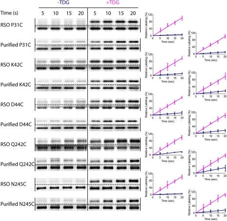
TMRM labeling of periplasmic single-Cys LacY mutants P31C, K42C, D44C, Q242C, and N245C (Figs. 1 and 4) was carried out both in RSO membrane vesicles and with purified proteins in DDM without or with TDG. The rate of TMRM labeling of each mutant is linear for at least 20 s, and TDG increases the rate in RSO membrane vesicles by ~5- to ~15-fold and with the purified protein in DDM by ~3- to ~9-fold, depending upon the mutant (Fig. 4 and Table 1). The results indicate that in both RSO membrane vesicles and in DDM, there is a low probability of opening of the periplasmic cleft in the absence of sugar and that TDG binding induces opening.
Fig. 4.
TMRM labeling of periplasmic single-Cys mutants in RSO membrane vesicles or as the purified proteins in DDM. Labeling of periplasmic single-Cys LacY mutants Q31C, K42C, D44C, Q242C, and N245C (Fig. 1) was performed with 40 μM TMRM (RSO membrane vesicles) or 4 μM TMRM (with purified proteins in DDM) for given times at 0 °C in the absence of TDG (−TDG; blue ◆) or after 10 min preincubation with TDG (+TDG; pink ■). Relative TMRM labeling rates were calculated as described in Experimental Procedures. The data are plotted relative to the 20-s points in the presence of TDG.
Other Cytoplasmic Single-Cys Mutants.
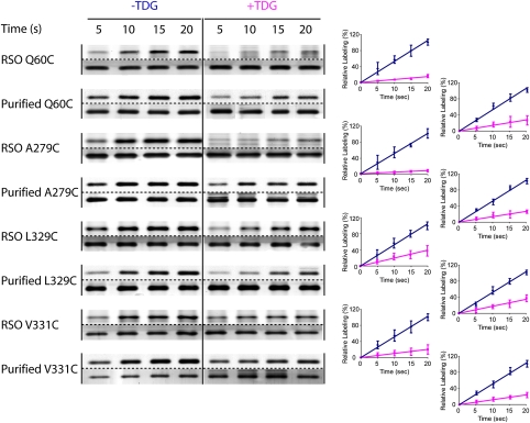
As expected, rates of alkylation of single-Cys mutants on the cytoplasmic side of the sugar-binding site exhibit the opposite behavior. Thus, the rate of TMRM labeling of single-Cys LacY mutants Q60C, A279C, L329C, and V331C is decreased upon TDG binding both with RSO membrane vesicles (~3- to ~15-fold) and with the purified mutant proteins in DDM (~3- to ~8-fold), depending upon the mutant (Fig. 5 and Table 1). Thus, LacY in the native bacterial membrane or as the purified protein in DDM is in an inward-open conformation in the absence of sugar, and sugar binding induces closing of the cytoplasmic cavity.
Fig. 5.
TMRM labeling of cytoplasmic single-Cys mutants in RSO membrane vesicles and as the purified proteins in DDM. Labeling of cytoplasmic single-Cys LacY mutants Q60C, S67C, A279C, L329C, and V331C (Fig. 1) was performed with 40 μM TMRM (RSO membrane vesicles) or 4 μM TMRM (with purified proteins in DDM) for given times at 0 °C in the absence of TDG (−TDG; blue ◆) or were preincubated for 10 min with TDG before addition of TMRM (+TDG; pink ■). Relative TMRM labeling rates were calculated as described in Experimental Procedures. The data are plotted relative to the 20-s points in the absence of TDG.
Discussion
In this study, a simple but sensitive alkylation method with a fluorescent thiol reagent (20, 21, 26) was used to examine the effect of sugar binding on alkylation of single-Cys LacY mutants in RSO membrane vesicles or with purified proteins in DDM micelles. The experiments were carried out at 0 °C, where thermal motion is restricted (27–29) and linear rates of labeling are readily obtained. TMRM labeling at 0 °C is almost negligible with LacY containing each of five single-Cys residues at positions on the periplasmic side of the sugar-binding site in RSO membrane vesicles or with purified protein in DDM micelles (Figs. 2 and 4 and Table 1). Therefore, each of these single-Cys replacements is unreactive and/or inaccessible to the alkylating agent. Although data are not shown, similar but less marked effects of TDG on TMRM labeling are observed at room temperature, caused primarily by increased rates of labeling in the absence of sugar. The observations are consistent with the interpretation that LacY in the native bacterial membrane is in a conformation similar to that of the x-ray crystal structures in the absence of ligand. The periplasmic side is tightly closed, and an open cavity is present facing the cytoplasm in this inward-facing conformation (8, 9, 11).
As stipulated by the alternating access model, on the cytoplasmic side of the sugar-binding site each of five single-Cys replacement mutants labels at a rapid rate in the absence of TDG both in RSO membrane vesicles and with purified protein in DDM. Moreover, TDG decreases the rate of TMRM labeling either in the membrane or with purified protein in DDM (Figs. 3 and 5 and Table 1). The findings agree with a variety of other measurements (6, 7, 19, 22, 23), showing that sugar binding induces closing of the cytoplasmic cavity and reduced reactivity/accessibility to alkylating agents.
The average increase in TMRM labeling observed in the presence of TDG in RSO vesicles is ~10-fold, and the average decrease in the presence of TDG is very similar (~9-fold) (Table 1). With purified single-Cys proteins in DDM, the comparable averages are ~6-fold and ~5-fold, respectively. Thus, the change in TMRM labeling induced by sugar on opposite faces of LacY appears to be about the same in RSO vesicles or with the purified single-Cys mutants in DDM. Therefore, the data provide further evidence not only that sugar binding markedly increases the open probability on the periplasmic side but also that sugar binding increases the probability of closing on the inside, the implication being that opening and closing may be reciprocal. However, reciprocity may not be obligatory, because evidence has been presented showing that the periplasmic pathway is fixed in an open conformation in the C154G mutant, whereas the cytoplasmic cavity is able to close and open (6, 7, 21). In addition, it has been demonstrated recently (25) that replacement of aspartate 68 with glutamic acid at the cytoplasmic end of helix II blocks sugar-induced opening of the periplasmic cleft but has little or no effect on closing of the cytoplasmic cavity.
Although the sugar-induced changes in the global conformation of LacY are qualitatively similar in RSO membrane vesicles and with the purified mutants in DDM, it is notable that that the magnitude of the effects is somewhat smaller with the purified proteins. Thus, the increases and decreases in TMRM labeling observed with the purified proteins upon addition of TDG are on average ~60% of those observed with RSO membrane vesicles. This difference is not surprising, because it is known that a lipid bilayer (30) and its composition (31) are important constraints on the structure of LacY.
Maximum effects of sugar on labeling of the single-Cys mutants in RSO membrane vesicles are observed when the vesicles are preincubated with TDG before addition of TMRM. When both reagents are added simultaneously to either N245C (periplasmic) or S67C (cytoplasmic) LacY, the effect of sugar on labeling is decreased [i.e., TDG causes slower labeling with N245C (Fig. 2A) and more rapid labeling with S67C (Fig. 3A)]. The effect of preincubation with both mutants likely reflects a delay in the time needed for TDG to gain access to the sugar-binding site, which is open only to the cytoplasmic side in RSO vesicles. Support for this interpretation is provided by the observations that the effect of preincubation is abrogated either when DDM is added to the vesicles or with protein purified in DDM. Furthermore, with the N245C mutant in RSO membrane vesicles, valinomycin and nigericin do not alter the effect of preincubation with TDG. Therefore, the effect is unlikely to be caused by the generation of a proton electrochemical gradient (interior positive and/or acid) secondary to downhill lactose/H+ symport (32).
In conclusion, the findings presented here support and extend previous conclusions regarding dynamic aspects of LacY derived from the use of a wide variety of biochemical/biophysical approaches. Taken as a whole, the following conclusions seem justified: (i) whether in the native bacterial membrane or as the purified protein in DDM, the predominant population of LacY is in an inward-facing conformation with a cavity open to the cytoplasm and a tightly packed periplasmic side, a model similar to that obtained by x-ray diffraction; and (ii) sugar binding in wild-type LacY induces opening of a hydrophilic periplasmic cavity and closing of the cytoplasmic cavity.
Experimental Procedures
Materials.
TMRM (T-6027) was obtained from Molecular Probes, Invitrogen Corp. ImmunoPure immobilized monomeric avidin was obtained from Pierce. All other materials were reagent grade and obtained from commercial sources.
Plasmid Construction.
All plasmids encoding single-Cys mutants in the Cys-less LacY background with a C-terminal biotin acceptor domain were constructed as described (20, 21).
Growth of Bacteria.
E. coli T184 (lacY−Z−) transformed with plasmid pT7-5 encoding a given mutant was grown aerobically at 37 °C in LB containing ampicillin (100 μg/mL). Fully grown cultures were diluted 10-fold and grown for another 2 h. After induction with 1 mM isopropyl 1-thio-β-D-galactopyranoside for 2 h, cells were harvested and used for the preparation of RSO membrane vesicles.
Preparation of RSO Membrane Vesicles.
RSO membrane vesicles were prepared from E. coli T184 expressing a specific mutant by lysozyme-EDTA treatment and osmotic lysis (33, 34). The vesicles were resuspended to a protein concentration of 10 mg/mL in 100 mM potassium phosphate (KPi; pH 7.2)/10 mM MgSO4, frozen in liquid nitrogen, and stored at −80 °C until use.
Labeling with TMRM.
Labeling with TMRM was carried out following a protocol developed previously (20, 21) with minor modifications. RSO membrane vesicles [0.1 mg of total protein in 50 μL 100 mM KPi (pH 7.2)/10 mM MgSO4 (RSO buffer)] containing a single-Cys mutant or single-Cys LacY purified from the same amount of RSO membrane vesicles as described below, were incubated with 40 μM (vesicles) or 4 μM TMRM (purified LacY) in the absence or presence of 10 mM TDG at 0 °C. At given times, DTT (10 mM, final concentration) was added to terminate the reaction. The vesicles then were solubilized in 2.0% DDM, and biotinylated LacY was purified by immobilized monomeric avidin Sepharose chromatography. Purified proteins (10 μL out of a total of 50 μL) were subjected to SDS/PAGE. The wet gels were imaged directly on an Amersham Typhoon 9410 Workstation (λex = 532 nm and λem = 580 nm for TMRM). The gels then were silver stained to quantify protein.
Fluorescent signals from TMRM labeling or silver-stained LacY bands were quantified by determining the density and area of each band by using ImageQuant (Molecular Dynamics, GE Healthcare Bio-Sciences Corp). Maximum TMRM labeling at 20 or 60 s, as indicated, was calculated by dividing the TMRM signal by the protein signal, which was then normalized to 100%. TMRM labeling at each time point was divided by the protein concentration for that sample and expressed as a percentage of the maximum TMRM labeling. Each set of data was fit by using a linear regression program. The slope of the fit data was used to estimate the relative initial rates of TMRM labeling. For each mutant, the ratio of the estimated initial rate of TMRM labeling in the presence of TDG relative to that observed in the absence of TDG then was calculated. The error bars represent the SEM for three samples at each of the times given.
Acknowledgments
We thank the members in the laboratory for advice and discussion and thank Irina Smirnova, in particular, for critically reading the manuscript. This work was supported by National Institutes of Health Grants DK051131, DK069463, GM073210, and GM074929 and National Science Foundation Grant 0450970 (to H.R.K.).
Footnotes
The authors declare no conflict of interest.
References
- 1.Viitanen P, Newman MJ, Foster DL, Wilson TH, Kaback HR. Purification, reconstitution, and characterization of the lac permease of Escherichia coli. Methods Enzymol. 1986;125:429–452. doi: 10.1016/s0076-6879(86)25034-x. [DOI] [PubMed] [Google Scholar]
- 2.Menick DR, Sarkar HK, Poonian MS, Kaback HR. cys154 Is important for lac permease activity in Escherichia coli. Biochem Biophys Res Commun. 1985;132:162–170. doi: 10.1016/0006-291x(85)91002-2. [DOI] [PubMed] [Google Scholar]
- 3.Smirnova IN, Kaback HR. A mutation in the lactose permease of Escherichia coli that decreases conformational flexibility and increases protein stability. Biochemistry. 2003;42:3025–3031. doi: 10.1021/bi027329c. [DOI] [PubMed] [Google Scholar]
- 4.Ermolova NV, Smirnova IN, Kasho VN, Kaback HR. Interhelical packing modulates conformational flexibility in the lactose permease of Escherichia coli. Biochemistry. 2005;44:7669–7677. doi: 10.1021/bi0502801. [DOI] [PubMed] [Google Scholar]
- 5.Nie Y, Smirnova I, Kasho V, Kaback HR. Energetics of ligand-induced conformational flexibility in the lactose permease of Escherichia coli. J Biol Chem. 2006;281:35779–35784. doi: 10.1074/jbc.M607232200. [DOI] [PMC free article] [PubMed] [Google Scholar]
- 6.Majumdar DS, et al. Single-molecule FRET reveals sugar-induced conformational dynamics in LacY. Proc Natl Acad Sci USA. 2007;104:12640–12645. doi: 10.1073/pnas.0700969104. [DOI] [PMC free article] [PubMed] [Google Scholar]
- 7.Smirnova I, et al. Sugar binding induces an outward facing conformation of LacY. Proc Natl Acad Sci USA. 2007;104:16504–16509. doi: 10.1073/pnas.0708258104. [DOI] [PMC free article] [PubMed] [Google Scholar]
- 8.Abramson J, et al. Structure and mechanism of the lactose permease of Escherichia coli. Science. 2003;301:610–615. doi: 10.1126/science.1088196. [DOI] [PubMed] [Google Scholar]
- 9.Mirza O, Guan L, Verner G, Iwata S, Kaback HR. Structural evidence for induced fit and a mechanism for sugar/H+ symport in LacY. EMBO J. 2006;25:1177–1183. doi: 10.1038/sj.emboj.7601028. [DOI] [PMC free article] [PubMed] [Google Scholar]
- 10.Guan L, et al. Manipulating phospholipids for crystallization of a membrane transport protein. Proc Natl Acad Sci USA. 2006;103:1723–1726. doi: 10.1073/pnas.0510922103. [DOI] [PMC free article] [PubMed] [Google Scholar]
- 11.Guan L, Mirza O, Verner G, Iwata S, Kaback HR. Structural determination of wild-type lactose permease. Proc Natl Acad Sci USA. 2007;104:15294–15298. doi: 10.1073/pnas.0707688104. [DOI] [PMC free article] [PubMed] [Google Scholar]
- 12.Smirnova IN, Kasho VN, Sugihara J, Choe JY, Kaback HR. Residues in the H+ translocation site define the pKa for sugar binding to LacY. Biochemistry. 2009;48(37):8852–8860. doi: 10.1021/bi9011918. [DOI] [PMC free article] [PubMed] [Google Scholar]
- 13.le Coutre J, Kaback HR, Patel CK, Heginbotham L, Miller C. Fourier transform infrared spectroscopy reveals a rigid alpha-helical assembly for the tetrameric Streptomyces lividans K+ channel. Proc Natl Acad Sci USA. 1998;95:6114–6117. doi: 10.1073/pnas.95.11.6114. [DOI] [PMC free article] [PubMed] [Google Scholar]
- 14.Patzlaff JS, Moeller JA, Barry BA, Brooker RJ. Fourier transform infrared analysis of purified lactose permease: A monodisperse lactose permease preparation is stably folded, alpha-helical, and highly accessible to deuterium exchange. Biochemistry. 1998;37:15363–15375. doi: 10.1021/bi981142x. [DOI] [PubMed] [Google Scholar]
- 15.Sayeed WM, Baenziger JE. Structural characterization of the osmosensor ProP. Biochim Biophys Acta. 2009;1788:1108–1115. doi: 10.1016/j.bbamem.2009.01.010. [DOI] [PubMed] [Google Scholar]
- 16.Kaback HR, Sahin-Tóth M, Weinglass AB. The kamikaze approach to membrane transport. Nat Rev Mol Cell Biol. 2001;2:610–620. doi: 10.1038/35085077. [DOI] [PubMed] [Google Scholar]
- 17.Kaback HR. Structure and mechanism of the lactose permease. C R Biol. 2005;328:557–567. doi: 10.1016/j.crvi.2005.03.008. [DOI] [PubMed] [Google Scholar]
- 18.Guan L, Kaback HR. Lessons from lactose permease. Annu Rev Biophys Biomol Struct. 2006;35:67–91. doi: 10.1146/annurev.biophys.35.040405.102005. [DOI] [PMC free article] [PubMed] [Google Scholar]
- 19.Kaback HR, et al. Site-directed alkylation and the alternating access model for LacY. Proc Natl Acad Sci USA. 2007;104:491–494. doi: 10.1073/pnas.0609968104. [DOI] [PMC free article] [PubMed] [Google Scholar]
- 20.Nie Y, Ermolova N, Kaback HR. Site-directed alkylation of LacY: Effect of the proton electrochemical gradient. J Mol Biol. 2007;374:356–364. doi: 10.1016/j.jmb.2007.09.006. [DOI] [PMC free article] [PubMed] [Google Scholar]
- 21.Nie Y, Sabetfard FE, Kaback HR. The Cys154—>Gly mutation in LacY causes constitutive opening of the hydrophilic periplasmic pathway. J Mol Biol. 2008;379:695–703. doi: 10.1016/j.jmb.2008.04.015. [DOI] [PMC free article] [PubMed] [Google Scholar]
- 22.Zhou Y, Guan L, Freites JA, Kaback HR. Opening and closing of the periplasmic gate in lactose permease. Proc Natl Acad Sci USA. 2008;105:3774–3778. doi: 10.1073/pnas.0800825105. [DOI] [PMC free article] [PubMed] [Google Scholar]
- 23.Smirnova I, Kasho V, Sugihara J, Kaback HR. Probing of the rates of alternating access in LacY with Trp fluorescence. Proc Natl Acad Sci USA. 2009;106:21561–21566. doi: 10.1073/pnas.0911434106. [DOI] [PMC free article] [PubMed] [Google Scholar]
- 24.Zhou Y, Nie Y, Kaback HR. Residues gating the periplasmic pathway of LacY. J Mol Biol. 2009;394:219–225. doi: 10.1016/j.jmb.2009.09.043. [DOI] [PMC free article] [PubMed] [Google Scholar]
- 25.Liu Z, Madej MG, Kaback HR. Helix dynamics in LacY: Helices II and IV. J Mol Biol. 2010;396:617–626. doi: 10.1016/j.jmb.2009.12.044. [DOI] [PMC free article] [PubMed] [Google Scholar]
- 26.Nie Y, Zhou Y, Kaback HR. Clogging the periplasmic pathway in LacY. Biochemistry. 2009;48:738–743. doi: 10.1021/bi801976r. [DOI] [PMC free article] [PubMed] [Google Scholar]
- 27.Venkatesan P, Kwaw I, Hu Y, Kaback HR. Site-directed sulfhydryl labeling of the lactose permease of Escherichia coli: Helix VII. Biochemistry. 2000;39:10641–10648. doi: 10.1021/bi000438b. [DOI] [PubMed] [Google Scholar]
- 28.Venkatesan P, Kaback HR. The substrate-binding site in the lactose permease of Escherichia coli. Proc Natl Acad Sci USA. 1998;95:9802–9807. doi: 10.1073/pnas.95.17.9802. [DOI] [PMC free article] [PubMed] [Google Scholar]
- 29.Venkatesan P, Liu Z, Hu Y, Kaback HR. Site-directed sulfhydryl labeling of the lactose permease of Escherichia coli: N-ethylmaleimide-sensitive face of helix II. Biochemistry. 2000;39:10649–10655. doi: 10.1021/bi0004394. [DOI] [PubMed] [Google Scholar]
- 30.le Coutre J, Narasimhan LR, Patel CK, Kaback HR. The lipid bilayer determines helical tilt angle and function in lactose permease of Escherichia coli. Proc Natl Acad Sci USA. 1997;94:10167–10171. doi: 10.1073/pnas.94.19.10167. [DOI] [PMC free article] [PubMed] [Google Scholar]
- 31.Bogdanov M, Heacock PN, Dowhan W. A polytopic membrane protein displays a reversible topology dependent on membrane lipid composition. EMBO J. 2002;21:2107–2116. doi: 10.1093/emboj/21.9.2107. [DOI] [PMC free article] [PubMed] [Google Scholar]
- 32.Garcia-Celma JJ, Smirnova IN, Kaback HR, Fendler K. Electrophysiological characterization of LacY. Proc Natl Acad Sci USA. 2009;106:7373–7378. doi: 10.1073/pnas.0902471106. [DOI] [PMC free article] [PubMed] [Google Scholar]
- 33.Kaback HR. Bacterial membranes. Methods Enzymol. 1971;32:99–120. [Google Scholar]
- 34.Short SA, Kaback HR, Kohn LD. Localization of D-lactate dehydrogenase in native and reconstituted Escherichia coli membrane vesicles. J Biol Chem. 1975;250:4291–4296. [PubMed] [Google Scholar]


