Figure 3.—
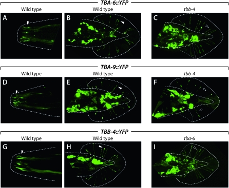
TBA-6, TBA-9, and TBB-4 fusion proteins localize to sensory cilia. A–I show the localization of the indicated tubulin∷YFP fusion protein expressed under the control of its own promoter. Genotypes, either wild-type or tubulin mutant, are shown above each image. Dotted lines indicate the outline of the body and the fan, the cuticular layer in which the rays are embedded. Thin dotted lines in C, F, and I indicate regions in which the fan is folded over onto itself. A, C, and E show nose cilia, while other panels show ventral or dorsal views of the male tail. All images are flattened stacks derived from multiple confocal sections spanning planes containing nose or ray cilia. Solid arrowheads indicate examples of the localization of tubulin fusion proteins to cilia. Open arrowheads highlight examples of dendritic endings that do not show significant tubulin localization. The dashed arrowhead in F indicates the beads-on-a-string appearance of TBA-9∷YFP accumulation in a tbb-4 mutant.

