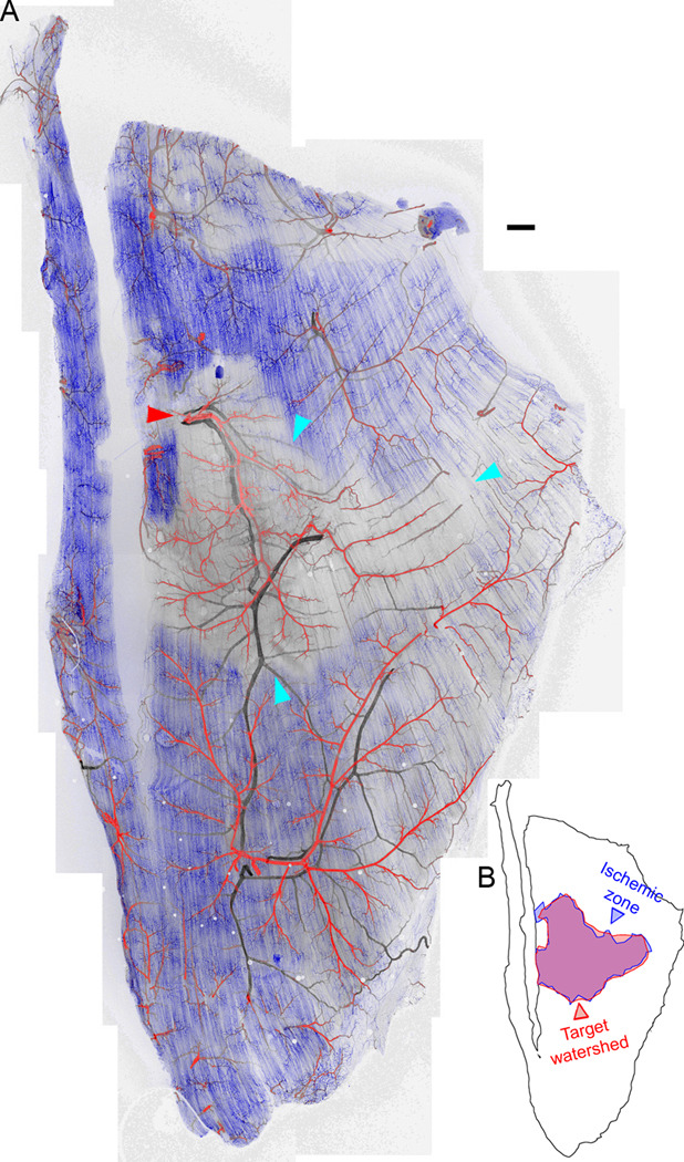
Figure 2. Loss of perfusion immediately following arteriolar ligation in Balb/c mice.
A, Perfusion of capillaries (blue, fluorescent intravenous lectin) was lost in part of the Balb/c spinotrapezius immediately post-ligation. The ligation site is indicated with a red arrowhead. The perfusion-poor volume is outlined with cyan arrowheads. This loss of perfusion was not observed in the contralateral muscle, or in ligated muscles of C57Bl/6 mice (Supplemental Figure S6). Red, arteries; black, veins. B, The perfusion-poor region is almost exactly contiguous with the watershed of the ligated arteriole. Scale bar for A, 250 µm.

