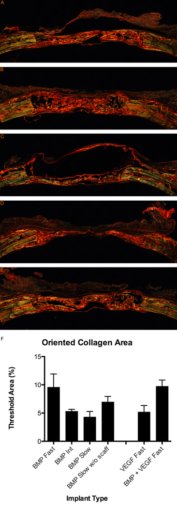
Figure 7.
Picrosirius red stain showing collagen orientation in regenerating bone at 6 weeks. Histological sections showing cross-section of entire defect with native bone at edges. (A) Fast degrading with 5µg BMP-2. (B) Intermediate degrading with 5µg BMP-2. (C) Slow degrading with 5µg BMP-2. (D) Fast degrading with 25µg VEGF. (E) Fast degrading with 25µg VEGF and 5µg BMP-2. (F) Quantification of organized bone in picrosirius red stained sections. Results are shown as mean and standard error of a minimum n = 4.

