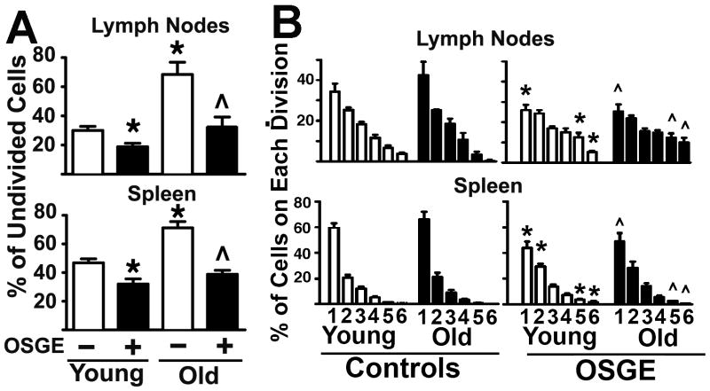
Figure 2. The number of cells that can enter cell cycle division declines with age, but does not affect the rate of cell division among cells that do enter the mitotic cycle, while ex-vivo OSGE treatments can reverse this defect.
A- The untreated and OSGE treated Trg+ cells corresponding to the peak 0 of cell division were quantified from matched sets of 3 young (open bars) and 1 old (filled bars) in each of 5 experiments. Each bar shows mean percentage ± SEM. The * indicates statistical significance with respect to young cells not exposed to OSGE, and the ^ represents significant differences with respect to old cells not exposed to OSGE. B-Untreated (−) and OSGE treated (+) Trg+ cells corresponding to peaks 1 to 6 of the CFSE cell division profile were counted from matched sets of at least 2 young (open bars) and 1 old (filled bars) in each of 7 experiments. The bars show mean percentage ± SEM for each peak in the CFSE cell division pattern. The * indicates significant differences with respect to untreated cells from young mice, and the ^ represents significant differences with respect to untreated of old mice (control) from the same division peak.

