Abstract
Myxococcus xanthus moves by gliding motility powered by Type IV pili (S-motility) and a second motility system, A-motility, whose mechanism remains elusive despite the identification of ~40 A-motility genes. In this study, we used biochemistry and cell biology analyses to identify multi-protein complexes associated with A-motility. Previously, we showed that the N-terminal domain of FrzCD, the receptor for the frizzy chemosensory pathway, interacts with two A-motility proteins, AglZ and AgmU. Here we characterized AgmU, a protein that localized to both the periplasm and cytoplasm. On firm surfaces, AgmU-mCherry co-localized with AglZ as distributed clusters that remained fixed with respect to the substratum as cells moved forward. Cluster formation was favored by hard surfaces where A-motility is favored. In contrast, AgmU-mCherry clusters were not observed on soft agar surfaces or when cells were in large groups, conditions that favor S-motility. Using glutathione-S-transferase (GST) affinity chromatography, AgmU was found to interact either directly or indirectly with multiple A-motility proteins including AglZ, AglT, AgmK, AgmX, AglW and CglB. These proteins, important for the correct localization of AgmU and AglZ, appear to be organized as a motility complex, spanning the cytoplasm, inner membrane and the periplasm. Identification of this complex may be important for uncovering the mechanism of A-motility.
Keywords: M. xanthus, A-motility, AgmU clusters, surface hardness, protein complex
Introduction
Myxococcus xanthus is a rod-shaped Gram-negative soil bacterium with a complex life cycle that includes predation, vegetative growth, and development (fruiting body formation). During vegetative growth, M. xanthus cells move in organized groups known as swarms, and feed on lysed microorganisms or organic matter by secreting hydrolytic enzymes and antimicrobials. When nutrients or prey are scarce, M. xanthus cells enter a developmental pathway that results in cellular aggregation in which 105 to 106 cells form fruiting bodies that contain spores (Kaiser, 2006, Berleman & Kirby, 2009, Mauriello & Zusman, 2007, Zusman et al., 2007, Shimkets, 1999). Directed motility is essential for vegetative swarming, predation, and development.
M. xanthus cells move by gliding motility and do not have flagella (Henrichsen, 1972). Hodgkin and Kaiser, over thirty years ago, showed that M. xanthus cells utilize two genetically distinct motility systems (Hodgkin, 1979). One system, called social (S)-motility, is required for the movement of cells in groups and is now known to be powered by the retraction of polar Type IV pili, similar to twitching motility in Pseudomonas aeruginosa (Li et al., 2003, Sun et al., 2000, Henrichsen, 1983). The other system, called adventurous (A)-motility, is required for the movement of isolated cells (Zusman et al., 2007, Hodgkin, 1979). However, the mechanism of A-motility is still unknown, although several hypotheses have been proposed (Mignot et al., 2007, Wolgemuth et al., 2002). These two motility systems enable M. xanthus cells to move selectively on different agar surfaces: A-motility works best on relatively hard, dry surfaces, whereas S-motility is favored on soft moist agar surfaces (Shi & Zusman, 1993) or when cells are submerged in methylcellulose (Sun et al., 2000). With both motility systems, cells periodically reverse their gliding direction. During reversals, the polarity of the cells is inverted so that the leading cell pole becomes the lagging cell pole and the old lagging cell pole becomes the new leading cell pole. The coordination of the two motility systems is essential for directed motility. The reversal frequencies of both the S- and A-motilities are regulated by the frizzy (Frz) chemosensory pathway (Zusman et al., 2007). Little is known about how these two motility systems are coordinated or how cells switch between these systems as they encounter different surfaces.
Several models of A-motility have been proposed based on experimental observations: (i) In the “slime gun” model, cells actively secrete a polyelectrolyte gel (slime) through “slime nozzles” located at the lagging cell pole; according to this model, the hydration of the slime propels cells forward (Wolgemuth et al., 2002). (ii) Another model, proposed by our laboratory, is the “focal adhesion” model, based on the cytological observations of the A-motility protein AglZ (Yang et al., 2004, Mignot et al., 2007). Mignot et al. reported that when cells move forward on 1.5% agar, AglZ clusters remain stationary with respect to the substratum (Mignot et al., 2007). This observation suggested a model in which the A-motility engines push against “focal adhesion” complexes that span the cell envelope and connect to an internal cytoskeleton (Mignot, 2007). Although these two models are very different, both require a protein complex spanning the cell envelope to power A-motility. Characterization of this complex will be a crucial step towards uncovering the mechanism of A-motility. About 40 genes have been identified as playing a role in A-motility (Yu & Kaiser, 2007, Youderian et al., 2003, Hartzell et al., 2008). However, it is still unknown how the proteins encoded by these genes are organized into functional A-motility complexes. In this study, we report that AgmU, a protein that interacts with FrzCD, is part of an envelope-spanning complex with AglZ, AglT, AgmK, AgmX, AglW and CglB. This work provides the basis for assigning function to several important A-motility proteins.
Results
AgmU interacts with the N-terminal region of FrzCD
In a previous study, we used affinity chromatography with GST-tagged FrzCD as bait to identify proteins that interact with FrzCD, the receptor for the Frz pathway. In that study, we identified two A-motility proteins, AglZ and AgmU as interacting partners with the N-terminal domain of FrzCD (Mauriello et al., 2009b). In the present study, the interaction between AgmU and FrzCD was characterized.
Figure 1A shows that AgmU, a large protein of 1218 amino acids, contains two clusters of tetratricopeptide repeats (TPR) in its N-terminal domain. The first cluster contains five TPR motifs (residues 163–400) and the second, four repeats (residues 620–810) (Figure 1A). TPR domains have been previously shown to be important in protein-protein interactions (D'Andrea & Regan, 2003), suggesting that these TPR clusters in AgmU might play a similar role together with other motility proteins. The AgmU C-terminal domain lacks a predicted function.
Figure 1. The two TPR clusters of AgmU interact with the N-terminus of FrzCD.
A) Schematic representation of the different AgmU fragments used in the in vitro cross-linking shown in (C). Each protein is fused to a N-terminal His6 tag. The first 60 amino acids of AgmU are shown in the inset. The possible recognition sites for the Tat secretion pathway (twin arginines) are shown in bold characters. B) Schematic representation of the N-terminal and C-terminal regions of FrzCD used in the in vitro cross-linking shown in (C). Each protein is fused to a N-terminal His6 tag. C) Anti-FrzCD immunoblotting of the in vitro formaldehyde cross-linking experiments with AgmU and FrzCD fragments. Purified FrzCD N-terminal (FrzCD-N) or C-terminal (FrzCD-C) regions and/or purified AgmU fragments (Roman numerals indicate AgmU domains shown in (A) incubated in the presence of the cross-linker (10 mM formaldehyde). Lanes 1–6, FrzCD or AgmU proteins incubated alone; lanes 7–10, FrzCD-N co-incubated with different AgmU fragments; lanes 11–14, FrzCD-C co-incubated with different AgmU fragments. The arrowheads indicate bands only seen after co-incubating FrzCD-N with full length AgmU or the TPR clusters of AgmU and cross-linker.
In order to investigate the interaction between AgmU and FrzCD, the two TPR clusters and the C-terminal domain of AgmU, and full length AgmU were expressed and purified in E. coli (TPR I, TPR II, C-ter, and AgmU in Figure 1A). We then examined their interactions with purified FrzCD by in vitro formaldehyde cross-linking. Figure 1B shows a Western blot in which anti-FrzCD antibodies were used to show that full-length AgmU and both TPR clusters of AgmU interacted with the N-terminal domain of FrzCD. In contrast, no evidence for an interaction between AgmU and the C-terminal domain of FrzCD was observed (Figure 1C).
AgmU is an essential component of the A-motility machinery in M. xanthus
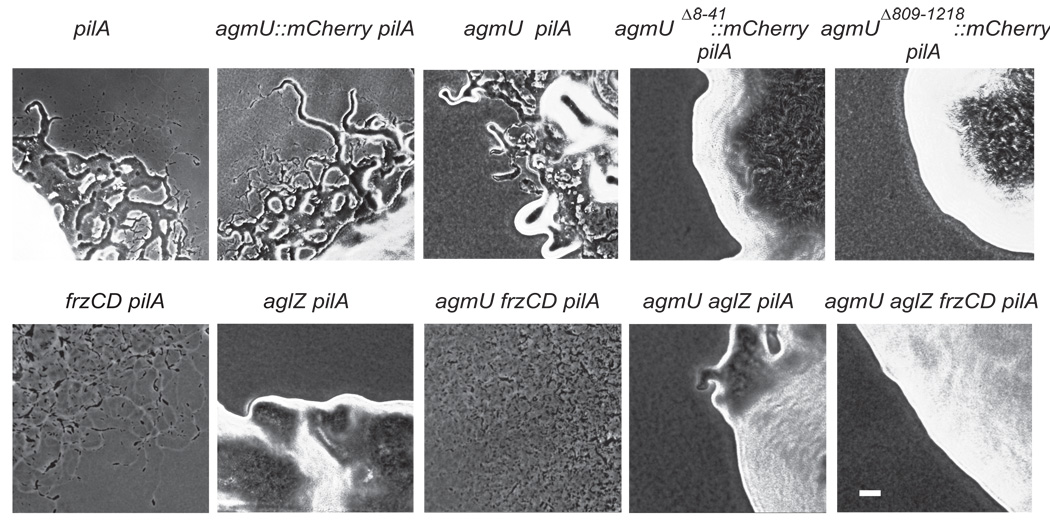
The agmU gene was first identified by Youderian et al. (Youderian et al., 2003) as an A-motility related gene through a genome wide screen using the transposon, magellan-4. In this study, we constructed an agmU in-frame deletion mutant that removed the coding region from amino acids 72 to 1206. The agmU deletion mutant, constructed in a strain lacking S-motility because of a pilA∷tet insertion, showed very few single cells at the edge of colonies on 1.5% agar. This indicates that it has a defect in A-motility (Hodgkin, 1979) (Figure 2). However, agmU pilA+ cells showed wild type S-motility swarming on soft (0.5%) agar, indicating that S-motility was not defective in the mutant (data not shown). To study the biological function of the AgmU-FrzCD interaction, an agmU frzCD pilA∷tet triple mutant was constructed. Figure 2 shows that in this strain, A-motility was restored, suggesting that AgmU, like AglZ, negatively regulates A-motility through its interaction with FrzCD (Mauriello et al., 2009b).
Figure 2. A-motility analysis of M xanthus strains DZ2 (wt), frzCD, aglZ and agmU mutants.
In the S− background (pilA), single cells swarming out from the edge of colonies on 1.5% agar (or agarose) were monitored as an indicator of A-motility (Hodgkin, 1979). Cells (10 µl), at a concentration of 4 × 10⁹ cfu ml−1, were spotted on CYE plates containing 1.5% (w/v) agar (or agarose), incubated at 32°C and photographed after 48 h with a WTI charge-coupled device (CCD)-72 camera, using a Nikon Labphot-2 microscope. Scale bar, 40 µm.
Since agmU frzCD pilA∷tet and aglZ frzCD pilA∷tet triple mutants both showed restored A-motility, we constructed an agmU aglZ frzCD pilA∷tet quadruple mutant. To our surprise, this quadruple mutant showed no A-motility (Figure 2). The phenotype of the agmU aglZ frzCD quadruple mutant indicates that either AgmU or AglZ is absolutely required for A-motility. This result suggests that AgmU and AglZ belong to the same A-motility machinery. We note that both AgmU and AglZ are proteins of more than 1000 amino acids and have multiple domains, suggesting that they could have both regulatory and structural functions.
AgmU has two distinct localization patterns in vivo
In order to further investigate the localization of AgmU in vivo, an agmU∷mCherry strain was constructed that encoded a mCherry tag fused to the C-terminus of AgmU. The gene fusion was inserted at the endogenous locus of agmU; this strain showed no defect in A-motility (Figure 2), S-motility or fruiting body formation (data not shown). The localization of AgmU-mCherry on 1.5% (w/v) agar (or agarose) was then monitored by fluorescence microscopy. Interestingly, we observed that AgmU-mCherry localized in two distinct patterns depending on the environment of the cells: i) in large groups of cells (usually more than 100 cells per group), AgmU-mCherry was localized (Figure 3A–3C, movie S1). This observation was confirmed by trans-section scans of 20 individual cells, which showed two fluorescence peaks that correspond to the location of the cell envelope (Figure 3B). In these cells, AgmU appeared to be slightly more concentrated towards the lagging cell pole (Figure 3C). ii) In small cell groups (usually less than 20 cells per group) or isolated cells, AgmU-mCherry was again observed near the envelope, but was also present as distributed clusters (Figure 3D–3F, movie S2). These AgmU-mCherry clusters appeared to be very dynamic, frequently appearing and disappearing (data not shown). Trans-section scans taken between these clusters showed two peaks, corresponding to the location of the cell envelope (Figure 3E), while scans across clusters gave an additional peak in the center (Figure 3F).
Figure 3. AgmU-mCherry shows two distinct localization patterns.
A) In large cell groups on 1.5% (w/v) agar (or agarose), AgmU-mCherry concentrates near the cell envelope. Images were taken with an Olympus IX70 DeltaVision microscope. B) Statistical analysis of the trans-section fluorescence scans in the same condition as panel A gives two fluorescence peaks at the location of the membrane. Twenty individual cells were scanned with ImageQuant software (GE healthcare) and the highest fluorescence density of each scan was normalized to 255 (same below). The inset shows a typical scan position in panel A. C) fluorescence scan along the long axis of one typical cell in panel A, shows the relative high concentration of AgmU-mCherry in the posterior half of the cell. D) In small groups or isolated cells on 1.5% (w/v) agar (or agarose), AgmU-mCherry shows two localization patterns. Besides the envelope-associated localization, protein clusters distributed along the cells were also seen. E) Statistical analysis of the scans between the clusters in the condition of panel D. F) Statistical analysis of the scans across clusters in the condition panel D. G) When the signal sequence of AgmU is deleted, AgmUΔ8--41-mCherry only forms cytoplasmic clusters. H) Statistical analysis of the scans between clusters in the condition of panel G. I) Statistical analysis of across the clusters in the condition of panel G. J) On 1.5% agar (or agarose), no clusters formed by AgmUΔ809-1218-mCherry are evident in either grouped or isolated cells, while the periplasmic localization is retained in this mutant. K) Statistical analysis of the scans in the condition of panel J. Scale bars, 5 µm.
AgmU localizes in both the periplasmic and cytoplasmic fractions
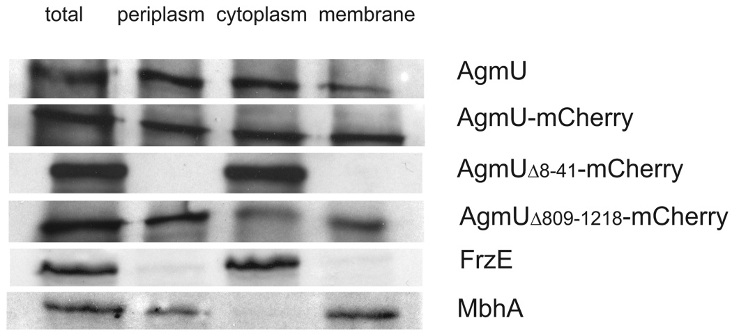
In order to study the unexpected localization pattern of AgmU, we subjected M. xanthus cells to osmotic shock and investigated the localization of AgmU in the various cell fractions. We used the shock procedure, described by Nelson et al. (Nelson et al., 1981), which involves treating M. xanthus cells with buffer containing 25% (w/v) sucrose, and then rapidly resuspending the cells in buffer lacking sucrose. The whole cell, periplasmic, cytoplasmic, and membrane fractions were then analyzed by SDS polyacrylamide gel electrophoresis and Western immunoblotting using anti-AgmU, anti- FrzE and anti-MbhA (Nelson et al., 1981) antibodies. Figure 4 shows the detected bands of AgmU, FrzE and MbhA cut from each Western blot. AgmU from wild type cells was found in the periplasmic, cytoplasmic and membrane fractions. Since AgmU lacks a transmembrane domain, the above localization pattern suggests that some of the AgmU molecules might be secreted and anchored to the cytoplasmic or outer membrane, and other molecules remain in the cytoplasm. In contrast, FrzE, a cytoplasmic histidine kinase, was only found in the cytoplasmic fraction, indicating that there was very little lysis of protoplasts during the shock procedure (Figure 4). Additionally, the periplasmic hemagglutinin, MbhA (Nelson et al., 1981), was observed in the periplasmic and membrane fractions, but not in the cytoplasmic fraction, indicating that the osmotic shock seperated cytoplasmic and periplasmic proteins effectively (Figure 4). The observation that AgmU-mCherry shows a dual localization pattern consistent with our fractionation experiments in which cells cultured in liquid showed AgmU to be present in both the periplasmic and cytoplasmic fractions.
Figure 4. AgmU localizes to both the periplasm and cytoplasm.
Western immunoblots of cell fractions following osmotic shock show that wild type AgmU, AgmU-mCherry and AgmUΔ809-1218-mCherry (AgmU C-terminus deletion) localize in both cytoplasmic and periplasmic fractions. While the protein with the hypothetical signal sequence deleted (AgmUΔ8--41-mCherry), only localizes in the cytoplasm. The cytoplasmic protein FrzE and lipoprotein MbhA are also shown as controls.
Analysis of the AgmU clusters
The N-terminal sequence of AgmU shows some similarity with the signal sequences of lipoproteins (Figure 1 A). To investigate the dual periplasmic and cytoplasmic localization pattern of AgmU, we constructed strains in which the amino acids 8–41 of AgmU or the AgmU C-terminal domain (amino acids 809–1218) were deleted from the agmU∷mCherry strain. The agmUΔ8–41∷mCherry pilA− and agmUΔ809–1218∷mCherry pilA− mutants showed dramatic defects in A-motility, defects that were more severe than deletions that spanned the entire agmU gene (Figure 2). We speculate that the truncated AgmUΔ8–41 and AgmUΔ809–1218 proteins interfere with overlaping functions of other A-motility proteins, such as AglZ, thereby causing a more severe defect in A-motility. Cell fractionation experiments showed that AgmUΔ8–41-mCherry is found only in the cytoplasm, while AgmUΔ809–1218-mCherry is found in both the periplasm, cytoplasm and membrane, indicating that the deletion of the C-terminal domain does not change the localization patterns of these proteins (Figure 4). We also followed the localization of AgmUΔ8–41-mCherry in living cells on 1.5% (w/v) agar (or agarose) by fluorescence microscopy. In these cells, AgmUΔ8–41-mCherry no longer localized near the cell envelope but instead formed larger centrally located clusters within the cell (Figures 3G–3I). We analyzed the clusters by trans-section scanning between clusters (Figure 3H) or across clusters (Figure 3I). In both cases, the fluorescence peaks corresponding to the cell envelope disappeared (Figures 3H, 3I). These observations confirm that the N-terminal sequence is required for the proper localization of AgmU in the periplasmic space. In contrast, AgmUΔ809–1218-mCherry, which contains the N-terminal sequence, was found concentrated near the cell envelope (Figure 3J, 3K). However, AgmUΔ809–1218-mCherry did not form fluorescence clusters in the center of cells (Figure 3K), although many cells showed high fluorescence intensity near the cell poles (Figure 3J). Since AgmUΔ809–1218-mCherry is found in the periplasm, cytoplasm and membrane (Figure 4), it is possible that the cytoplasmic fraction of AgmUΔ809–1218-mCherry concentrates near the cell poles. These results suggest that the C-terminal domain of AgmU is required for the formation of distributed cytoplasmic clusters.
AgmU-mCherry clusters co-localize with AglZ-GFP but not with FrzCD-GFP
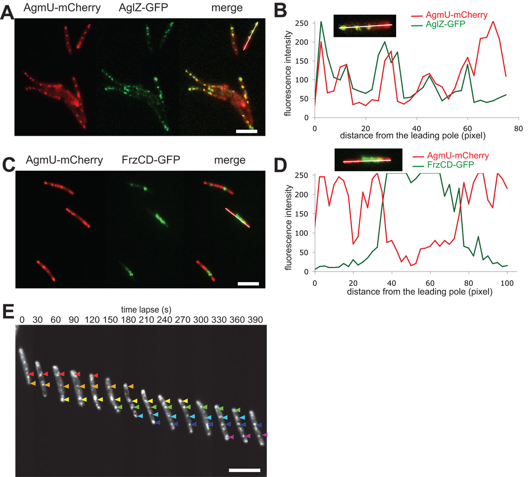
Since agmU pilA and aglZ pilA strains showed similar A-motility phenotypes and both AgmU and AglZ proteins form clusters, we were interested in determining whether these two proteins interact with the same A-motility machinery. To explore this possibility, a double labeled agmU∷mCherry aglZ∷gfp strain was constructed that showed wild type motility (Figure 7B). As shown in Figure 5A and movie S3, the clusters of AgmU and AglZ overlapped with each other. Scans of fluorescence density along the long axis of cells indicated that the fluorescence peaks of AgmU and AglZ were located at the same positions, except for the lagging cell pole, where the fluorescence intensity of AglZ-GFP was usually lower (Mignot et al., 2007) while the fluorescence intensity of AgmU-mCherry was higher (Figure 3A, 3C). In contrast, AgmU and FrzCD did not appear to co-localize. A doubly labeled agmU∷mCherry frzCD∷gfp strain showed the AgmU-mCherry clusters localized in distributed positions along the cell length and at the cell poles, while the FrzCD-GFP clusters localized in different positions and were always non-polar, as previously described (Mauriello et al., 2009a). Figures 5C and 5D show that in merged images, the two proteins occupied mutually exclusive positions similar to AglZ and FrzCD (Mauriello et al., 2009b).
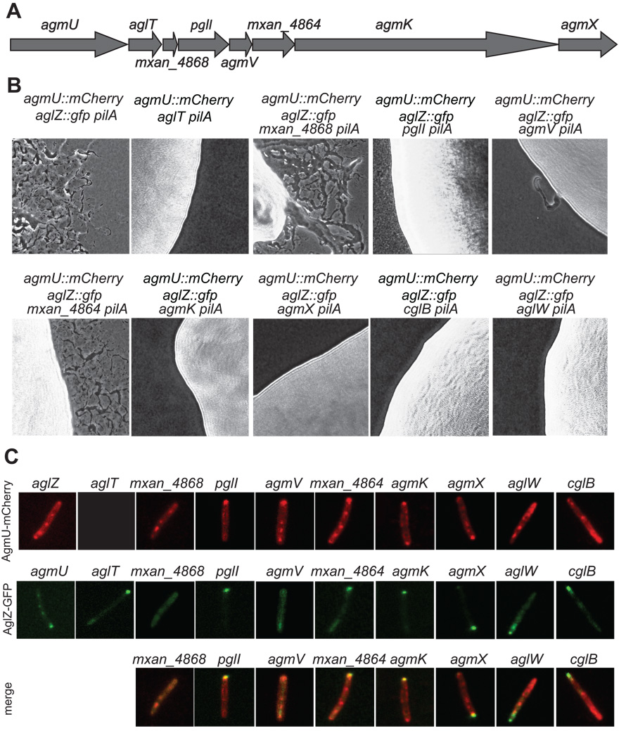
Figure 7. The genes that encode proteins interacting with AgmU are all required for A-motility.
A) agmU is the first gene of a large A-motility related gene cluster. B) A-motility analysis of the parental strain agmU∷mCherry aglZ-gfp∷kan, the additional deletion mutants of the genes downstream of agmU, and the additional deletion mutants of cglB and aglW. The movement of (pilA) cells that lack S-motility were monitored as an indicator of A-motility on 1.5% agar (or agarose) (Hodgkin, 1979). Samples were cultured and photographed as described in Figure 2. Note that the strains aglT agmU-mCherry∷kan and aglT aglZ-gfp∷kan have similar phenotypes and only the phenotype of aglT agmU-mCherry∷kan is shown. Scale bar, 40 µm. C) The products of a series of genes effect the localization of AgmU and AglZ. All the gene products regulate the localization of AglZ while only AglT, PglI and AgmK affect the localization of AgmU. In the aglT background, almost no AgmU-mCherry is detectable (see Figure S2). Scale bar, 5 µm.
Figure 5. The clusters of AgmU identify the same “focal adhesion” sites as AglZ.
A) The localization of AgmU-mCherry clusters overlaps with those of AglZ-GFP in vivo. B) Fluorescence scans along the cell length in panel A. mCherry and GFP signals are scanned separately with ImageQuant (GE Healthcare). The highest fluorescence density of each scan was normalized to 255. The inset shows the 2-fold magnification of the cell scanned in panel A. C) AgmU-mCherry clusters localize differently from that of FrzCD-GFP in vivo. D) A fluorescence scan along the cell length in panel C. The inset shows the two-fold magnification of the cell scanned in panel C. E) When cells move forward, AgmU-mCherry clusters remain fixed relative to the substratum. The fluorescent signal of mCherry in a single cell on 1.5% (w/v) agar (or agarose) is monitored with an Olympus DeltaVision IX70 microscope and recorded every 30 s. The positions of each cluster during a 390 s time course are marked with arrows in different colors. Scale bars, 5 µm.
To determine whether AgmU shows a localization pattern that is consistent with focal adhesion sites as described previously for AglZ (Mignot et al., 2007), we imaged agmU∷mCherry cells every 30 s for 10 min by fluorescence microscopy. By analyzing the images, we found that when cells were moving, the AgmU clusters appeared to remain relatively fixed with respect to the substratum, rather than moving forward with the cell. Figure 5E and movie S4 show typical time-lapse image sequence of AgmU-mCherry in a moving cell. These results suggest that AgmU is localized at the same focal adhesion sites as AglZ (Mignot et al., 2007).
Surface hardness influences AgmU localization
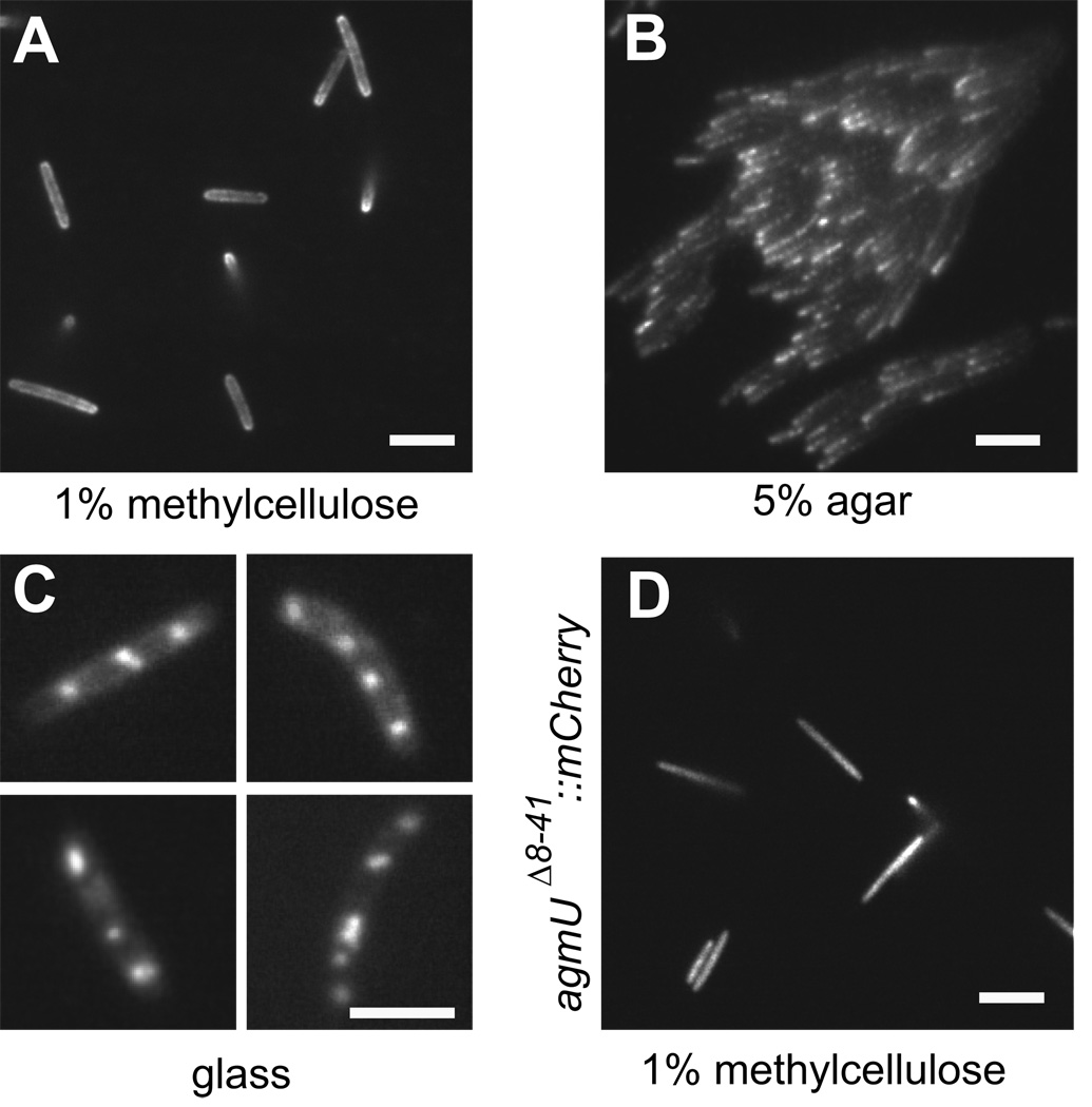
As described above and in Figure 3A, clusters of AgmU-mCherry on 1.5% agar (or agarose) were only observed in isolated cells or in small cell groups, but never observed in large groups. These observations suggest that AgmU cluster formation is regulated by signals from the environment or by cell-cell contact. To test the effect of the environment, we floated isolated cells in 1% methylcellulose (Figure 6A), CYE liquid medium or water (data not shown): none of these cells formed AgmU clusters. The distinguishing difference between these media and 1.5% agar or agarose is the physical hardness of the environment around the cells. To test the effect of substratum hardness on the formation of AgmU clusters, we placed cells on 5% agar or agarose. On these surfaces, almost every isolated cell showed AgmU-mCherry clusters (data not shown). Surprisingly, under these conditions, almost every cell in large groups also showed AgmU-mCherry clusters (Figure 6B; note that only grouped cells in monolayers were imaged); in contrast, these clusters were never seen when large cell groups were examined on 1.5% agar or agarose (Figure 3A, movie S1). These results indicate that the hardness of the substratum serves as a physical signal that regulates the localization of AgmU. To further verify the effect of substratum hardness on AgmU localization, we followed about 50 cells spotted directly onto glass microscope slides. On this surface, cells did not move. However, very large AgmU-mCherry clusters were observed in almost every cell. Figure 6C shows four typical cells.
Figure 6. The formation of AgmU clusters is regulated by the hardness of the substratum.
A) AgmU never formed clusters in 1% (w/v) methylcellulose. B) On 5% (w/v) agar (or agarose), AgmU-mCherry formed clusters even in large cell groups. C) Super-sized AgmU-mCherry clusters are found when cells are placed on a glass surface. Four individual cells are shown, but more than 50 cells were observed. D) AgmUΔ8–41-mCherry did not form clusters in 1% (w/v) methylcellulose. Scale bars, 5 µm.
Previously, it was shown that AglZ forms distributed clusters in isolated cells, while in large cell groups or methylcellulose, AglZ was diffuse along the cell length, only forming dominant clusters at the leading cell pole (Mauriello et al., 2009b). To test whether the localization of AglZ is also regulated by the hardness of the substratum, we spotted the aglZ∷gfp cells on hard (1.5%) or very hard (5%) agar or agarose. We observed no significant difference in cluster formation between 1.5% and 5% agar or agarose, suggesting that the localization of AglZ was not directly regulated by substratum hardness (data not shown).
Genes downstream of agmU are required for functional localization of AgmU and AglZ
As shown in Figure 7A, agmU is the first gene of an 8-gene cluster (http://www.wikimods.org/organism/myxococcus-xanthus/gene-page?zoom=5&offset=0&locus=MXAN_4870). The downstream genes aglT (Youderian et al., 2003), agmK (Youderian et al., 2003), agmX (Youderian et al., 2003), agmV (Hartzell et al., 2008) and pglI (Yu & Kaiser, 2007) were previously reported to be required for A-motility during the characterization of a collection of mutants with transposon insertions; the function of the genes mxan_4868 and mxan_4864 were not reported. For some genes encoding multiple domains (i.e. agmX and agmK), different insertions caused different phenotypes (Youderian et al., 2003). In this study, to more carefully analyze the function of each gene, we constructed in-frame deletions in all of the seven genes downstream of agmU in strains carrying the agmU∷mCherry aglZ∷gfp and pilA∷tet fusions. These constructs allowed us to determine the motility phenotypes of the various mutants as well as the effect of each mutation on the localization of AgmU and AglZ.
Figure 7B shows that all seven of the deletion mutants showed A-motility defects to some degree. Figure 7C shows the effect of these mutations on the localization of AgmU and AglZ. AgmU∷mCherry localization was clearly defective in the aglT, pglI and agmK mutants, but relatively unchanged in the other mutants and in the aglZ mutant. Western immunoblot analysis using anti-AgmU antibodies showed that the aglT mutant produced very little AgmU (Figure S2). Since AglT may be a lipoprotein, we speculate that AglT regulates the folding of AgmU and/or protecting mature AgmU from digestion by periplasmic proteases. The pglI and agmK mutants showed AgmU localized near the membrane and in aberrant clusters at the leading pole or at both the leading and lagging poles (Figure 7C). However, these mutants showed no significant change in the amount of AgmU found in the periplasm or cytoplasm (data not shown), suggesting that the cytoplasmic AgmU was concentrated at the cell poles. Interestingly, the pglI and agmK mutants failed to form distributed clusters along the cell length. Additionally, they did not form clusters on 5% agar (Figure S3). The localization of AgmU in these mutants suggests that PglI and AgmK serve to sense the hardness of the substratum or function in the positioning of the “focal adhesion” sites. PglI is a TonB homologue, while AgmK is a large TPR protein with unknown function.
Figure 7C (second row) shows that all of the seven genes downstream of agmU were required for AglZ to localize normally: (i) deletions in aglT, pglI, mxan_4864 and agmK caused AglZ to form a single cluster at the leading cell pole; (ii) deletions in mxan_4868 and agmV showed diffuse AglZ-GFP that did not form clusters; and (iii) deletions in agmU and agmX caused AglZ-GFP clusters to be more diffused than in the wild type (Figure 7C).
AgmU and AglZ are components of the same A-motility complex
The altered localization of AgmU and AglZ in the various A-motility mutants described above suggests that many proteins interact to control the localization of the adhesion complexes associated with A-motility. In order to identify these interacting proteins, a series of GST affinity chromatography experiments were performed using GST-tagged fragments of AgmU, AglT, AgmX and AgmK as baits, which were heterologously over-expressed and purified from E. coli. Since the GST-tagged full length AgmU was difficult to express and purify, we used the N and C-terminus of AgmU (AgmU-N, amino acids 52–860, including both the TPR clusters but not the signal sequence; AgmU-C, amino acids 800–1218) mixed in the ratio of 1:1 as bait. We also used the GST-tagged full length AglT only lacking the putative signal sequence (amino acids 33–478); the N-terminus of AglX (amino acids 2–373); and an AgmK fragment (amino acids 2681–3364) containing five TPR motifs.
The proteins that interacted with the baits were purified by affinity chromatography from wild type (strain DZ2) lysates and identified with MS/MS mass spectrometry (MS/MS, Proteomics/Mass Spectrometry Laboratory, UC Berkeley) as described (Mauriello et al., 2009b). The chromatography experiment with each GST-tagged bait was performed twice in parallel. Mass spectormetry identified as many as 100 proteins that co-purified with each bait (data not shown). Only the annotated A-motility proteins which were identified in both the two parallel chromatographic experiments were listed in Table 1. AgmU interacted with FrzCD as expected, but also with AglT, AgmK, AgmX and AglZ. Among these proteins, AgmK and AgmX were predicted to contain transmembrane fragments. We were concerned that some membrane vesicles containing AgmK or AgmX might be co-purified with AgmU, yielding false positive results. This possibility was excluded by using soluble fragments of AgmK and AgmX as baits, confirming the fidelity of the interacting proteins (Table 1). Additionally, two other A-motility related lipoproteins were identified with AgmU and other baits: i) AglW (Youderian et al., 2003), a TolB homologue that co-purified with GST-tagged AgmU, AglT, AgmK and AgmX; and ii) CglB (Rodriguez & Spormann, 1999), a contact stimulatable motility protein that co-purified with GST-tagged AgmU, AglT and AgmK.
Table 1.
Summary of the GST affinity chromatography.
| bait prey |
AgmU | AglT | AgmX | AgmK (AA 2681–3364) |
|---|---|---|---|---|
| FrzCD | √ | × | × | × |
| AglT | √ | − | × | × |
| AglW | √ | √ | √ | √ |
| AglZ | √ | √ | √ | √ |
| AgmK | √ | √ | √ | − |
| AgmU | − | √ | √ | √ |
| AgmX | √ | × | − | × |
| CglB | √ | √ | × | √ |
“√”, co-purified with the bait; “-”, not determined; “×”, not co-purified with the bait.
To study the function of AglW and CglB in A-motility, we constructed in-frame deletions in aglW and cglB in strains carrying the agmU∷mCherry, aglZ∷gfp and pilA∷tet fusions. Both the aglW and cglB deletion mutants showed defective A-motility (Figure 7B). In these strains, AglZ formed single clusters at the cell poles rather than distributed clusters (Figure 7C). AgmU localization was unaffected in both mutants (Figure 7C). Interestingly, the aglW strain showed the same growth rate as wild type (data not shown), but the cells formed accordion waves, also known as ripples (Berleman et al., 2006, Shimkets & Kaiser, 1982, Welch & Kaiser, 2001) on rich media (Figure S4). Since these waves have been shown to be associated with feeding on lysed prey cells or macromolecules, their presence suggests that aglW cells release macromolecules into the environment (Berleman et al., 2006).
Discussion
M. xanthus glides on surfaces by two distinct motility mechanisms, A-motility and S-motility. To achieve efficient locomotion, these motility systems must be coordinated. The Frz chemosensory pathway controls reversals for both motility systems, but it is not known how this pathway interacts with the motility engines. FrzCD, the methyl-accepting chemotaxis receptor for the Frz pathway, was previously shown to interact with two A-motility proteins, AglZ and AgmU (Mauriello et al., 2009b, Youderian et al., 2003, Yang et al., 2004). In this study, we examined the role of AgmU in controlling A-motility and showed that, like AglZ, it acts as a negative regulator of FrzCD activity, coupling A-motility to the Frz chemosensory pathway. For example, although agmU and aglZ mutants are defective in A-motility, agmU frzCD and aglZ frzCD double mutants show restored A-motility. However, agmU aglZ frzCD triple mutants show no A-motility, indicating that while the regulatory activities of AgmU and AglZ appear to be redundant with respect to the Frz pathway, together they are essential for A-motility. We speculate that both AgmU and AglZ have other functions in addition to the regulation of the Frz pathway, because in vivo fluorescence microscopy showed that both AgmU and AglZ did not co-localize with FrzCD; this suggests that only a small portion of AgmU and AglZ function as regulators through the interactions with FrzCD (Figure 5) (Mauriello et al., 2009b). This hypothesis is consistent with our in vitro cross-linking experiments, which show that only a small proportion of AgmU and AglZ could be directly cross-linked with FrzCD (Figure 1 and Mauriello et al., 2009b).
The data presented suggest that AgmU and AglZ work together as partners. Indeed, AgmU and AglZ both interact directly with the N-terminus of FrzCD coupling the activity of the Frz pathway in the regulation of A-motility (Mauriello et al., 2009b) (Figure 1C). Moreover, these two proteins co-localize in the previously described “focal adhesion” sites associated with A-motility; these sites remain fixed with respect to the substratum rather than with their cellular positions as cells move forward (Figure 5). To identify additional interaction partners in these “focal adhesion” sites, we used GST affinity chromatography with AgmU-GST as bait. These pull down experiments, summarized in Table 1, show that AgmU interacted with six proteins: AglT, AglW, AglZ, AgmK, AgmX, CglB, in an A-motility complex that spans the cytoplasm, inner membrane and periplasm. These interactions were further confirmed by additional pull down experiments with GST-tagged AglT, AgmK and AgmX fragments as baits. A schematic representation of the A-motility complex formed by AgmU and the other six A-motility proteins is presented in Figure 8. MreB, an actin-like protein (Carballido-Lopez, 2006), and MglA, a Ras family GTPase, which also interact with AglZ (Mauriello et al., 2010, Yang et al., 2004), are also involved in this complex. As another 30 proteins have been shown to be associated with A-motility (Hodgkin, 1979, Youderian et al., 2003, MacNeil et al., 1994), the complex that we propose may represent just a small piece of the A-motility complex. Figure 8 also suggests putative functions for the different components of the A-motility complex:
AglZ is a cytoplasmic protein which contains a N-terminal pseudo-receiver domain and a long C-terminal coiled-coil domain, showing similarity with FrzS, a S-motility protein (Mignot et al., 2007, Ward et al., 2000). AglZ-YFP forms distributed clusters that remain stationary with respect to the substratum as cells move forward. The localization of AglZ clusters requires direct interactions with the cytoskeleton protein, MreB, and the Ras-like GTPase, MglA (Yang et al., 2004, Mauriello et al., 2010).
AglT is a putative lipoprotein with six tandem TPR motifs. The amount of AgmU in aglT cell lysates was very low compared to the wild type. This result suggests that AglT serves to maintain AgmU in the correct conformation or to protect it from periplasmic proteolytic activities. AglT and AgmU might directly interact through their TPR motifs.
PglI is a TonB-like transporter with a forkhead-associated (FHA) domain at its N-terminus and a collagen domain near its C-terminus. The presence of the FHA and collagen domains suggests additional functions in mediating protein-protein interactions in the A-motility complex (Durocher & Jackson, 2002, Heino, 2007).
AgmK is a protein of extraordinary size (3812 amino acids) with two potential transmembrane fragments near its C-terminus (amino acids 3491–3506, 3550–3570) and at least 17 TPR motifs. The structural complexity and the putative transmembrane topology of AgmK make it an ideal candidate for a structural scaffold that anchors AgmU at “focal adhesion” sites and/or as a sensor for the hardness of the substratum.
AgmX shows limited similarity with plant extensins, which are usually found in the extracellular matrix (Kieliszewski & Lamport, 1994).
CglB is a lipoprotein required for A-motility. A-motility of a cglB mutant could be transiently restored by extracellular complementation upon mixing with cglB+ cells (Rodriguez & Spormann, 1999, Spormann & Kaiser, 1999).
AglW is a TolB homologue containing the Trp-Asp (WD) repeats, which form a β-propeller structure that is a common binding site for TPR motifs (Neer et al., 1994, Smith, 2008). In E. coli, TolB proteins participate in maintaining the integrity of the outer membrane through interactions with the peptidoglycan-associated lipoprotein (PAL) (Lazzaroni et al., 1999, Bonsor et al., 2009). The function of AglW might be supplying an assembly anchor for the A-motility complex at the peptidoglycan layer. We note that aglW is the last gene of an A-motility related operon. The genes upstream of aglW, aglX and aglV encode a TolQ/TolR pair (Youderian et al., 2003), homologous to the flagella motor MotA/MotB (Cascales et al., 2001).
MglA is a Ras-like GTPase which is required for both A- and S-motility (Hodgkin, 1979). MglA was reported to regulate the localization of AglZ and FrzS through direct interactions. The localization of MglA is dependent on the cytoskeleton protein MreB (Mauriello et al., 2010).
MreB is an actin-like cytoskeleton protein which forms helical filaments in rod shaped bacteria and is involved in several important cellular processes such as cell shape determination, cell polarity, and DNA segregation (Shih & Rothfield, 2006, Gitai et al., 2005, Gitai et al., 2004, Jones et al., 2001, Madabhushi & Marians, 2009, Kruse et al., 2003, Carballido-Lopez, 2006). In M. xanthus, perturbation of the MreB cytoskeleton blocks both A- and S-motility (Mauriello et al., 2010). MreB is required for the functional localization of AglZ, FrzS and MglA, it forms a double helix with the same periodicity as the AglZ clusters and directly interacts with AglZ (Mauriello et al., 2010).
Figure 8. A working model for the A-motility complex.
The proteins identified in this report and previous studies are summarized in this model. The outer membrane, pepidoglycan layer and the inner membrane near the “focal adhesion” site are shown in fragments. Previously, FrzCD was shown to interact with both AgmU and AglZ (Mauriello et al., 2009b); AglZ was shown to interact with the cytoskeleton protein MreB and the GTPase MglA (Hartzell & Kaiser, 1991, Mauriello et al., 2010). Data from E. coli TolB protein (AglW in this study) suggest an interaction of this protein with peptidoglycan-associated lipoprotein PAL (Bonsor et al., 2009), whose homologue in M. xanthus is MXAN_4581. Note that the proteins and structures in this model are not represented proportionally to their actual sizes.
Among the identified A-motility proteins, AglT, AgmK, AgmX, AglZ, AglW and CglB were all found to interact with AgmU (Table 1). They are predicted to form a large complex that spans the cytoplasm, membrane and periplasm. Although not identified by mass spectrometry, the other four proteins, MXAN_4868, PglI, AgmV and MXAN_4864 may play roles in conjunction with this complex, because they are required to form functional AglZ “focal adhesion” clusters; PglI is also required for AgmU cluster localization (Figure 7C). All the genes in the agmU gene cluster are well conserved among myxobacteria species (e.g. Stigmatella aurantiaca, Anaeromyxobacter dehalogenans and Sorangium cellulosum), suggesting an essential function of this A-motility complex. We note that it is difficult to judge from our pull down experiments whether the interactions between proteins are direct or indirect. Additionally, some proteins may interact with this A-motility complex but fail to be pulled down, since detergent was not used in our experiments and some membrane proteins might therefore have been excluded due to their insolubility.
AgmU, unlike AglZ, is found in the periplasmic, cytoplasmic and membrane fractions of cells. In vivo experiments with AgmU-mCherry confirmed this dual localization. Since agmU mutants that lacked the N-terminal sequence (agmUΔ8–41-mCherry) or the C-terminal domain (AgmUΔ809–1218-mCherry) showed severe A-motility defects, both the periplasmic and cytoplasmic localizations of AgmU are required for functional A-motility. AgmUΔ8–41-mCherry was not present in the periplasmic fraction (Figure 4), but still formed clusters observable by fluorescence microscopy (Figure 3G–3I), indicating that transport of the protein to the periplasm is not essential for cluster formation. In contrast, AgmUΔ809–1218-mCherry maintained periplasmic, cytoplasmic and membrane localization in fractionation assays, but lost the ability to form distributed clusters (Figures 3J, 3K, 4, and S1), suggesting that the C-terminal domain is required for cluster formation.
It is still not clear if AgmU is a lipoprotein or how AgmU is secreted. The N-terminal sequence of AgmU contains the positively charged N-terminus (n-region) and the hydrophobic core (h-region) of a typical Sec pathway signal sequence, but lacks the polar C-terminus (c-region) and the conserved cystine residue (Natale et al., 2008). This N-terminal sequence also contains two pairs of arginine residues (Figure 1A), which are potential recognition sites for the twin-arginine translocation (Tat) system. However, both of the arginine pairs lack the hydrophobic residues that normally follow and the Sec “avoidance signal” (usually a positively charged residue in the c-region) (Natale et al., 2008). Taken together, the poorly-conserved “signal sequence” may delay the secretion of AgmU and generate the formation of cytoplasmic clusters. The mechanism for dual periplasmic and cytoplasmic localization of AgmU is unknown. Similar dual localization patterns were reported for the Helicobacter pylori KatA protein (Harris & Hazell, 2003) and the E. coil GroESx protein (Lee & Ahn, 2000).
An unexpected finding in this study was that the formation of AgmU clusters is regulated by the physical hardness of the substratum. On 1.5% agar (or agarose), a substrate that facilitates both A- and S-motility, AgmU clusters were only observed in isolated cells or in small cell groups (Figures 4D–4F). In contrast, in large cell groups, where S-motility is dominant, clusters were never observed (Figures 4A–4C), except for some cells at the very “edge” of the group (data not shown). But when cells were spotted on 5% agar (or agarose), AgmU clusters were present in almost every cell, even in large cell groups (Figure 6B). This observation suggests that the formation of AgmU clusters is regulated by the hardness of the surface on which cells are gliding and it may play an important role in the switch favoring A-motility. This hypothesis is consistent with three additional experiments: First, in 1% methylcellulose, a condition that favors S-motility, distributed AgmU clusters were never observed (Figure 6A). This experiment ruled out the possibility that cell-cell contact may inhibit AgmU cluster formation. Second, in CYE liquid culture or water, where no gliding motility is present, AgmU clusters were never observed, indicating that the formation of AgmU clusters was not inhibited by any chemical content of the substratum. (data not shown). Third, on a glass surface, AgmU formed extraordinary large clusters (Figure 6C).
In large groups, cells are enveloped in a soft extracellular matrix, which inhibits the formation of AgmU clusters. In contrast, there is very little extracellular matrix around single cells and small cell groups, where the AgmU clusters appear. We note that AgmU clusters were not always observed in isolated cells on 1.5% agar (or agarose), possibly due to the following factors: First, under the experimental conditions used for imaging cells (liquid cell culture dropped on an agar pad), it is difficult to make the environment of each cell (especially the hardness of the surface) absolutely identical. However, with 5% agar (or agarose) or glass, the effect of surface hardness stands out and AgmU clusters were observed in every single cell. Second, AgmU clusters appear to form in a thin layer right above the surface of the substrate (data not shown), which makes it impossible to focus on the clusters of every cell in the same focal plane.
The experiments reported in this paper support the hypothesis that A-motility is regulated and powered, at least in part, by distributed motility proteins that act together as part of a complex. This complex could also work together with a proposed “slime secretion” motility system (Kaiser, 2009) or act as a “motility sensor” for the Frz pathway. We have identified many of these proteins but clearly many additional proteins remain to be characterized. Of particular interest are the elusive motor proteins that are presumed to power cell movement. We note that the M. xanthus genome encodes eight MotAB/TolQR homologues. These homologues are excellent candidates for motor proteins as MotAB from E. coli powers flagellar rotation (Minamino et al., 2008).
Material and Methods
Strains and growth conditions
Bacterial strains and plasmids are listed in Table 2. M. xanthus strains were cultured in CYE medium, which contains 10 mM MOPS pH 7.6, 1% (w/v) Bacto Casitone (BD Biosciences), 0.5% Bacto yeast extract and 4 mM MgSO4 (Campos et al., 1978). For A-motility assays, 10 µl cells of each strain in the concentration of 4×10⁹ colony forming units (cfu) ml−1 were spotted on CYE plates containing 1.5% (w/v) agar (or agarose), incubated at 32°C for 48 h and photographed with a WTI charge-coupled device (CCD)-72 camera, on a Nikon Labphot-2 microscope.
Table 2.
Strains and plasmids used in this study.
| strains/plasmids | genotype | reference source |
|---|---|---|
| M. xanthus strains | ||
| DZ2 | Wild type | (Campos et al., 1978) |
| DZ4469 | pilA∷tet | (Vlamakis et al., 2004) |
| TM7 | aglZ∷kan | (Mignot et al., 2007) |
| DZ4725 | aglZ∷kan pilA∷tet | (Mauriello et al., 2009b) |
| DZ4760 | aglZ-gfp∷kan | Mignot et al. unpublished |
| DZ4769 | frzCD pilA∷tet | this study |
| DZ4771 | ΔagmU pilA∷tet | this study |
| DZ4772 | agmU∷mCherry pilA∷tet | this study |
| DZ4773 | agmUΔ8–41∷mCherry pilA∷tet | this study |
| DZ4774 | agmUΔ809–1218∷mCherry pilA∷tet | this study |
| DZ4775 | ΔagmU ΔfrzCD pilA∷tet | this study |
| DZ4776 | ΔagmU aglZ∷kan pilA∷tet | this study |
| DZ4777 | ΔagmU ΔfrzCD aglZ∷kan pilA∷tet | this study |
| DZ4778 | agmU∷mCherry aglZ-gfp∷kan | this study |
| DZ4779 | agmU∷mCherry frzCD∷gfp | this study |
| DZ4780 | agmU∷mCherry aglZ-gfp∷kan pilA∷tet | this study |
| DZ4781 | ΔaglT agmU-mCherry∷kan pilA∷tet | this study |
| DZ4782 | ΔaglT aglZ-gfp∷kan pilA∷tet | this study |
| DZ4783 | Δmxan_4868 agmU∷mCherry aglZ-gfp∷kan pilA∷tet | this study |
| DZ4784 | ΔpglI_agmU∷mCherry aglZ-gfp∷kan pilA∷tet | this study |
| DZ4785 | ΔagmV agmU∷mCherry aglZ-gfp∷kan pilA∷tet | this study |
| DZ4786 | Δmxan_4864 agmU∷mCherry aglZ-gfp∷kan pilA∷tet | this study |
| DZ4787 | ΔagmK agmU∷mCherry aglZ-gfp∷kan pilA∷tet | this study |
| DZ4788 | ΔagmX agmU∷mCherry aglZ-gfp∷kan pilA∷tet | this study |
| DZ4789 | ΔaglW agmU∷mCherry aglZ-gfp∷kan pilA∷tet | this study |
| DZ4790 | ΔcglB agmU∷mCherry aglZ-gfp∷kan pilA∷tet | this study |
| E. coli strains | ||
| DH5α | host strain for molecular cloning | Invitrogen |
| BL21 (DE3) Tuner | host strain for protein expression | Novagen |
| plasmids | ||
| pET28a | His-tagged protein expression vector | Novagen |
| pGEX-KG | GST-tagged protein expression vector | (Guan & Dixon, 1991) |
| pGEX-2TK | GST-tagged protein expression vector | GE Healthcare |
| pBJ113 | Plasmid for gene deletions/insertions, galKS, kanR | (Julien et al., 2000) |
| pBN3 | pET28a with his6∷frzCD (2–135 AA) | (Mauriello et al., 2009b) |
| pBN4 | pET28a with his6∷frzCD (132–417 AA) | (Mauriello et al., 2009b) |
| pBN9 | pBJ113 with agmU deletion cassette | this study |
| pBN10 | pBJ113 with agmU∷mCherry insertion cassette | this study |
| pBN11 | pBJ113 with agmUΔ8–41 deletion cassette | this study |
| pBN12 | pBJ113 with agmUΔ809–1218 deletion cassette | this study |
| pBN13 | pBJ113 with aglT deletion cassette | this study |
| pBN14 | pBJ113 with mxan_4868 deletion cassette | this study |
| pBN15 | pBJ113 with pglI deletion cassette | this study |
| pBN16 | pBJ113 with agmV deletion cassette | this study |
| pBN17 | pBJ113 with mxan_4864 deletion cassette | this study |
| pBN18 | pBJ113 with agmK deletion cassette | this study |
| pBN19 | pBJ113 with agmX deletion cassette | this study |
| pBN20 | pBJ113 with aglW deletion cassette | this study |
| pBN21 | pBJ113 with cglB deletion cassette | this study |
| pBN22 | pET28a with his6∷agmU | this study |
| pBN23 | pET28a with his6∷agmU (TPRI, AA120–420) | this study |
| pBN24 | pET28a with his6∷agmU (TPRII, AA560–860) | this study |
| pBN25 | pET28a with his6∷agmU (C-terminal, AA800-–1218) | this study |
| pBN26 | pGEX-KG with gst∷agmU (N-terminal, AA52–860) | this study |
| pBN27 | pGEX-KG with gst∷agmU (C-terminal, AA800–1218) | this study |
| pBN28 | pGEX-KG with gst∷aglT (AA33–478) | this study |
| pBN29 | pGEX-2TK with gst∷agmX (AA2–373) | this study |
| pBN30 | pGEX-KG with gst∷agmK (AA2681–3364) | this study |
Double and triple mutants were constructed by electroporating M. xanthus cells with 4 µg of plasmid DNA or 1 µg of chromosomal DNA. Transformed cells were plated on CYE plates supplemented with 100 mg ml−1 sodium kanamycin sulfate. To construct the in-frame deletion or insertion strains, in-frame deletion or insertion cassettes were amplified with polymerase chain reaction (PCR) using chromosomal DNA as template, digested and inserted into plasmid pBJ113. All constructs were confirmed by DNA sequencing. Transformants were obtained by homologous recombination as previously described (Bustamante et al., 2004). To construct pilA∷tet and aglZ-gfp∷kan insertions, chromosomal DNA of strain TM7 (Mignot et al., 2007) and DZ4760 (which carries an aglZ-gfp∷kan insertion, Mignot et al., unpublished; Table 2) were electroporated into the parental strains. The primers used in the constructions of the in-frame deletions and insertions are summarized in table S1.
Protein expression and purification
The coding sequence of each protein or protein fragment was amplified by PCR from genomic DNA of DZ2 strain, digested and inserted into pET28a (Navagen), pGEX-KG (Guan & Dixon, 1991) or pGEX-2TK (GE Healthcare) vectors. The primers used in the cloning for protein expression are listed in Table S2. All constructs were confirmed by DNA sequencing. Expression and purification of the recombinant proteins were performed as described (Mauriello et al., 2009b), except that CHAPS and glycerol were only used for purification of FrzCD fragments.
In vitro protein cross-linking
In vitro protein cross-linking reactions were performed as described (Mauriello et al., 2009b). Proteins were diluted to the following concentrations to keep them at a 1:1 molar ratio: FrzCDN-ter, 2.5 µg ml−1; FrzCDC-ter, 2.5 µg ml−1; AgmU, 25 µg ml−1; AgmU-TPR1, 5 µg ml−1; AgmU-TPR2, 5 µg ml−1; AgmU-C-ter, 10 µg ml−1.
Osmotic shock fractionation
Osmotic shock fractionation was performed as described (Nelson et al., 1981). For AgmU and FrzE, cells were harvest from CYE liquid culture at the concentration of 4×10⁸ cfu ml−1 by centrifugation at 8,000 rpm at 4°C for 10 min. For MbhA, cells were harvest from CF plates after 48 hours of development (Nelson et al., 1981). The pellet was weighted and re-suspended into ice-cold 10 mM Tris-HCl pH 7.5 to a concentration of 10% (w/v). After 10 min incubation on ice, the buffer was changed to 10 mM Tris-HCl pH 7.5 25% (w/v) sucrose after 10 min of centrifugation at 8,000 rpm, 4°C. The cells were incubated on ice for 15 min with gentle shaking. The cells were collected with centrifugation (8,000 rpm, 4°C, 10 min) and shocked on ice with 10 mM Tris-HCl pH 7.5 for 10 min with gentle shaking. The cells were again subjected to centrifugation (8,000 rpm, 4°C, 10 min) and the supernatant was kept as the periplasmic fraction. The pellet was later sonicated and the supernatant of high-speed centrifugation (100, 000 g, 4°C, 30 min) was collected as the cytoplasmic fraction. 10 µl of each fraction was loaded into 10% SDS PAGE and Western blot was performed using polyclonal anti-AgmU antibodies.
Immunoblot analysis
Immunoblotting were performed as described (Mauriello et al., 2009b). AgmU polyclonal antibodies were produced from Covance Co. using 10 mg of protein mixture containing AgmU-TPR1, AgmU-TPR2 and AgmU-C-ter in the molar ration of ~1:1:1.
GST affinity chromatography and mass spectrometry
GST affinity chromatography was performed as described (Mauriello et al., 2009b), except that CHAPS and glycerol were not added to the PBS buffer. 0.1 mg of each purified GST-tagged protein was injected into a 1 ml GSTrap™ HP column (GE Heathcare); in the case of AgmU, 50 µg GST tagged AgmU N- and C-terminal domains were mixed and injected. MS/MS was performed in the Proteomics/Mass Spectrometry Laboratory of UC Berkeley as described (Mauriello et al., 2009b, Scott et al., 2008).
Time-lapse fluorescence microscopy
Time-lapse fluorescence microscopy was performed as described (Mauriello et al., 2009b). For the fluorescence microscopy on a glass surface, 3 µl cells from liquid CYE culture in the concentration of 4×10⁸ cfu ml−1 were spotted on glass slide and covered with cover slide immediately. In order to avoid the evaporation of water, nail polish was used to seal the cover slide.
Supplementary Material
Acknowlegements
We thank Tam Mignot for the DZ4760 strain. We thank Lori Kohlstaedt and Daniela Mavrici for their help with mass spectormetry. We thank Venassa Fan and Kailin Mesa for their excellent technical assistance. We thank Eva Campodonico, James Berleman and Juan-Jesus Vicente for insightful comments on the manuscript. This study is funded by a grant from the National Institutes of Health to DRZ (GM20509).
Footnotes
Competing interest
The authors declare no competing interest.
References
- Berleman JE, Chumley T, Cheung P, Kirby JR. Rippling is a predatory behavior in Myxococcus xanthus. J Bacteriol. 2006;188:5888–5895. doi: 10.1128/JB.00559-06. [DOI] [PMC free article] [PubMed] [Google Scholar]
- Berleman JE, Kirby JR. Deciphering the hunting strategy of a bacterial wolfpack. FEMS Microbiol Rev. 2009;33:942–957. doi: 10.1111/j.1574-6976.2009.00185.x. [DOI] [PMC free article] [PubMed] [Google Scholar]
- Bonsor DA, Hecht O, Vankemmelbeke M, Sharma A, Krachler AM, Housden NG, Lilly KJ, James R, Moore GR, Kleanthous C. Allosteric beta-propeller signalling in TolB and its manipulation by translocating colicins. EMBO J. 2009;28:2846–2857. doi: 10.1038/emboj.2009.224. [DOI] [PMC free article] [PubMed] [Google Scholar]
- Bustamante VH, Martinez-Flores I, Vlamakis HC, Zusman DR. Analysis of the Frz signal transduction system of Myxococcus xanthus shows the importance of the conserved C-terminal region of the cytoplasmic chemoreceptor FrzCD in sensing signals. Mol Microbiol. 2004;53:1501–1513. doi: 10.1111/j.1365-2958.2004.04221.x. [DOI] [PubMed] [Google Scholar]
- Campos JM, Geisselsoder J, Zusman DR. Isolation of bacteriophage MX4, a generalized transducing phage for Myxococcus xanthus. J Mol Biol. 1978;119:167–178. doi: 10.1016/0022-2836(78)90431-x. [DOI] [PubMed] [Google Scholar]
- Carballido-Lopez R. The bacterial actin-like cytoskeleton. Microbiol Mol Biol Rev. 2006;70:888–909. doi: 10.1128/MMBR.00014-06. [DOI] [PMC free article] [PubMed] [Google Scholar]
- Cascales E, Lloubes R, Sturgis JN. The TolQ-TolR proteins energize TolA and share homologies with the flagellar motor proteins MotA-MotB. Mol Microbiol. 2001;42:795–807. doi: 10.1046/j.1365-2958.2001.02673.x. [DOI] [PubMed] [Google Scholar]
- D'Andrea LD, Regan L. TPR proteins: the versatile helix. Trends Biochem Sci. 2003;28:655–662. doi: 10.1016/j.tibs.2003.10.007. [DOI] [PubMed] [Google Scholar]
- Durocher D, Jackson SP. The FHA domain. FEBS Lett. 2002;513:58–66. doi: 10.1016/s0014-5793(01)03294-x. [DOI] [PubMed] [Google Scholar]
- Gitai Z, Dye N, Shapiro L. An actin-like gene can determine cell polarity in bacteria. Proc Natl Acad Sci U S A. 2004;101:8643–8648. doi: 10.1073/pnas.0402638101. [DOI] [PMC free article] [PubMed] [Google Scholar]
- Gitai Z, Dye NA, Reisenauer A, Wachi M, Shapiro L. MreB actin-mediated segregation of a specific region of a bacterial chromosome. Cell. 2005;120:329–341. doi: 10.1016/j.cell.2005.01.007. [DOI] [PubMed] [Google Scholar]
- Guan KL, Dixon JE. Eukaryotic proteins expressed in Escherichia coli: an improved thrombin cleavage and purification procedure of fusion proteins with glutathione S-transferase. Anal Biochem. 1991;192:262–267. doi: 10.1016/0003-2697(91)90534-z. [DOI] [PubMed] [Google Scholar]
- Harris AG, Hazell SL. Localisation of Helicobacter pylori catalase in both the periplasm and cytoplasm, and its dependence on the twin-arginine target protein, KapA, for activity. FEMS Microbiol Lett. 2003;229:283–289. doi: 10.1016/S0378-1097(03)00850-4. [DOI] [PubMed] [Google Scholar]
- Hartzell P, Kaiser D. Function of MglA, a 22-kilodalton protein essential for gliding in Myxococcus xanthus. J Bacteriol. 1991;173:7615–7624. doi: 10.1128/jb.173.23.7615-7624.1991. [DOI] [PMC free article] [PubMed] [Google Scholar]
- Hartzell P, Shi W, Youderian P. Gliding Motility of Myxococcus xanthus. In: Whitworth DE, editor. Myxobacteria: Multicellularity and Differentiation. ASM Press; 2008. pp. 103–122. [Google Scholar]
- Heino J. The collagen family members as cell adhesion proteins. Bioessays. 2007;29:1001–1010. doi: 10.1002/bies.20636. [DOI] [PubMed] [Google Scholar]
- Henrichsen J. Bacterial surface translocation: a survey and a classification. Bacteriol Rev. 1972;36:478–503. doi: 10.1128/br.36.4.478-503.1972. [DOI] [PMC free article] [PubMed] [Google Scholar]
- Henrichsen J. Twitching motility. Annu Rev Microbiol. 1983;37:81–93. doi: 10.1146/annurev.mi.37.100183.000501. [DOI] [PubMed] [Google Scholar]
- Hodgkin J, Kaiser D. Genetics of gliding motility in Myxococcus xanthus (Myxobacterales): Two gene systems control movement. Mol. Gen. Genet. 1979;171:177–191. [Google Scholar]
- Jones LJ, Carballido-Lopez R, Errington J. Control of cell shape in bacteria: helical, actin-like filaments in Bacillus subtilis. Cell. 2001;104:913–922. doi: 10.1016/s0092-8674(01)00287-2. [DOI] [PubMed] [Google Scholar]
- Julien B, Kaiser AD, Garza A. Spatial control of cell differentiation in Myxococcus xanthus. Proc Natl Acad Sci U S A. 2000;97:9098–9103. doi: 10.1073/pnas.97.16.9098. [DOI] [PMC free article] [PubMed] [Google Scholar]
- Kaiser D. A microbial genetic journey. Annu Rev Microbiol. 2006;60:1–25. doi: 10.1146/annurev.micro.60.080805.142209. [DOI] [PubMed] [Google Scholar]
- Kaiser D. Are there lateral as well as polar engines for A-motile gliding in myxobacteria? J Bacteriol. 2009;191:5336–5341. doi: 10.1128/JB.00486-09. [DOI] [PMC free article] [PubMed] [Google Scholar]
- Kieliszewski MJ, Lamport DT. Extensin: repetitive motifs, functional sites, post-translational codes, and phylogeny. Plant J. 1994;5:157–172. doi: 10.1046/j.1365-313x.1994.05020157.x. [DOI] [PubMed] [Google Scholar]
- Kruse T, Moller-Jensen J, Lobner-Olesen A, Gerdes K. Dysfunctional MreB inhibits chromosome segregation in Escherichia coli. EMBO J. 2003;22:5283–5292. doi: 10.1093/emboj/cdg504. [DOI] [PMC free article] [PubMed] [Google Scholar]
- Lazzaroni JC, Germon P, Ray MC, Vianney A. The Tol proteins of Escherichia coli and their involvement in the uptake of biomolecules and outer membrane stability. FEMS Microbiol Lett. 1999;177:191–197. doi: 10.1111/j.1574-6968.1999.tb13731.x. [DOI] [PubMed] [Google Scholar]
- Lee JE, Ahn TI. Periplasmic localization of a GroES homologue in Escherichia coli transformed with groESx cloned from Legionella-like endosymbionts in Amoeba proteus. Res Microbiol. 2000;151:605–618. doi: 10.1016/s0923-2508(00)90133-5. [DOI] [PubMed] [Google Scholar]
- Li Y, Sun H, Ma X, Lu A, Lux R, Zusman D, Shi W. Extracellular polysaccharides mediate pilus retraction during social motility of Myxococcus xanthus. Proc Natl Acad Sci U S A. 2003;100:5443–5448. doi: 10.1073/pnas.0836639100. [DOI] [PMC free article] [PubMed] [Google Scholar]
- MacNeil SD, Mouzeyan A, Hartzell PL. Genes required for both gliding motility and development in Myxococcus xanthus. Mol Microbiol. 1994;14:785–795. doi: 10.1111/j.1365-2958.1994.tb01315.x. [DOI] [PubMed] [Google Scholar]
- Madabhushi R, Marians KJ. Actin homolog MreB affects chromosome segregation by regulating topoisomerase IV in Escherichia coli. Mol Cell. 2009;33:171–180. doi: 10.1016/j.molcel.2009.01.001. [DOI] [PMC free article] [PubMed] [Google Scholar]
- Mauriello EM, Mouhamar F, Nan B, Ducret A, Dai D, Zusman DR, Mignot T. Bacterial motility complexes require the actin-like protein, MreB and the Ras homologue, MglA. EMBO J. 2010;29:315–326. doi: 10.1038/emboj.2009.356. [DOI] [PMC free article] [PubMed] [Google Scholar]
- Mauriello EM, Zusman DR. Polarity of motility systems in Myxococcus xanthus. Curr Opin Microbiol. 2007 doi: 10.1016/j.mib.2007.09.012. [DOI] [PMC free article] [PubMed] [Google Scholar]
- Mauriello EMF, Astling DP, Sliusarenko O, Zusman DR. Localization of a bacterial cytoplasmic receptor is dynamic and changes with cell-cell contacts. Proc Natl Acad Sci U S A. 2009a;106:4852–4857. doi: 10.1073/pnas.0810583106. [DOI] [PMC free article] [PubMed] [Google Scholar]
- Mauriello EMF, Nan B, Zusman DR. AglZ regulates adventurous (A-) motility in Myxococcus xanthus through its interaction with the cytoplasmic receptor, FrzCD. Mol Microbiol. 2009b;72:964–977. doi: 10.1111/j.1365-2958.2009.06697.x. [DOI] [PMC free article] [PubMed] [Google Scholar]
- Mignot T. The elusive engine in Myxococcus xanthus gliding motility. Cell Mol Life Sci. 2007;64:2733–2745. doi: 10.1007/s00018-007-7176-x. [DOI] [PMC free article] [PubMed] [Google Scholar]
- Mignot T, Shaevitz JW, Hartzell PL, Zusman DR. Evidence that focal adhesion complexes power bacterial gliding motility. Science. 2007;315:853–856. doi: 10.1126/science.1137223. [DOI] [PMC free article] [PubMed] [Google Scholar]
- Minamino T, Imada K, Namba K. Molecular motors of the bacterial flagella. Curr Opin Struct Biol. 2008;18:693–701. doi: 10.1016/j.sbi.2008.09.006. [DOI] [PubMed] [Google Scholar]
- Natale P, Bruser T, Driessen AJ. Sec- and Tat-mediated protein secretion across the bacterial cytoplasmic membrane--distinct translocases and mechanisms. Biochim Biophys Acta. 2008;1778:1735–1756. doi: 10.1016/j.bbamem.2007.07.015. [DOI] [PubMed] [Google Scholar]
- Neer EJ, Schmidt CJ, Nambudripad R, Smith TF. The ancient regulatory-protein family of WD-repeat proteins. Nature. 1994;371:297–300. doi: 10.1038/371297a0. [DOI] [PubMed] [Google Scholar]
- Nelson DR, Cumsky MG, Zusman DR. Localization of myxobacterial hemagglutinin in the periplasmic space and on the cell surface of Myxococcus xanthus during developmental aggregation. J Biol Chem. 1981;256:12589–12595. [PubMed] [Google Scholar]
- Rodriguez AM, Spormann AM. Genetic and molecular analysis of cglB, a gene essential for single-cell gliding in Myxococcus xanthus. J Bacteriol. 1999;181:4381–4390. doi: 10.1128/jb.181.14.4381-4390.1999. [DOI] [PMC free article] [PubMed] [Google Scholar]
- Scott AE, Simon E, Park SK, Andrews P, Zusman DR. Site-specific receptor methylation of FrzCD in Myxococcus xanthus is controlled by a tetratrico peptide repeat (TPR) containing regulatory domain of the FrzF methyltransferase. Mol Microbiol. 2008;69:724–735. doi: 10.1111/j.1365-2958.2008.06323.x. [DOI] [PMC free article] [PubMed] [Google Scholar]
- Shi W, Zusman DR. The two motility systems of Myxococcus xanthus show different selective advantages on various surfaces. Proc Natl Acad Sci U S A. 1993;90:3378–3382. doi: 10.1073/pnas.90.8.3378. [DOI] [PMC free article] [PubMed] [Google Scholar]
- Shih YL, Rothfield L. The bacterial cytoskeleton. Microbiol Mol Biol Rev. 2006;70:729–754. doi: 10.1128/MMBR.00017-06. [DOI] [PMC free article] [PubMed] [Google Scholar]
- Shimkets LJ. Intercellular signaling during fruiting-body development of Myxococcus xanthus. Annu Rev Microbiol. 1999;53:525–549. doi: 10.1146/annurev.micro.53.1.525. [DOI] [PubMed] [Google Scholar]
- Shimkets LJ, Kaiser D. Induction of coordinated movement of Myxococcus xanthus cells. J Bacteriol. 1982;152:451–461. doi: 10.1128/jb.152.1.451-461.1982. [DOI] [PMC free article] [PubMed] [Google Scholar]
- Smith TF. Diversity of WD-repeat proteins. Subcell Biochem. 2008;48:20–30. doi: 10.1007/978-0-387-09595-0_3. [DOI] [PubMed] [Google Scholar]
- Spormann AM, Kaiser D. Gliding mutants of Myxococcus xanthus with high reversal frequencies and small displacements. J Bacteriol. 1999;181:2593–2601. doi: 10.1128/jb.181.8.2593-2601.1999. [DOI] [PMC free article] [PubMed] [Google Scholar]
- Sun H, Zusman DR, Shi W. Type IV pilus of Myxococcus xanthus is a motility apparatus controlled by the frz chemosensory system. Curr Biol. 2000;10:1143–1146. doi: 10.1016/s0960-9822(00)00705-3. [DOI] [PubMed] [Google Scholar]
- Vlamakis HC, Kirby JR, Zusman DR. The Che4 pathway of Myxococcus xanthus regulates type IV pilus-mediated motility. Mol Microbiol. 2004;52:1799–1811. doi: 10.1111/j.1365-2958.2004.04098.x. [DOI] [PubMed] [Google Scholar]
- Ward MJ, Lew H, Zusman DR. Social motility in Myxococcus xanthus requires FrzS, a protein with an extensive coiled-coil domain. Mol Microbiol. 2000;37:1357–1371. doi: 10.1046/j.1365-2958.2000.02079.x. [DOI] [PubMed] [Google Scholar]
- Welch R, Kaiser D. Cell behavior in traveling wave patterns of myxobacteria. Proc Natl Acad Sci U S A. 2001;98:14907–14912. doi: 10.1073/pnas.261574598. [DOI] [PMC free article] [PubMed] [Google Scholar]
- Wolgemuth C, Hoiczyk E, Kaiser D, Oster G. How myxobacteria glide. Curr Biol. 2002;12:369–377. doi: 10.1016/s0960-9822(02)00716-9. [DOI] [PubMed] [Google Scholar]
- Yang R, Bartle S, Otto R, Stassinopoulos A, Rogers M, Plamann L, Hartzell P. AglZ is a filament-forming coiled-coil protein required for adventurous gliding motility of Myxococcus xanthus. J Bacteriol. 2004;186:6168–6178. doi: 10.1128/JB.186.18.6168-6178.2004. [DOI] [PMC free article] [PubMed] [Google Scholar]
- Youderian P, Burke N, White DJ, Hartzell PL. Identification of genes required for adventurous gliding motility in Myxococcus xanthus with the transposable element mariner. Mol Microbiol. 2003;49:555–570. doi: 10.1046/j.1365-2958.2003.03582.x. [DOI] [PubMed] [Google Scholar]
- Yu R, Kaiser D. Gliding motility and polarized slime secretion. Mol Microbiol. 2007;63:454–467. doi: 10.1111/j.1365-2958.2006.05536.x. [DOI] [PubMed] [Google Scholar]
- Zusman DR, Scott AE, Yang Z, Kirby JR. Chemosensory pathways, motility and development in Myxococcus xanthus. Nat Rev Microbiol. 2007;5:862–872. doi: 10.1038/nrmicro1770. [DOI] [PubMed] [Google Scholar]
Associated Data
This section collects any data citations, data availability statements, or supplementary materials included in this article.


