Abstract
OBJECTIVE
Cross-sectional studies have reported increased levels of urinary type IV collagen in diabetic patients with progression of diabetic nephropathy. The aim of this study was to determine the role of urinary type IV collagen in predicting development and progression of early diabetic nephropathy and deterioration of renal function in a longitudinal study.
RESEARCH DESIGN AND METHODS
Japanese patients with type 2 diabetes (n = 254, 185 with normoalbuminuria and 69 with microalbuminuria) were enrolled in an observational follow-up study. The associations of urinary type IV collagen with progression of nephropathy and annual decline in estimated glomerular filtration rate (eGFR) were evaluated.
RESULTS
At baseline, urinary type IV collagen levels were higher in patients with microalbuminuria than in those with normoalbuminuria and correlated with urinary β2-microglobulin (β = 0.57, P < 0.001), diastolic blood pressure (β = 0.15, P < 0.01), eGFR (β = 0.15, P < 0.01), and urinary albumin excretion rate (β = 0.13, P = 0.01) as determined by multivariate regression analysis. In the follow-up study (median duration 8 years), urinary type IV collagen level at baseline was not associated with progression to a higher stage of diabetic nephropathy. However, the level of urinary type IV collagen inversely correlated with the annual decline in eGFR (γ = −0.34, P < 0.001). Multivariate regression analysis identified urinary type IV collagen, eGFR at baseline, and hypertension as factors associated with the annual decline in eGFR.
CONCLUSIONS
Our results indicate that high urinary excretion of type IV collagen is associated with deterioration of renal function in type 2 diabetic patients without overt proteinuria.
Diabetic nephropathy is characterized structurally by accumulation of mesangial matrix and thickening of the basement membrane in the glomeruli (1), as well as renal tubular hypertrophy and associated basement membrane alterations in the tubulointerstitium, which precede tubulointerstitial fibrosis (2). These abnormalities are associated with renal overproduction of the extracellular matrix proteins such as type IV collagen (3). We and others reported previously that the production of type IV collagen is enhanced in glomerular mesangial cells (4,5), podocytes (6), and proximal tubular cells (7,8) cultured under a high-glucose condition. Cross-sectional studies reported a stepwise increase in urinary type IV collagen excretion in diabetic patients, in parallel with increased levels of urinary albumin (9–14). Thus, a high level of urinary type IV collagen has been proposed as a marker of development and progression of early diabetic kidney disease. However, there is little information on whether the increase in type IV collagen excretion in urine is a predictor of progression of diabetic nephropathy or deterioration of renal function in type 2 diabetic patients.
The aim of this study was to determine whether the urinary levels of type IV collagen can predict the progression of early diabetic kidney disease. For this purpose, we conducted a prospective observational follow-up study involving type 2 diabetic patients with normoalbuminuria and microalbuminuria and investigated the relationship between urinary levels of type IV collagen and the progression of diabetic nephropathy stage as well as the annual decline in estimated glomerular filtration rate (eGFR) during the follow-up period.
RESEARCH DESIGN AND METHODS
The participants with type 2 diabetes were recruited from patients who regularly visited the outpatient clinic of the Department of Medicine, Shiga University of Medical Science, between 1997 and 1999. Type 2 diabetes in these patients had been diagnosed in accordance with the criteria of the World Health Organization. Patients with cancer, liver disease, infectious disease, collagen disease, and nondiabetic kidney disease confirmed by renal biopsy were excluded from the study. In each of the first 2 years (baseline period), each individual provided urine collected over a 24-h period once a year for the measurements of urinary albumin excretion rate (AER), blood samples for biochemical measurements, and a spot urine sample for type IV collagen. The serum and urine samples were kept at −80°C if not analyzed immediately. Based on two annual measurements of urinary AER by immunoturbidimetry assay (7070E; Hitachi High-Technologies Co., Tokyo, Japan), patients were classified according to different stages of nephropathy (regardless of duration of diabetes) as having normoalbuminuria (AER <20 μg/min), microalbuminuria (20 μg/min ≤ AER <200 μg/min), or overt proteinuria (AER ≥200 μg/min) if the stages of the two annual measurements were identical. Patients who showed different stages of nephropathy in the first 2 years were excluded from this study. Finally, 254 patients with normoalbuminuria (n = 185) and microalbuminuria (n = 69) were enrolled in this study. The participants underwent a standardized clinical examination, biochemical tests, and a measurement of AER in a 24-h urine collection once annually, during the follow-up period. The follow-up period lasted until the end of 2007 or death. The study protocol and informed consent procedure were approved by the Ethics Committee of Shiga University of Medical Science.
Measurement of urinary type IV collagen
The urinary concentrations of type IV collagen were measured using a highly sensitive one-step sandwich enzyme immunoassay kit (Daiichi Fine Chemical Co., Toyama, Japan). This assay uses two monoclonal antibodies against the 7S domain and the triple helix domain of human placental type IV collagen (15). The procedure was performed according to the instruction provided by the manufacturer. The sensitivity of this assay is >0.8 ng/ml. The intra- and interassay coefficients of variation were 4.1 and 5.7%, respectively. The urinary concentrations of creatinine were simultaneously measured by the Jaffe method. The urinary excretion levels of type IV collagen were expressed as micrograms per gram of creatinine. The geometric mean of urinary type IV collagen was used as the baseline value of each individual for the analysis because the levels of urinary type IV collagen were measured twice in each individual.
Definitions of outcome
Changes in the stage of diabetic nephropathy represented transition from any stage to a more advanced stage in 2-year consecutive measurements. Intermittent transition to advanced stages in subjects was not counted as progression. The annual decline in eGFR over the course of the study was determined from the slope of the plot of all measurements of eGFR for each individual (median 8 times; range 3–9 times) calculated by linear regression analysis and was expressed as milliliters per minute per 1.73 m2 per year. In this study, eGFR was calculated using the simplified prediction equation proposed by the Japanese Society of Nephrology (16): eGFR (milliliters per minute per 1.73 m2) = 194 × [age (years)]−0.287 × [serum creatinine (milligrams per deciliter)]−1.094 × 0.739 (for females). The serum concentration of creatinine was measured using the enzymatic method.
Statistical analysis
Data are expressed as means ± SD or medians (interquartile range [IQR]). Categorical variables were compared between two groups using the χ2 test, whereas the unpaired Student's t test was used for normally distributed variables, and the Mann-Whitney U test was used for variables with skewed distribution. For the comparison among four groups, one-way ANOVA with a Scheffé test was used to compare the differences between groups. The Spearman correlation coefficient was used to analyze the association between two variables. A multivariate regression model with a stepwise forward method was applied to evaluate the independence of factors, using logarithmic transformed values of nonnormally distributed variables. Hypertension in this study was defined as blood pressure ≥140/90 mmHg or current use of antihypertensive drugs. The association of parameters considered to be involved in the worsening of diabetic nephropathy was estimated using the Kaplan-Meier procedure and was compared by the log-rank test. The follow-up time was censored if microalbuminuria developed or progressed or if the patient was unavailable for follow-up. All data were analyzed using SPSS (version 11; SPSS, Chicago, IL). P < 0.05 was considered statistically significant.
RESULTS
Table 1 summarizes the clinical characteristics of the participants according to the stage of diabetic nephropathy at baseline. The median level of urinary type IV collagen in patients with microalbuminuria was significantly higher than that in those with normoalbuminuria. Univariate regression analysis showed that urinary type IV collagen correlated with urinary β2-microglobulin (Spearman coefficient γ = 0.55, P < 0.001) and weakly with AER (γ = 0.25, P < 0.001), systolic blood pressure (γ = 0.18, P < 0.01), diastolic blood pressure (γ = 0.14, P = 0.03), and triglycerides (γ = 0.13, P = 0.03). Multivariate regression analysis with a stepwise forward method identified log urinary β2-microglobulin (β = 0.57, P < 0.001), diastolic blood pressure (β = 0.15, P < 0.01), eGFR (β = 0.15, P < 0.01), and log AER (β = 0.13, P = 0.01) as significant independent determinants of log urinary type IV collagen. Furthermore, the level of urinary type IV collagen was higher in patients with hypertension than in normotensive subjects (7.27 μg/g Cr [IQR 5.38–9.49] vs. 5.78 μg/g Cr [4.56–7.34], P < 0.001). Urinary type IV collagen levels were not influenced by sex.
Table 1.
Clinical characteristics of study subjects at baseline according to the stage of diabetic nephropathy
| Normoalbuminuria | Microalbuminuria | |
|---|---|---|
| n | 185 | 69 |
| Sex (male/female) | 80/105 | 45/24* |
| Age (years) | 60 ± 9 | 62 ± 7 |
| Duration of diabetes (years) | 13 ± 8 | 14 ± 7 |
| BMI (kg/m2) | 23.1 ± 3.4 | 24.1 ± 2.9 |
| A1C (%) | 7.1 ± 0.8 | 7.3 ± 1.0 |
| Diabetes treatment (diet/oral agents/insulin) | 33/117/35 | 5/44/20* |
| Systolic blood pressure (mmHg) | 133 ± 15 | 137 ± 16 |
| Diastolic blood pressure (mmHg) | 76 ± 8 | 79 ± 8 |
| Hypertension (%) | 47 | 68* |
| Taking renin-angiotensin system inhibitors (%) | 15 | 26* |
| Total cholesterol (mg/dl) | 216 ± 29 | 206 ± 25 |
| Triglycerides (mg/dl) | 97 (66–134) | 106 (78–145) |
| HDL cholesterol (mg/dl) | 60 (49–72) | 53 (44–61)* |
| Urinary AER (μg/min) | 7.5 (5.3–10.1) | 44 (33–66)* |
| eGFR (ml/min per 1.73 m2) | 84 ± 15 | 80 ± 14* |
| Urinary β2-microglobulin (μg/g Cr) | 125 (81–195) | 138 (94–365)* |
| Urinary type IV collagen (μg/g Cr) | 5.96 (4.61–7.70) | 7.47 (5.52–11.96)* |
Data are mean ± SD for normally distributed variables and median (25th–75th interquartiles) for skewed variables unless otherwise indicated.
*P < 0.05 vs. normoalbuminuria.
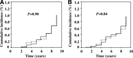
We explored the predictive effect of urinary type IV collagen on worsening of diabetic nephropathy. During the follow-up period (median 8 years; range 3–9 years), 16 of the 185 diabetic subjects with normoalbuminuria and 9 of the 69 with microalbuminuria progressed to more advanced stages of diabetic nephropathy. Among subjects with normoalbuminuria at baseline, urinary type IV collagen levels were not different between those who progressed to microalbuminuria (6.04 μg/g Cr [IQR 4.73–7.67 μg/g Cr]) and those who did not (5.62 μg/g Cr [3.06–8.07 μg/g Cr], P = 0.46, by Mann-Whitney U test). In contrast, the median urinary AER was significantly higher in those who progressed (10.6 μg/min [8.0–13.0 μg/min]) than in those who did not (7.1 μg/min [5.1–9.8 μg/min], P = 0.002). Among the subjects with microalbuminuria, the urinary type IV collagen level was not different between those who progressed to overt proteinuria (5.52 μg/g Cr [4.33–8.25 μg/g Cr]) and those who did not (7.68 μg/g Cr [5.86–12.21 μg/g Cr], P = 0.11), whereas urinary AER tended to be different in these two subgroups (55.7 [35.8–131.3] vs. 42.9 μg/min [31.9–64.5 μg/min], respectively, P = 0.07). Furthermore, urinary type IV collagen above the median cutoff point (≥6.36 μg/g Cr) was neither predictive for future development of microalbuminuria in patients with normoalbuminuria nor for progression to overt proteinuria in those with microalbuminuria, as examined with the Kaplan-Meier method (log-rank test: P = 0.90 for normoalbuminuria, P = 0.84 for microalbuminuria) (Fig. 1).
Figure 1.
Kaplan-Meier curves for development of microalbuminuria in patients with normoalbuminuria (A) and progression of overt proteinuria in those with microalbuminuria (B) grouped according to the median cutoff value of urinary type IV collagen level. The difference between two subgroups was compared by a log-rank test. ——, patients above the value (6.36 μg/g Cr; n = 127); – – –, patients below the value (<6.36 μg/g Cr; n = 127).
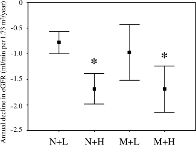
Next, we analyzed the relationship between urinary type IV collagen and deterioration of renal function. Univariate regression analysis showed that the level of urinary type IV collagen inversely correlated with the annual decline in eGFR (γ = −0.34, P < 0.001) (Fig. 2), whereas urinary AER correlated weakly with this decline (γ = −0.14, P = 0.03). Multivariate regression analysis with a stepwise forward method identified log urinary type IV collagen (β = −0.28, P < 0.001), eGFR at baseline (β = −0.18, P = 0.002), and hypertension (β = −0.13, P = 0.03) as factors associated with the annual decline in eGFR. When the patients were divided according to the median cutoff level of urinary type IV collagen, the mean value of eGFR at the end of study was significantly lower in the patients above the median cutoff level (≥6.36 μg/g Cr) than in those below the median cutoff level (69.6 ± 16.3 vs. 73.8 ± 15.8 ml/min per 1.73 m2; P = 0.039), whereas the mean value of eGFR at baseline was not different in the two subgroups (83.2 ± 15.4 vs. 81.9 ± 13.8 ml/min per 1.73 m2; P = 0.50). Interestingly, those above the median cutoff level showed a rapid decline of eGFR in the follow-up compared with those who were below (−1.69 ± 1.40 vs. −0.82 ± 1.15 ml/min per 1.73 m2/year; P < 0.001). Finally, the annual decline in eGFR was compared in four subgroups of patients according to the stage of diabetic nephropathy and the median cutoff level of urinary type IV collagen (Fig. 3). In patients with normoalbuminuria, the annual decline in eGFR was significantly larger in those with urinary type IV collagen level above the median cutoff level than in those with who were below the median value. Similarly, the annual rate in eGFR of those with microalbuminuria and above the median cutoff value was larger than that in those with normoalbuminuria below the median value, although the annual decline in those with microalbuminuria and below the median value was not different.
Figure 2.
Correlation between urinary type IV collagen and annual decline in eGFR (Spearman coefficient γ = −0.34, P < 0.001). The log-transformed values of urinary type IV collagen were used in this plot because the raw values showed a skewed distribution.
Figure 3.
Annual decline in eGFR. Subjects were categorized as being above or below the median cutoff of urinary type IV collagen level (6.36 μg/g Cr) and urinary AER (20 μg/min). N+L, patients with normoalbuminuria and less than the median cutoff value of urinary type IV collagen (n = 103, −0.78 ± 0.11 ml/min per 1.73 m2/year); N+H, patients with normoalbuminuria and above the median cutoff value (n = 82, −1.68 ± 0.15 ml/min per 1.73 m2/year); M+L, patients with microalbuminuria and less than the median cutoff value (n = 24, −0.97 ± 0.27 ml/min per 1.73 m2/year); M+H, patients with microalbuminuria and above the median cutoff value (n = 45, −1.69 ± 0.22 ml/min per 1.73 m2/year). Data are mean ± SEM. *P < 0.01 vs. patients with normoalbuminuria and below the median value (ANOVA with Scheffé test).
CONCLUSIONS
The present study showed that the level of urinary type IV collagen was higher in patients with microalbuminuria than in those with normoalbuminuria and correlated with the level of urinary β2-microglobulin, diastolic blood pressure, eGFR, and AER at baseline. In addition, the level of urinary type IV collagen at baseline inversely correlated with the annual decline in eGFR during the follow-up period (median 8 years), whereas these levels were not associated with worsening of the stage of diabetic nephropathy. Furthermore, the large annual decline in eGFR of patients with increased urinary type IV collagen excretion was observed regardless of the stage of diabetic nephropathy.
In this study, the median level of urinary type IV collagen was higher in diabetic patients with microalbuminuria than in those with normoalbuminuria, in agreement with previous studies (9–14). Several cross-sectional studies reported that a substantial number of diabetic patients with normoalbuminuria had high levels of urinary type IV collagen (10,14). Based on these results, it was proposed that urinary type IV collagen could increase before any increase in albuminuria in diabetic patients and could be a useful marker for the identification of early stages of diabetic nephropathy. However, our follow-up study did not show any predictive role of urinary type IV collagen in the development and progression of diabetic nephropathy, although urinary AER is a highly variable quantity. Our result suggests that the increase in urinary type IV collagen in the diabetic patients without overt proteinuria could be considered to reflect other renal damage processes not directly related with the degree of albuminuria.
A recent study identified patients whose renal function deteriorated rapidly without any increase in urinary albumin excretion (17,18). In this study, the level of urinary type IV collagen at baseline correlated with the decline in eGFR during the follow-up. This predictive power of urinary type IV collagen was observed even in patients with normoalbuminuria as well as in those with microalbuminuria. Interestingly, multivariate regression analysis in this study showed that urinary levels of type IV collagen correlated with the levels of urinary β2-microglobulin, in agreement with the finding of a previous study (12). The increase in urinary β2-microglobulin is considered to be associated with impaired renal tubular cell function and a marker of renal tubulointerstitial injury. Okonogi et al. (19) reported that urinary IV collagen excretion in type 2 diabetic patients was associated with histopathological changes in glomeruli and tubulointerstitium. Although diabetic nephropathy was traditionally considered to cause primarily glomerular damage, it is now widely accepted that deterioration of renal function in diabetic patients correlates with the degree of tubulointerstitial fibrosis (20). Taken together with these findings, increased levels of urinary type IV collagen could reflect the degree of renal impairment associated with renal glomerular and tubulointerstitial injuries. Thus, measurement of urinary type IV collagen may be useful for the identification of diabetic patients whose renal function could decline rapidly without worsening of albuminuria.
Urinary type IV collagen levels did not correlate with A1C levels in our population. This finding is in agreement with those reported by two groups in Japanese type 2 diabetic patients (11,19). Cohen et al. (13) also reported no such correlation in type 1 and 2 diabetic patients. These results suggest that other factors apart from hyperglycemia contribute to the increase in urinary type IV collagen. In this study, systolic and diastolic blood pressure correlated weakly with urinary type IV collagen; the latter was significantly higher in patients with hypertension than in those without hypertension. Similarly, Tomino et al. (14) reported significant correlations between urinary type IV collagen and systolic and diastolic blood pressure, but not with fasting glucose levels, in an Asian multicenter study. Kotajima et al. (11) reported that urinary type IV collagen was increased in hypertensive type 2 diabetic patients at the 6-month follow-up compared with baseline, but did not increase in those without hypertension. Thus, these results suggest that urinary type IV collagen reflects the degree of hypertension-related renal injury, in addition to the abnormal metabolic conditions in diabetic patients.
Our study has some limitations. Urinary AER is highly variable and is not a relatively reproducible quantity like eGFR. In this study, we did not enroll the patients whose stages of nephropathy in two measurements of urinary AER at baseline were not identical. These patients may rapidly progress to more advanced stages, and, thus, their inclusion might underestimate the predictive power of urinary type IV collagen on development and progression of diabetic nephropathy. In this study we did not evaluate the time-dependent changes in urinary type IV collagen levels during the follow-up period. The levels of urinary type IV collagen could be modified by a variety of medications (21). Recently, we reported frequent remission of microalbuminuria in type 2 diabetic patients, which was associated with improvement of cardiorenal outcome (22). Also, the rapid annual decline in eGFR in those with higher levels of urinary type IV collagen may be due in part to regression to the mean. Further studies are required to determine whether the urinary type IV collagen level is a suitable therapeutic index of improving renal outcomes. At this stage, the source of increased urinary type IV collagen in diabetic patients is unknown. Urinary type IV collagen measured in this study is considered to be the intact form with a molecular weight of ∼540 kDa, which is detected as a single peak in gel filtration analysis (23). Thus, it is unlikely that urinary type IV collagen was filtered through the glomeruli from blood in the early stages of diabetic nephropathy. Upregulation of type IV collagen in podocytes (6), proximal tubular cells (7,8), and/or infiltrating fibroblasts in the tubulointerstitium (24) observed in diabetes may be responsible for the increased urinary excretion. Furthermore, reduced degradation of type IV collagen in diabetes may be responsible for the increased excretion of urinary type IV collagen (25).
In summary, the present study demonstrated that increased urinary excretion of type IV collagen is a predictor of deterioration of renal function rather than progression of nephropathy in type 2 diabetic patients without overt proteinuria and that this relationship may be associated with the degree of glomerular and tubulointerstitial impairment. Thus, measurement of urinary type IV collagen may be useful for the identification of diabetic patients without overt proteinuria who have a higher risk for rapid deterioration of renal function. Further intervention study is required to clarify whether a reduction in urinary type IV collagen is therapeutically useful in diabetic patients.
Acknowledgments
No potential conflicts of interest relevant to this article were reported.
S.A. researched data, contributed to discussion, wrote the manuscript, and review/edited the manuscript. M.H., D.K., and A.K. contributed to discussion and reviewed/edited the manuscript. K.I., S.K., T.S., H.K., and Y.N. researched data and reviewed/edited the manuscript. T.U. and H.M. researched data, contributed to discussion, and reviewed/edited the manuscript.
Parts of this study were presented in abstract form at the 70th Scientific Sessions of the American Diabetes Association, Orlando, Florida, 25–29 June 2010.
We acknowledge Shigehisa Matuki and Hidekuni Shima, from Daiichi Fine Chemical Co., Ltd., for the measurement of urinary type IV collagen levels and Mayumi Yamanaka for data management.
Footnotes
The costs of publication of this article were defrayed in part by the payment of page charges. This article must therefore be hereby marked “advertisement” in accordance with 18 U.S.C. Section 1734 solely to indicate this fact.
References
- 1. Najafian B, Mauer M: Progression of diabetic nephropathy in type 1 diabetic patients. Diabetes Res Clin Pract 2009;83:1–8 [DOI] [PubMed] [Google Scholar]
- 2. Ziyadeh FN: Significance of tubulointerstitial changes in diabetic renal disease. Kidney Int Suppl 1996;54:S10–S13 [PubMed] [Google Scholar]
- 3. Suzuki D, Miyazaki M, Jinde K, Koji T, Yagame M, Endoh M, Nomoto Y, Sakai H: In situ hybridization studies of matrix metalloproteinase-3, tissue inhibitor of metalloproteinase-1 and type IV collagen in diabetic nephropathy. Kidney Int 1997;52:111–119 [DOI] [PubMed] [Google Scholar]
- 4. Ayo SH, Radnik RA, Garoni JA, Glass WF, 2nd, Kreisberg JI: High glucose causes an increase in extracellular matrix proteins in cultured mesangial cells. Am J Pathol 1990;136:1339–1348 [PMC free article] [PubMed] [Google Scholar]
- 5. Haneda M, Kikkawa R, Horide N, Togawa M, Koya D, Kajiwara N, Ooshima A, Shigeta Y: Glucose enhances type IV collagen production in cultured rat glomerular mesangial cells. Diabetologia 1991;34:198–200 [DOI] [PubMed] [Google Scholar]
- 6. Iglesias-de la Cruz MC, Ziyadeh FN, Isono M, Kouahou M, Han DC, Kalluri R, Mundel P, Chen S: Effects of high glucose and TGF-β1 on the expression of collagen IV and vascular endothelial growth factor in mouse podocytes. Kidney Int 2002;62:901–913 [DOI] [PubMed] [Google Scholar]
- 7. Razzaque MS, Koji T, Horita Y, Nishihara M, Harada T, Nakane PK, Taguchi T: Synthesis of type III collagen and type IV collagen by tubular epithelial cells in diabetic nephropathy. Pathol Res Pract 1995;191:1099–1104 [DOI] [PubMed] [Google Scholar]
- 8. Ziyadeh FN, Simmons DA, Snipes ER, Goldfarb S: Effect of myo-inositol on cell proliferation and collagen transcription and secretion in proximal tubule cells cultured in elevated glucose. J Am Soc Nephrol 1991;1:1220–1229 [DOI] [PubMed] [Google Scholar]
- 9. Kado S, Aoki A, Wada S, Katayama Y, Kugai N, Yoshizawa N, Nagata N: Urinary type IV collagen as a marker for early diabetic nephropathy. Diabetes Res Clin Pract 1996;31:103–108 [DOI] [PubMed] [Google Scholar]
- 10. Yagame M, Suzuki D, Jinde K, Saotome N, Sato H, Noguchi M, Sakai H, Kuramoto T, Sekizuka K, Iijima T, Suzuki S, Tomino Y: Significance of urinary type IV collagen in patients with diabetic nephropathy using a highly sensitive one-step sandwich enzyme immunoassay. J Clin Lab Anal 1997;11:110–116 [DOI] [PMC free article] [PubMed] [Google Scholar]
- 11. Kotajima N, Kimura T, Kanda T, Obata K, Kuwabara A, Fukumura Y, Kobayashi I: Type IV collagen as an early marker for diabetic nephropathy in non-insulin-dependent diabetes mellitus. J Diabetes Complications 2000;14:13–17 [DOI] [PubMed] [Google Scholar]
- 12. Watanabe H, Sanada H, Shigetomi S, Katoh T, Watanabe T: Urinary excretion of type IV collagen as a specific indicator of the progression of diabetic nephropathy. Nephron 2000;86:27–35 [DOI] [PubMed] [Google Scholar]
- 13. Cohen MP, Lautenslager GT, Shearman CW: Increased collagen IV excretion in diabetes. A marker of compromised filtration function. Diabetes Care 2001;24:914–918 [DOI] [PubMed] [Google Scholar]
- 14. Tomino Y, Suzuki S, Azushima C, Shou I, Iijima T, Yagame M, Wang LN, Chen HC, Lai KN, Tan SY, Kim MJ: Asian multicenter trials on urinary type IV collagen in patients with diabetic nephropathy. J Clin Lab Anal 2001;15:188–192 [DOI] [PMC free article] [PubMed] [Google Scholar]
- 15. Obata K, Iwata K, Ichida T, Inoue K, Matsumoto E, Muragaki Y, Ooshima A: One step sandwich enzyme immunoassay for human type IV collagen using monoclonal antibodies. Clin Chim Acta 1989;181:293–303 [DOI] [PubMed] [Google Scholar]
- 16. Matsuo S, Imai E, Horio M, Yasuda Y, Tomita K, Nitta K, Yamagata K, Tomino Y, Yokoyama H, Hishida A: Collaborators developing the Japanese equation for estimated GFR. Revised equations for estimated GFR from serum creatinine in Japan. Am J Kidney Dis 2009;53:982–992 [DOI] [PubMed] [Google Scholar]
- 17. Perkins BA, Ficociello LH, Ostrander BE, Silva KH, Weinberg J, Warram JH, Krolewski AS: Microalbuminuria and the risk for early progressive renal function decline in type 1 diabetes. J Am Soc Nephrol 2007;18:1353–1361 [DOI] [PubMed] [Google Scholar]
- 18. Yokoyama H, Kanno S, Takahashi S, Yamada D, Itoh H, Saito K, Sone H, Haneda M: Determinants of decline in glomerular filtration rate in nonproteinuric subjects with or without diabetes and hypertension. Clin J Am Soc Nephrol 2009;4:1432–1440 [DOI] [PMC free article] [PubMed] [Google Scholar]
- 19. Okonogi H, Nishimura M, Utsunomiya Y, Hamaguchi K, Tsuchida H, Miura Y, Suzuki S, Kawamura T, Hosoya T, Yamada K: Urinary type IV collagen excretion reflects renal morphological alterations and type IV collagen expression in patients with type 2 diabetes mellitus. Clin Nephrol 2001;55:357–364 [PubMed] [Google Scholar]
- 20. Taft JL, Nolan CJ, Yeung SP, Hewitson TD, Martin FI: Clinical and histological correlations of decline in renal function in diabetic patients with proteinuria. Diabetes 1994;43:1046–1051 [DOI] [PubMed] [Google Scholar]
- 21. Sato A, Tabata M, Hayashi K, Saruta T: Effects of the angiotensin II type 1 receptor antagonist candesartan, compared with angiotensin-converting enzyme inhibitors, on the urinary excretion of albumin and type IV collagen in patients with diabetic nephropathy. Clin Exp Nephrol 2003;7:215–220 [DOI] [PubMed] [Google Scholar]
- 22. Araki S, Haneda M, Koya D, Hidaka H, Sugimoto T, Isono M, Isshiki K, Chin-Kanasaki M, Uzu T, Kashiwagi A: Reduction in microalbuminuria as an integrated indicator for renal and cardiovascular risk reduction in patients with type 2 diabetes. Diabetes 2007;56:1727–1730 [DOI] [PubMed] [Google Scholar]
- 23. Takizawa H, Satoh T, Kurusu A, Hishiki T, Shike T, Gohda T, Maeda A, Makita Y, Suzuki S, Fukui M, Tomino Y: Increase of urinary type IV collagen in normoalbuminuric patients with impaired glucose tolerance. Nephron 1998;79:474–475 [DOI] [PubMed] [Google Scholar]
- 24. Lam S, van der Geest RN, Verhagen NA, Daha MR, van Kooten C: Secretion of collagen type IV by human renal fibroblasts is increased by high glucose via a TGF-β-independent pathway. Nephrol Dial Transplant 2004;19:1694–1701 [DOI] [PubMed] [Google Scholar]
- 25. Phillips AO, Steadman R, Morrisey K, Martin J, Eynstone L, Williams JD: Exposure of human renal proximal tubular cells to glucose leads to accumulation of type IV collagen and fibronectin by decreased degradation. Kidney Int 1997;52:973–984 [DOI] [PubMed] [Google Scholar]


