Abstract
Silymarin, an extract from milk thistle (Silybum marianum), and its purified flavonolignans have been recently shown to inhibit HCV infection, both in vitro and in vivo. In the current study, we further characterized silymarin's antiviral actions. Silymarin had antiviral effects against HCVcc infection that included inhibition of virus entry, RNA and protein expression, and infectious virus production. Silymarin did not block HCVcc binding to cells, but inhibited the entry of several viral pseudoparticles (pp), and fusion of HCVpp with liposomes. Silymarin but not silibinin inhibited JFH-1 genotype 2a NS5B-dependent RNA polymerase activity at concentrations 5–10 times higher than required for anti-HCVcc effects. Furthermore, silymarin had inefficient activity on the genotype 1b BK and four 1b RDRPs derived from HCV-infected patients. Moreover, silymarin did not inhibit HCV replication in 5 independent genotype 1a, 1b, and 2a replicon cell lines that did not produce infectious virus. Silymarin inhibited microsomal triglyceride transfer protein activity, apolipoprotein B secretion, and infectious virion production into culture supernatants. Silymarin also blocked cell-to-cell spread of virus. While inhibition of in-vitro NS5B polymerase activity is demonstrable, the mechanisms of silymarin's antiviral action appear to include blocking of virus entry and transmission, possibly by targeting the host cell.
Keywords: milk thistle, JFH-1, HCVcc, virology, liver disease
Chronic hepatitis C is a serious global medical problem necessitating novel, effective, inexpensive, and less toxic treatments. Hepatitis C virus (HCV), infects an estimated 130 million people throughout the world, leading to a half million deaths per year due to liver disease (1).
Pegylated interferon (IFN) plus ribavirin therapy is the current treatment for the patient with chronic hepatitis C (2). However, 50% of treated patients do not clear viremia during treatment, which is costly and has significant side effects. As a result, many patients use natural products to supplement or circumvent IFN-based regimens, with silymarin being the most common botanical medicine (3).
Silymarin is an extract from the of Silybum marianum, that consists of at least seven flavonolignans and the flavonoid taxifolin (4). Silibinin is a partially purified mixture of two of flavonolignans, silybin A and silybin B. Silymarin has been used to treat a range of liver disorders including hepatitis, cirrhosis, and poisoning from wild mushrooms (5). Recently, we showed silymarin inhibits HCV infection of Huh7 and Huh7.5.1 cells (6), and Ferenci's group showed that intravenous silibinin administration reduces viral loads in previous non-responders to IFN therapy (7). Therefore, in the current study, we determined the stages in the HCV lifecycle that are blocked by silymarin.
Experimental Procedures
Reagents
Human hepatoma Huh7 cells were grown in Huh7 medium as described (6). HepG2 cells and Huh7.5.1 cells (8) were cultured in Huh7 medium. BB7 and FL-NEO cells are Huh7 cell lines that contain subgenomic and genomic length genotype 1b replicons(9). JFH-1 subgenomic genotype 2a replicon cell lines in Huh7 or Huh7.5 backgrounds were generated by transfecting in-vitro transcribed SGR-JFH1 replicon RNA into Huh7 or Huh7.5 and selecting with 800 μg/mL G418. Single colonies expressing the highest level of HCV protein were designated SGR7 and SGR7.5. Luc-ubi-neo/ET is a subgenomic genotype 1b derived replicon cell line (10). The HCV genotype 1a H77s subgenomic replicon cell line has been described previously (11). All replicon lines were maintained in Huh7 medium containing 400 μg/ml G418. Primary human hepatocytes were kindly provided by Dr. Stephen Strom, University of Pittsburgh and maintained in hepatocyte culture medium (Lonza, Walkersville, MD).
JFH-1 viral stock preparation, cell infection and titration was performed as described (6).
Silymarin from US Pharmacopeia (Rockville, MD) was used in all experiments except for the MTP (figure 4A) and apoB (Figure 4E) assays, where silymarin from Sigma. Sigma silymarin contains similar levels of the major flavonolignans as USP silymarin, as described in (12). Stocks were prepared in DMSO at a concentration of 50 mg/ml. Silibinin was purified from silymarin as described (13).
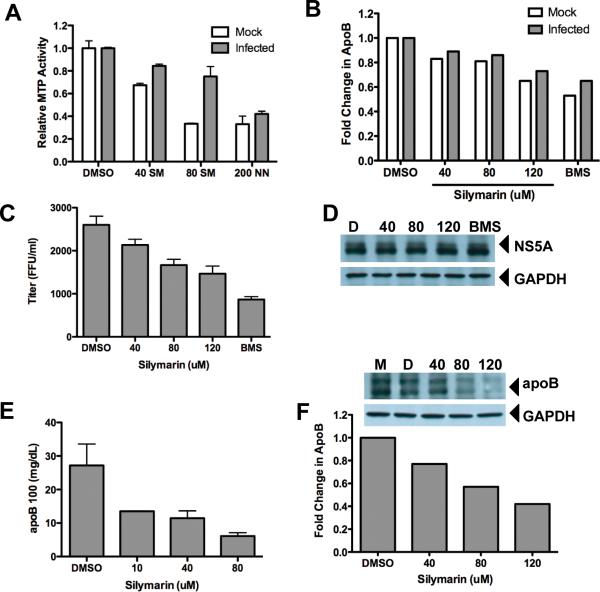
Figure 4. Silymarin inhibits microsomal triglyceride transfer protein (MTP) activity, apolipoprotein B (apoB) secretion, and infectious virus production.
A, Silymarin inhibits MTP activity in Huh7.5.1 cells. Chronically infected Huh7.5.1 cells (14 days post-infection) and uninfected cells were treated with the indicated μM doses of silymarin (SM) or as a positive control, 200 μg/ml naringenin (NN), and 24 hours later MTP activity was measured as described in the Materials and Methods. B, Silymarin inhibits apoB secretion from mock and HCV-infected cells. Huh7.5.1 cells were infected or mock-infected with JFH-1 at an m.o.i. of 0.01 for 96 hours before treatment with fresh medium containing DMSO, 10μM BMS-200150 (a small molecule inhibitor of MTP), or silymarin at the indicated μM doses for 5 hours. Culture supernatants were harvested, and apoB measured by ELISA. BMS=BMS-200150. C, De novo infectious virion production into culture supernatants is blocked by silymarin. Supernatants from panel B were diluted 1:20 and infectious titers were determined by focus forming unit assay on naïve Huh7.5.1 cells. BMS=BMS-200150. D, Treatment of infected cells for 5 hours does not inhibit intracellular HCV replication. Protein lysates were harvested from cultures described in panel B and equal amounts of total protein were blotted for NS5A. The positions of HCV NS5A and loading control GAPDH are indicated. D is the DMSO control, 40, 80, and 120 are the doses of silymarin in μM, and BMS is the MTP inhibitor BMS-200150. E, Silymarin blocks apoB secretion from primary human hepatocytes. Cells were treated with the indicated concentrations of SM for 24 hours before supernatants were harvested and apoB measured by ELISA. F, Silymarin blocks apoB secretion in HepG2 cells. Cells were treated with the indicated μM concentrations of silymarin for 5 hours before supernatants were harvested and apoB measured by ELISA. Inset, Silymarin blocks intracellular apoB levels. Cell lysates from HepG2 cells treated in panel F were probed for apoB by western blot. apoB100 and loading control GAPDH are indicated with arrows. M is the media control, D is the DMSO control, and 40, 80, and 120 are the doses of silymarin in μM.
Egg yolk phosphatidylcholine (PC), cholesterol and Triton X-100 were purchased from Sigma. Octadecyl rhodamine B chloride (R18) was from Fluoprobes.
Addition of compounds to cultures
Silymarin was further diluted in DMSO prior to use and the final concentration of DMSO in culture media was always between 0.2–0.4%. Silymarin treatment involved exposing cells to a single administration of silymarin for various times. DMSO was included as a solvent control. To focus on the effects of silymarin on infectious virus production, cells were first infected at a m.o.i. of 0.01 and cells were passaged at 72 hours post-infection, followed by addition of silymarin 24 hours after passaging, or 96 hours post-infection. Under these conditions, the culture was fully infected (Supplemental Figure S1).
Roferon, Intron-A, or Pegasys was used as positive controls for antiviral effects. Roferon (Roche, Pal Alto, CA) was used at 10 IU/ml, Pegasys was used at 10 ng/ml, and Intron-A was used at various concentrations. BMS-200150, a small molecule inhibitor of MTP, was provided by Pablo Gastaminza and Francis Chisari.
Western Blot Analysis
Western blots were performed as described (6). GAPDH, apoB, Stat1, NS5A in FL-NEO and BB7 cells, and core proteins were detected using commercial antiserum (Santa Cruz Biotechnology, Santa Cruz, CA; BioDesign/Meridian Life Science, Saco, ME; Affinity BioReagents, Golden, CO). NS5A in JFH-1 infected cells was detected with serum from patients infected with HCV genotype 2a.
HCV RNA Quantitation
HCV RNA was quantitated by real time RT-PCR, as previously described (14).
HCV luciferase replicon assay
Luc-ubi-neo cells were plated and grown for 24 hours. Medium was removed, the cells washed once and treated subsequently with given concentrations of silymarin or DMSO in triplicate. After 3 days of incubation at 37°C, cell culture medium was removed and cells were lysed by freeze-thawing in buffer and luciferase assays were performed.
HCV-1a Replicon Assay
Antiviral studies to determine the impact of silymarin treatment on HCV RNA levels with the genotype 1a replicon were conducted using real time RT-PCR and assay conditions previously described (15).
HCV NS5B Polymerase Assays
NS5BΔC21 C-terminally fused to a hexa-histidine tag was expressed and purified for HCV JFH1 and for the genotype 1b isolates as described (16, 17). All reaction components except nucleotides and template were preincubated for 15 minutes at room temperature, then the reaction was started by adding the nucleotide mixture and template and was incubated for 1.5 hours at room temperature. Reaction products were precipitated, passed through filters, washed five times, and air dried. Samples were subjected to liquid scintillation counting. All measurements were done in triplicate and the IC50 values were calculated with GraphPad Prism.
Viral Pseudoparticle Entry Assays
Pseudoviruses were generated as previously described (18). Huh-7.5 cells were pretreated for 1hr with 40, 80 and 160 μM silymarin (SM) or equivalent volume of DMSO, diluted in media. Cells were inoculated with an equal volume of pseudoparticles bringing the final concentration of SM to 20, 40 and 80 μM. 72 h post-infection, the medium was removed and cells lysed with cell lysis buffer (Promega, Madison, WI). Luciferase activity was then measured. Specific infectivity was calculated by subtracting the mean Env-pp signal from the HCVpp, MLVpp or VSVpp signals. Relative infectivity was then calculated as a percentage of untreated control cell infection, i.e. the mean luciferase value of the replicate untreated cells was defined as 100%.
Fusion assay
Fusion between HCVpp and liposomes was assayed as already described (19). Briefly, liposomes composed of PC, cholesterol and Octadecyl Rhodamine B chloride (R18) (65:30:5 mol%) were added at a 15 μM final concentration to a cuvette at 37°C containing HCVpp in PBS at pH 7.2. After equilibration, diluted HCl was added to pH 5.0 final and lipid mixing measured as the dequenching of R18 (excitation 560 nm, emission 590 nm), resulting in an increase in the fluorescence signal. Silymarin was preincubated with HCVpp and liposomes for 3 min at 37°C, and lipid mixing measured after medium acidification.
Infectious Virion Production
Culture supernatants were harvested at defined times points post-infection, and supernatants were clarified by centrifugation. Intracellular virus titers were determined after treatment with Brefeldin A (BFA), which has been shown to block HCV release by causing intracellular accumulation of virions. For this, treated cells were scraped into PBS and lysed by freeze-thawing as described (20). All supernatants were stored at −80C before dilution and titering on naïve Huh7.5.1 cells using standard focus-forming unit (FFU) assays as previously described (8).
Cell to Cell Spread
Cell to cell spread of virus was measured as previously described (21). Briefly, unlabelled naive `target' cells were incubated with HCV H77/JFH infected `producer' cells that were labeled with CMFDA (Molecular Probes, Invitrogen, Carlsbad, CA). A neutralizing antibody was added to the co-culture to abrogate the infectivity and transmission of cell-free virus particles within the culture media, allowing us to quantify antibody insensitive viral transmission. Infection was quantified by staining the co-cultures with an anti-NS5A antibody (9E10) and appropriate secondary antibody, followed by flow cytometry.
apoB ELISA
Huh7.5.1 cells were grown overnight in Huh7 media. The media was removed and cells were washed, and fresh medium with the appropriate compounds was added to cells. Culture supernatants were harvested at 24 and 48 hours later, clarified by centrifugation and stored at −80°C. apoB was measured by ELISA using manufacturer's protocol (ALerCHEK, Portland, ME).
MTP Assay
MTP activity was measured using a commercially available fluorescence assay using a commercial kit (Roar Biomedical, Inc., New York, NY) as described (22)
Statistics
Differences between means of readings were compared using a Student's T-test. A p-value of <0.05 was considered significant.
Results
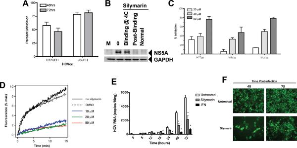
We previously showed that silymarin inhibits HCV RNA and protein expression in the HCVcc system with JFH-1 virus (6). Figure 1A demonstrates that in addition to wild type JFH-1 virus, silymarin also blocks replication of HCVcc chimeras, including constructs that contain the H77 (genotype 1a) and J6 (genotype 2a) structural genes in the JFH-1 non-structural gene backbone. Inhibition of HCVcc was 50% for H77/JFH and 75% for J6/JFH chimeras. Thus, silymarin has antiviral actions against multiple HCVcc infectious systems. To determine if silymarin could inhibit binding of HCV virions to cells, we performed virus-cell binding studies at 4°C under conditions where virus binds to but does not enter cells (23). As shown in Figure 1B, when silymarin was present only during virus-binding, there was little effect on HCV replication. However, if silymarin was added to cells immediately after binding and for the duration of the infection, HCV protein expression was severely impaired. The same effect was observed if silymarin was present during binding and for the duration of the experiment. Next, to determine if silymarin blocked virus entry, we tested the effect of silymarin on viral pseudoparticle entry including HCVpp, VSVpp, and MLVpp. Figure 1C demonstrates that silymarin inhibited the entry of all three pseudotyped viruses. We then examined the effect of silymarin on the fusion of HCVpp with fluorescent liposomes, which examines the effects of compounds on lipid mixing and membrane fusion (24). As shown in Figure 1D, silymarin drastically inhibited HCVpp-mediated fusion by 80% at 10 μM silymarin, while 20 μM led to a 90% reduction in fusion. DMSO, the solvent control, did not affect fusion. The IC50 of silymarin for membrane fusion inhibition was estimated at 5 μM, far below the doses of silymarin known to confer cytotoxicity in Huh7.5.1 cells (>80 μM, Supplemental Figure S2, Panel E). The data suggest that silymarin does not affect binding, but inhibits the entry of HCV at the fusion stage.
Figure 1. Antiviral effects of silymarin against HCVcc.
A, Silymarin inhibits virus infection in multiple HCVcc systems. Huh-7.5 cells were incubated with 80μM silymarin or DMSO for 1hr at 37C before inoculation with either H77/JFH or J6/JFH HCVcc viruses. The infection was allowed to proceed in the presence of the compounds for 48 and 72hrs before virus was quantitated by staining for NS5A-positive foci. Percent inhibition reflects inhibition of NS5A-positive foci by silymarin relative to the DMSO control. B, Silymarin does not block virus binding. Huh7.5.1 cells were incubated at 4°C for 5 hours in the presence of HCVcc (JFH-1, m.o.i. of 0.01) with or without 40μM silymarin. Cells were then washed extensively at 4°C and then cells were incubated for 72 hours at 37°C to allow the HCV lifecycle to continue in the presence or absence of additional silymarin. “M” denotes mock infected cells. “0” denotes cells that were infected but not treated with anything. “Binding @ 4C” denotes silymarin was only present during the 5-hour adsorption period. “Post-binding” denotes silymarin was added after the 5-hour adsorption period and for the duration of the infection. “Normal” denotes silymarin was present during the 5-hour adsorption and for the duration of the infection. C, Silymarin inhibits pseudoparticle entry. Huh-7.5 cells were treated with silymarin (SM) or equivalent volume of DMSO for 1 hour prior to infection with HCV, VSV, or MLV pseudoparticles (pp). 72 h post-infection, the medium was removed and luciferase activity was measured on cell lysates. D, Silymarin blocks HCVpp-mediated lipid mixing. HCVpp in PBS at pH 7.2 were incubated or not with indicated concentrations of silymarin in DMSO, for 3 min at 37°C, in the presence of PC:cholesterol:R18 liposomes. Acidification to pH 5.0 was performed by adding diluted HCl to the cuvette, and R18 dequenching was assayed for 15 min at excitation and emission wavelengths of 560 and 590 nm, respectively. Maximal dequenching was obtained after addition of 0.1% final Triton X-100 to the cuvette. Black, no silymarin; blue, 10 μM; green, 20 μM and red, 80 μM silymarin, respectively. Dotted curve, lipid mixing in the presence of 1% final DMSO (highest concentration used in the assay). E, Silymarin inhibits HCV RNA production. Huh7.5.1 cells were infected at an m.o.i. of 0.01 with JFH-1 and 24 hours later, silymarin (40μM) or IFN (10 units/ml) was added to cells, and thereafter, total RNA was isolated from cells at the indicated time points. HCV RNA was quantitated by real time RT-PCR. Asterisks indicate silymarin or IFN reduction of viral loads is significantly different than untreated cells (p<0.01). F, Silymarin reduces infectious virus production into culture supernatants. Huh7.5.1 cells were infected at an m.o.i. of 0.01 with JFH-1 in the presence of 40μM silymarin or DMSO and supernatants were harvested 48 and 72 hours post-infection and titered by FFU assay on naïve Huh7.5.1 cells.
Next, we examined the kinetics of inhibition of HCV RNA production. In this experiment we first infected cells for 24 hours, followed by silymarin administration, or IFN-α as a positive control. As shown in Figure 1E, relative to untreated cells, silymarin caused a significant (p<0.01) reduction in JFH-1 RNA production at 48 and 72 hours after treatment. IFN treatment also reduced viral loads. However, significant suppression (p<0.01) of HCV RNA production by IFN started at 18 hours post-treatment and was maintained until 72 hours of treatment. Thus, the kinetics of silymarin mediated suppression of HCV RNA replication were delayed as compared to IFN.
As shown in Figure 1F, silymarin reduced infectious virus yields (measured as FFU/ml) by 5- and 2-fold at 48 and 72 hours post-infection from Huh7.5.1 cells (and in Huh7 cells; data not shown). We can rule out the possibility of carry over silymarin from the initial culture because the supernatants were diluted 1:5–1:1000 before testing on naïve cells. Altogether, the data show that silymarin does not affect virus binding to cells, but inhibits virus entry and fusion, HCV protein and RNA synthesis, and production of progeny viruses in culture supernatants.
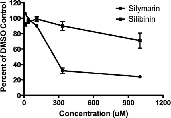
Inhibition of HCV RNA and protein expression by silymarin could be due to direct inhibition of viral enzymes, as recently shown for NS5B polymerase activity (25). Therefore, we tested whether silymarin and silibinin block HCV NS5B polymerase activity. Recombinant NS5B protein from JFH-1 (genotype 2a) lacking the C-terminal 21 amino acids was expressed in E. coli and purified (16). As shown in Figure 2, silymarin was able to inhibit JFH-1 NS5B polymerase activity, with an IC50 for silymarin around 300 μM. Silibinin had minimal effects on JFH-1 polymerase, but only at very high doses (IC50 >400μM), which were at least 5–10 fold higher than effective antiviral doses in vitro (6). At the doses required for inhibition of in-vitro NS5B polymerase activity, silymarin is toxic to cultured Huh7 (6) and Huh7.5.1 cells (Supplemental Figure S2).
Figure 2. Silymarin inhibits JFH-1 polymerase at high dose.
HCV NS5B polymerase from isolate JFH-1 (genotype 2a) was incubated with the indicated concentrations of Silymarin or Silibinin, respectively, in presence of [32P]GTP and polyC. Incorporation of radioactivity was quantified by TCA precipitation and liquid scintillation counting. Silymarin and to a lesser degree, silibinin, inhibited JFH-1 polymerase.
We next tested silymarin on RdRp activity of the genotype 1b BK strain and four patient-derived 1b RdRps from patients in the Virahep-C clinical study (26). The RNA polymerase activities of the patient-derived enzymes were variable (16–104% relative to the well-characterized BK enzyme; Table 1). Silymarin inhibited all five RdRps, with IC50 values ranging from 27.7 to 162 μM. However, in four of the five cases the inhibitory activity of silymarin rapidly plateaued, with maximal inhibition levels of 42.6 to 82.8% relative to the activity in the absence of silymarin (Supplemental Figure S3). The fifth enzyme (#242) had an inhibition profile that could not be fit to a single-phase exponential decay curve, but its maximal inhibition by silymarin was only 43% and its apparent IC50 was >1,000 μM. Therefore, the IC50 values for most of these subtype 1b RdRps were respectable below the plateau level, but they were poorly inhibited by silymarin at the concentrations employed in the cell culture experiments.
Table 1.
Inhibition Profiles of Genotype 1b RdRps by Silymarin. Polymerase assays were performed and analyzed as described in the Materials and Methods.
| Polymerase Function | POLYMERASE | ||||
|---|---|---|---|---|---|
| BK | #234 | #242 | #245 | #103 | |
| Relative RdRp Activity | 100 | 15.8 | 104 | 99 | 38.3 |
| IC50 (μM) | 27.7 | 45.4 | >1000 | 162 | 162 |
| Maximal Inhibition (%) | 48.5 | 42.6 | 43.0 | 53.3 | 82.8 |
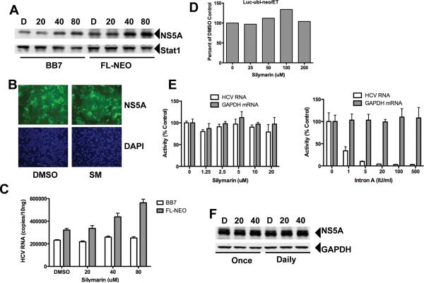
If silymarin truly inhibits NS5B polymerase activity, it should be able to inhibit HCV replication in replicon cell lines that do not produce infectious virus. Figure 3A–C depicts the effects of various doses of silymarin on HCV protein and RNA expression in genotype 1b BB7 subgenomic and FL-NEO genomic replicon cell lines. Silymarin did not significantly inhibit viral protein expression in either cell line when assessed by western blot (Figure 3A) or by immunofluorescence (Figure 3B). Silymarin did not inhibit HCV RNA expression in either cell line(Figure 3C). HCV replication was also not inhibited by silymarin in Luc-ubi-neo/ET cells, an independent genotype 1b replicon. (Figure 3D), or in a subgenomic genotype 1a replicon cell line (Figure 3E). In contrast, treatment with IFN-α caused robust suppression of HCV RNA production from the HCV-1a replicon. We tested concentrations of silymarin up to 1000 μM but failed to see any suppression of HCV RNA from the 1a replicon that was independent of cytotoxicity, measured as GAPDH mRNA levels (Supplemental Figure S4). NS5A protein expression was not affected by silymarin in JFH-1 derived genotype 2a SGR7 (Figure 3F) or SGR7.5 replicon cell lines (data not shown). Furthermore, extended treatment of FL-NEO replicon cells (or BB7 cells; data not shown) for 13 days did not affect the levels of HCV NS5A protein (Supplemental Figure S5). Therefore, silymarin had no antiviral activity against replicon cell lines that did not produce infectious virus. The data in Figures 2 and 3 suggest that silymarin inhibition of NS5B polymerase activity is not a significant component of silymarin's anti-HCV activity in the HCVcc system.
Figure 3. Silymarin does not block HCV replication in HCV replicon cell lines.
Cells were treated once with the indicated doses of silymarin and incubated for 72 hours before replication was assessed. A, protein expression in subgenomic BB7 and genomic FL-NEO HCV-1b replicons following treatment with 20–80 μM of silymarin. D=DMSO. Positions of HCV NS5A and cellular Stat1 proteins are indicated. B, Intracellular NS5A protein expression in DMSO and silymarin treated FL-NEO cells. Cells were treated as described above and NS5A protein detected by immunofluorescence as described in the Materials and Methods. NS5A positive cells are depicted in green while the blue cells represent nuclei counterstained by DAPI. C, HCV RNA levels in FL-NEO and BB7 cells. D, Effect of silymarin against Luc-ubi-neo/ET cells, a Con1-based genotype 1b replicon subgenomic replicon. Values represent percent change in luciferase light units relative to DMSO control. E, Effect on HCV-1a subgenomic replicon. Left panel shows HCV and GAPDH RNA levels following Silymarin treatment, while right panel shows RNA levels following treatment with Intron-A (recombinant IFN-α). F, Effect of silymarin on subgenomic genotype 2a JFH-1-derived Huh7 replicon cell line. Cells were treated with 20 or 40μM of silymarin. Positions of HCV NS5A and cellular GAPDH proteins are indicated.
HCV assembles at lipid droplets (27, 28), and the virus is thought to exit the infected liver cell by hitching a ride on the apolipoprotein assembly and secretion pathway, in particular MTP-dependent VLDL release (20, 29, 30). Because silymarin blocked infectious virus production (Figure 1), we determined whether silymarin also inhibits MTP activity and apoB secretion. In these studies, silymarin was added to cells that were either fully infected (96 hours post-infection) or chronically infected for 14 days. Thus, the experimental design effectively eliminated antiviral effects involving blockade of virus entry and instead allowed us focus on the effects of silymarin on production of progeny viruses. Silymarin inhibited MTP activity in a dose-dependent manner in 14-day chronically infected cells by 25±15% and in non-infected cells by 66±1% at 80 μM (Figure 4A). Naringenin, shown recently to block MTP-dependent virus release (22), also blocked MTP activity. Silymarin inhibition of MTP activity correlated with reduced apoB secretion in both mock and JFH-1 infected Huh7.5.1 cells (Figure 4B). The small molecule inhibitor of MTP, BMS-200150, served as a positive control for inhibition of apoB secretion. Silymarin inhibition of MTP activity and apoB secretion correlated with a reduction in de novo virion production from fully infected cultures treated for 5 hours (Figure 4C). Importantly, the reduction in infectious virus production was not due to a reduction in intracellular replication, as NS5A protein levels were not affected by the 5-hour treatments with DMSO, silymarin, or BMS-200150 (Figure 4D). Furthermore, the effect on apoB secretion was not unique to Huh7 cells as silymarin also caused dose-dependent suppression of apoB secretion from primary human hepatocytes (Figure 4E) and HepG2 cells, as measured by ELISA and western blot (Figure 4F). When we examined intracellular infectious virus as a measure of virus assembly, the general secretion inhibitor Brefeldin A (BFA) caused accumulation of intracellular infectious virus, which was inhibited by the MTP inhibitor BMS-200150, as described (20). However, silymarin had no effect on BFA-induced accumulation of infectious virus (Supplemental Figure S6). Collectively, the data demonstrate that silymarin blocks MTP-dependent apoB secretion and infectious virion production into culture supernatants, but does not appear to block virus assembly. We then determined if silymarin blocks other pathways of virus transmission.
It has been recently shown that in addition to releasing virus particles into culture medium, HCV is capable of direct cell-to-cell transmission (21). To examine effects of silymarin on this antibody insensitive route of transmission, we used a novel assay where fluorescently labeled infected producer cells were mixed with unlabeled naïve cells and HCV NS5A protein expression was detected using antibodies labeled in the red spectrum. Silymarin reduced both total and cell-to-cell transmission (Figure 5A). We also observed equal suppression of both total and cell-to-cell transmission (Figure 5B), suggesting that silymarin does not discriminate between routes of virus transmission.
Figure 5. Silymarin inhibits total virus transmission and cell-to-cell spread.
Naïve unlabelled Huh-7.5 cell targets were pre-incubated with 80μM Silymarin or equivalent volume of DMSO for 1h at 37C. The pre-treated cells were then co-cultured with CMFDA labeled H77/JFH infected Huh-7.5 producers for 48hrs. The cultures were performed in the presence of either mAb 9/27, an anti-E2 (HVR) neutralizing antibody or an anti-HIV gp120 irrelevant control, mAb 10/76B. Each treatment was performed in duplicate. The co-cultures were harvested, stained and analysed by flow cytometry and the percentage of infected cells was calculated. Cell culture media from each co-culture was tested for the presence of HCVcc particles. Released H77/JFH virus were readily detectably in media containing control antibody, no infectious particles were detected in the media containing mAb 9/27, indicating that extracellular spread of virus was not occuring and only cell-cell spread was operative. A, Silymarin (SM) reduces both total and cell:cell transmission. B, Silymarin inhibits both total and cell:cell transmission in comparable manner suggesting that it does not discriminate between the alternative routes.
Discussion
Despite global use for millenia, the detailed molecular mechanisms of silymarin-induced hepatoprotection are not known. In recent studies (6, 31) we have shown that silymarin displays antiviral, anti-inflammatory, and immunomodulatory functions. These activities, together with antioxidant functions of silymarin (32) could effectively comprise the hepatoprotection observed in many animal models of liver disease (33–35). Using HCVpp, HCVcc, and liposome mixing experiments, we demonstrate that silymarin inhibits virus entry and fusion, RNA and protein synthesis, and infectious virus production into culture supernatants and cell-to-cell spread. Silymarin but not silibinin inhibited NS5B polymerase activity. The lack of activity of silibinin in our study contrasts with a recent study showing silybin A, silybin B, and their water-soluble dihydrogen succinate forms found in Legalon-SIL, a commercial preparation of silibinin, inhibited HCV polymerase function, with IC50s in the 75–100 μM range(25). The water soluble versions of silybin A and silybin B found in Legalon-SIL contain two succinate moieties that increase the molecular weight of the compound by over 244 atomic mass units, from 482 to 726. Thus, the water soluble molecules are quite different chemically from the natural compounds, which are insoluble in water, and as a result the metabolism and biological effects of the compounds may differ. However, in our study, silymarin did not inhibit HCV RNA and protein expression in multiple independent replicon cell lines that did not produce infectious progeny viruses; in agreement with a recent study showing that silymarin did not inhibit HCV NS5A protein or RNA expression in a subgenomic replicon cell line(36). The data suggest that blockade of polymerase activity is not a major antiviral mechanism, at least in the HCVcc system. Instead, we provided evidence to suggest that inhibition virus entry and virus transmission contribute to the antiviral effects of silymarin.
Indeed, silymarin blocked the entry of three different enveloped pseudoviruses, and also potently inhibited the fusion of liposome membranes. Silymarin flavonolignans belong to the family of phytoestrogens and are composed of a phenylbenzopyrone structure (4). The structures of these molecules are relatively hydrophobic, so it is possible that silymarin may act by incorporating into lipid membranes of both viruses and target cells, or at least may display partition into lipid bilayers, similar to other plant flavonoids (37). This would lead to the stabilization of membranes by silymarin, which would in turn become less prone to fusion. This behaviour is reminiscent of arbidol, a broad-spectrum antiviral inhibiting HCV entry, membrane fusion and replication (24). This hypothesis is further corroborated by our observations that silymarin blocks cell entry of pseudotyped particles of other enveloped viruses such as VSVpp and MLVpp. Future studies will focus on further dissecting these mechanisms.
We also showed that silymarin inhibits MTP activity, apoB secretion, and production of infectious virus particles. In support of this argument and in agreement with the results obtained in the current report, the flavonoid taxifolin which is present in silymarin, has been shown to block MTP activity and apoB secretion (38). Silymarin has also been shown to alter lipid profiles (39), so it is possible that the botanical may block virus transmission by targeting multiple components of lipid metabolism.
Silymarin does many things to cells including modulation of signal transduction (40), the redox state (41), T-cell function (6, 31), and NF-κB (42). These studies suggest that direct effects of silymarin on cell functions is responsible for the prevention of liver disease in many animal models (33–35). We therefore hypothesize that silymarin's blockade of virus entry and transmission occurs by targeting the host cell. Studies are in progress to identify the cellular target(s) of silymarin that may explain the many activities elicited by this botanical.
Our demonstration of anti-HCV actions of silymarin (6) was initially at odds with clinical studies that found no effect of silymarin on HCV replication in vivo (43). However, daily intravenous administration of a soluble form of silibinin inhibits HCV viral loads by 3–4 logs in 1–2 weeks in previous IFN non-responder patients (7). This important study illustrates the clear differences in outcome based on route of administration and the type of silymarin-derived preparation being tested. Further clinical and in vitro studies are required to evaluate silymarin's hepatoprotective effects, metabolism and bioavailability. Moreover, since it is now clear that patients with chronic hepatitis C self-prescribe botanicals, especially silymarin (3), regardless of whether they receive standard of care therapy with pegylated IFN plus ribavirin, it will be important to design clinical trials that evaluate the effects and interactions of silymarin, given orally and intravenously, either by itself or with antivirals for HCV, including new STAT-C therapies, on reduction of viral load and improvement in liver function and/or prevention of liver disease. Because of its multiple actions on cells and hypothesized modulation of cellular targets, silymarin and silymarin-derived compounds may also prove relevant for liver diseases of non-viral origin.
Supplementary Material
Acknowledgements
We thank Xiaohong Cheng for technical assistance, and Pablo Gastaminza and Frank Chisari for BMS-200150.
Financial Support SJP is partially supported by NIH grant AT002895 from NCCAM. VL is supported by the Deutsche Forschungsgemeinschaft (DFG) grant Lo 1556/1-1. TNG and NHO were supported via NIH grant CA104286 from NCI. NB was supported by NIH contract AI-25488 from NIAID. YN is supported by NIH grant K01-DK080241 and P30-DK040561 through Harvard Clinical Nutrition Center. Research in the McKeating laboratory is supported by PHS grants AI50798 and AI40034-14, the MRC and The Wellcome Trust. JET is partially supported by NIH grant R21 CA125321. EIP was supported by grants from the ANRS, Agence Nationale de Recherche contre le SIDA et les hépatites virales.
List of Abbreviations
- apoB
apolipoprotein B
- BFA
Brefeldin A
- CAM
complementary and alternative medicine
- CMFDA
5-chloromethylfluorescein diacetate
- HCV
hepatitis C virus
- HCVcc
hepatitis C virus cell culture
- HCVpp
hepatitis C virus pseudoparticle
- IFN
interferon
- MTP
microsomal triglyceride transfer protein
- m.o.i.
multiplicity of infection
- NS
non-structural
- RdRp
RNA dependent RNA polymerase
- RT-PCR
reverse transcriptase polymerase chain reaction
- SB
silibinin
- SDS-PAGE
sodium dodecyl sulfate polyacrylamide gel electrophoresis
- SGR
subgenomic replicon
- SM
silymarin
- TNF-α
tumor necrosis factor alpha
Literature Cited
- 1.Alter MJ. Epidemiology of hepatitis C virus infection. World J Gastroenterol. 2007;13:2436–2441. doi: 10.3748/wjg.v13.i17.2436. [DOI] [PMC free article] [PubMed] [Google Scholar]
- 2.Feld JJ, Hoofnagle JH. Mechanism of action of interferon and ribavirin in treatment of hepatitis C. Nature. 2005;436:967–972. doi: 10.1038/nature04082. [DOI] [PubMed] [Google Scholar]
- 3.Seeff LB, Curto TM, Szabo G, Everson GT, Bonkovsky HL, Dienstag JL, Shiffman ML, et al. Herbal product use by persons enrolled in the hepatitis C Antiviral Long-Term Treatment Against Cirrhosis (HALT-C) Trial. Hepatology. 2008;47:605–612. doi: 10.1002/hep.22044. [DOI] [PubMed] [Google Scholar]
- 4.Kroll DJ, Shaw HS, Oberlies NH. Milk thistle nomenclature: why it matters in cancer research and pharmacokinetic studies. Integr Cancer Ther. 2007;6:110–119. doi: 10.1177/1534735407301825. [DOI] [PubMed] [Google Scholar]
- 5.Rainone F. Milk thistle. Am Fam Physician. 2005;72:1285–1288. [PubMed] [Google Scholar]
- 6.Polyak SJ, Morishima C, Shuhart MC, Wang CC, Liu Y, Lee DY. Inhibition of T-cell inflammatory cytokines, hepatocyte NF-kappaB signaling, and HCV infection by standardized silymarin. Gastroenterology. 2007;132:1925–1936. doi: 10.1053/j.gastro.2007.02.038. [DOI] [PubMed] [Google Scholar]
- 7.Ferenci P, Scherzer TM, Kerschner H, Rutter K, Beinhardt S, Hofer H, Schoniger-Hekele M, et al. Silibinin is a potent antiviral agent in patients with chronic hepatitis C not responding to pegylated interferon/ribavirin therapy. Gastroenterology. 2008;135:1561–1567. doi: 10.1053/j.gastro.2008.07.072. [DOI] [PubMed] [Google Scholar]
- 8.Zhong J, Gastaminza P, Cheng G, Kapadia S, Kato T, Burton DR, Wieland SF, et al. Robust hepatitis C virus infection in vitro. Proc Natl Acad Sci U S A. 2005;102:9294–9299. doi: 10.1073/pnas.0503596102. [DOI] [PMC free article] [PubMed] [Google Scholar]
- 9.Blight KJ, Kolykhalov AA, Rice CM. Efficient initiation of HCV RNA replication in cell culture. Science. 2000;290:1972–1974. doi: 10.1126/science.290.5498.1972. [DOI] [PubMed] [Google Scholar]
- 10.Frese M, Barth K, Kaul A, Lohmann V, Schwarzle V, Bartenschlager R. Hepatitis C virus RNA replication is resistant to tumour necrosis factor-alpha. J Gen Virol. 2003;84:1253–1259. doi: 10.1099/vir.0.18997-0. [DOI] [PubMed] [Google Scholar]
- 11.Yi M, Lemon SM. Adaptive mutations producing efficient replication of genotype 1a hepatitis C virus RNA in normal Huh7 cells. J Virol. 2004;78:7904–7915. doi: 10.1128/JVI.78.15.7904-7915.2004. [DOI] [PMC free article] [PubMed] [Google Scholar]
- 12.Wen Z, Dumas TE, Schrieber SJ, Hawke RL, Fried MW, Smith PC. Pharmacokinetics and metabolic profile of free, conjugated, and total silymarin flavonolignans in human plasma after oral administration of milk thistle extract. Drug Metab Dispos. 2008;36:65–72. doi: 10.1124/dmd.107.017566. [DOI] [PubMed] [Google Scholar]
- 13.Graf TN, Wani MC, Agarwal R, Kroll DJ, Oberlies NH. Gram-scale purification of flavonolignan diastereoisomers from Silybum marianum (Milk Thistle) extract in support of preclinical in vivo studies for prostate cancer chemoprevention. Planta Med. 2007;73:1495–1501. doi: 10.1055/s-2007-990239. [DOI] [PubMed] [Google Scholar]
- 14.Plumlee CR, Lazaro CA, Fausto N, Polyak SJ. Effect of ethanol on innate antiviral pathways and HCV replication in human liver cells. Virol J. 2005;2:89. doi: 10.1186/1743-422X-2-89. [DOI] [PMC free article] [PubMed] [Google Scholar]
- 15.Bourne N, Pyles RB, Yi M, Veselenak RL, Davis MM, Lemon SM. Screening for hepatitis C virus antiviral activity with a cell-based secreted alkaline phosphatase reporter replicon system. Antiviral Res. 2005;67:76–82. doi: 10.1016/j.antiviral.2005.03.006. [DOI] [PubMed] [Google Scholar]
- 16.Binder M, Quinkert D, Bochkarova O, Klein R, Kezmic N, Bartenschlager R, Lohmann V. Identification of determinants involved in initiation of hepatitis C virus RNA synthesis by using intergenotypic replicase chimeras. J Virol. 2007;81:5270–5283. doi: 10.1128/JVI.00032-07. [DOI] [PMC free article] [PubMed] [Google Scholar]
- 17.Cannon NA, Donlin MJ, Mayes LM, Lyra AC, Di Bisceglie AM, Tavis JE. Evidence for action of ribavirin through the hepatitis C virus RNA polymerase. J Viral Hepat. 2009;16:595–604. doi: 10.1111/j.1365-2893.2009.01109.x. [DOI] [PMC free article] [PubMed] [Google Scholar]
- 18.Hsu M, Zhang J, Flint M, Logvinoff C, Cheng-Mayer C, Rice CM, McKeating JA. Hepatitis C virus glycoproteins mediate pH-dependent cell entry of pseudotyped retroviral particles. Proc Natl Acad Sci U S A. 2003;100:7271–7276. doi: 10.1073/pnas.0832180100. [DOI] [PMC free article] [PubMed] [Google Scholar]
- 19.Lavillette D, Bartosch B, Nourrisson D, Verney G, Cosset FL, Penin F, Pecheur EI. Hepatitis C virus glycoproteins mediate low pH-dependent membrane fusion with liposomes. J Biol Chem. 2006;281:3909–3917. doi: 10.1074/jbc.M509747200. [DOI] [PubMed] [Google Scholar]
- 20.Gastaminza P, Cheng G, Wieland S, Zhong J, Liao W, Chisari FV. Cellular determinants of hepatitis C virus assembly, maturation, degradation, and secretion. J Virol. 2008;82:2120–2129. doi: 10.1128/JVI.02053-07. [DOI] [PMC free article] [PubMed] [Google Scholar]
- 21.Timpe JM, Stamataki Z, Jennings A, Hu K, Farquhar MJ, Harris HJ, Schwarz A, et al. Hepatitis C virus cell-cell transmission in hepatoma cells in the presence of neutralizing antibodies. Hepatology. 2008;47:17–24. doi: 10.1002/hep.21959. [DOI] [PubMed] [Google Scholar]
- 22.Nahmias Y, Goldwasser J, Casali M, van Poll D, Wakita T, Chung RT, Yarmush ML. Apolipoprotein B-dependent hepatitis C virus secretion is inhibited by the grapefruit flavonoid naringenin. Hepatology. 2008;47:1437–1445. doi: 10.1002/hep.22197. [DOI] [PMC free article] [PubMed] [Google Scholar]
- 23.Polyak SJ, Rawls WE, Harnish DG. Characterization of Pichinde virus infection of cells of the monocytic lineage. J Virol. 1991;65:3575–3582. doi: 10.1128/jvi.65.7.3575-3582.1991. [DOI] [PMC free article] [PubMed] [Google Scholar]
- 24.Pecheur EI, Lavillette D, Alcaras F, Molle J, Boriskin YS, Roberts M, Cosset FL, et al. Biochemical Mechanism of Hepatitis C Virus Inhibition by the Broad-Spectrum Antiviral Arbidol. Biochemistry. 2007;46:6050–6059. doi: 10.1021/bi700181j. [DOI] [PMC free article] [PubMed] [Google Scholar]
- 25.Ahmed-Belkacem A, Ahnou N, Barbotte L, Wychowski C, Pallier C, Brillet R, Pohl RT, et al. Silibinin and Related Compounds are Direct Inhibitors of Hepatitis C Virus RNA-Dependent RNA Polymerase. Gastroenterology. 2009 doi: 10.1053/j.gastro.2009.11.053. [DOI] [PubMed] [Google Scholar]
- 26.Conjeevaram HS, Fried MW, Jeffers LJ, Terrault NA, Wiley-Lucas TE, Afdhal N, Brown RS, et al. Peginterferon and ribavirin treatment in African American and Caucasian American patients with hepatitis C genotype 1. Gastroenterology. 2006;131:470–477. doi: 10.1053/j.gastro.2006.06.008. [DOI] [PubMed] [Google Scholar]
- 27.Boulant S, Targett-Adams P, McLauchlan J. Disrupting the association of hepatitis C virus core protein with lipid droplets correlates with a loss in production of infectious virus. J Gen Virol. 2007;88:2204–2213. doi: 10.1099/vir.0.82898-0. [DOI] [PubMed] [Google Scholar]
- 28.Miyanari Y, Atsuzawa K, Usuda N, Watashi K, Hishiki T, Zayas M, Bartenschlager R, et al. The lipid droplet is an important organelle for hepatitis C virus production. Nat Cell Biol. 2007;9:1089–1097. doi: 10.1038/ncb1631. [DOI] [PubMed] [Google Scholar]
- 29.Huang H, Sun F, Owen DM, Li W, Chen Y, Gale M, Jr., Ye J. Hepatitis C virus production by human hepatocytes dependent on assembly and secretion of very low-density lipoproteins. Proc Natl Acad Sci U S A. 2007;104:5848–5853. doi: 10.1073/pnas.0700760104. [DOI] [PMC free article] [PubMed] [Google Scholar]
- 30.Icard V, Diaz O, Scholtes C, Perrin-Cocon L, Ramiere C, Bartenschlager R, Penin F, et al. Secretion of hepatitis C virus envelope glycoproteins depends on assembly of apolipoprotein B positive lipoproteins. PLoS ONE. 2009;4:e4233. doi: 10.1371/journal.pone.0004233. [DOI] [PMC free article] [PubMed] [Google Scholar]
- 31.Morishima C, Shuhart MC, Wang CC, Paschal DM, Apodaca MC, Liu Y, Sloan DD, et al. Silymarin Inhibits in Vitro T Cell Proliferation and Cytokine Production in Hepatitis C Virus Infection. Gastroenterology. 2009 doi: 10.1053/j.gastro.2009.09.021. [DOI] [PMC free article] [PubMed] [Google Scholar]
- 32.Saller R, Brignoli R, Melzer J, Meier R. An updated systematic review with meta-analysis for the clinical evidence of silymarin. Forsch Komplementmed. 2008;15:9–20. doi: 10.1159/000113648. [DOI] [PubMed] [Google Scholar]
- 33.Schumann J, Prockl J, Kiemer AK, Vollmar AM, Bang R, Tiegs G. Silibinin protects mice from T cell-dependent liver injury. J Hepatol. 2003;39:333–340. doi: 10.1016/s0168-8278(03)00239-3. [DOI] [PubMed] [Google Scholar]
- 34.Pietrangelo A, Borella F, Casalgrandi G, Montosi G, Ceccarelli D, Gallesi D, Giovannini F, et al. Antioxidant activity of silybin in vivo during long-term iron overload in rats. Gastroenterology. 1995;109:1941–1949. doi: 10.1016/0016-5085(95)90762-9. [DOI] [PubMed] [Google Scholar]
- 35.Desplaces A, Choppin J, Vogel G, Trost W. The effects of silymarin on experimental phalloidine poisoning. Arzneimittelforschung. 1975;25:89–96. [PubMed] [Google Scholar]
- 36.Bonifaz V, Shan Y, Lambrecht RW, Donohue SE, Moschenross D, Bonkovsky HL. Effects of silymarin on hepatitis C virus and haem oxygenase-1 gene expression in human hepatoma cells. Liver Int. 2009;29:366–373. doi: 10.1111/j.1478-3231.2008.01833.x. [DOI] [PMC free article] [PubMed] [Google Scholar]
- 37.Sengupta B, Banerjee A, Sengupta PK. Investigations on the binding and antioxidant properties of the plant flavonoid fisetin in model biomembranes. FEBS Lett. 2004;570:77–81. doi: 10.1016/j.febslet.2004.06.027. [DOI] [PubMed] [Google Scholar]
- 38.Theriault A, Wang Q, Van Iderstine SC, Chen B, Franke AA, Adeli K. Modulation of hepatic lipoprotein synthesis and secretion by taxifolin, a plant flavonoid. J Lipid Res. 2000;41:1969–1979. [PubMed] [Google Scholar]
- 39.Skottova N, Krecman V. Silymarin as a potential hypocholesterolaemic drug. Physiol Res. 1998;47:1–7. [PubMed] [Google Scholar]
- 40.Agarwal R, Agarwal C, Ichikawa H, Singh RP, Aggarwal BB. Anticancer potential of silymarin: from bench to bed side. Anticancer Res. 2006;26:4457–4498. [PubMed] [Google Scholar]
- 41.Comelli MC, Mengs U, Schneider C, Prosdocimi M. Toward the definition of the mechanism of action of silymarin: activities related to cellular protection from toxic damage induced by chemotherapy. Integr Cancer Ther. 2007;6:120–129. doi: 10.1177/1534735407302349. [DOI] [PubMed] [Google Scholar]
- 42.Dhanalakshmi S, Singh RP, Agarwal C, Agarwal R. Silibinin inhibits constitutive and TNFalpha-induced activation of NF-kappaB and sensitizes human prostate carcinoma DU145 cells to TNFalpha-induced apoptosis. Oncogene. 2002;21:1759–1767. doi: 10.1038/sj.onc.1205240. [DOI] [PubMed] [Google Scholar]
- 43.Liu J, Manheimer E, Tsutani K, Gluud C. Medicinal herbs for hepatitis C virus infection: a Cochrane hepatobiliary systematic review of randomized trials. Am J Gastroenterol. 2003;98:538–544. doi: 10.1111/j.1572-0241.2003.07298.x. [DOI] [PubMed] [Google Scholar]
Associated Data
This section collects any data citations, data availability statements, or supplementary materials included in this article.


