Summary
Dietary restriction (DR) results in a robust increase in lifespan while maintaining the physiology of much younger animals in a wide range of species. Here, we examine the role of drr-2, a DR-responsive gene recently identified, in determining the longevity of C. elegans. Inhibition of drr-2 has been shown to increase longevity. However, the molecular mechanisms by which drr-2 influence longevity remain unknown. We report here that drr-2 encodes an ortholog of human eukaryotic translation initiation factor 4H (eIF4H), whose function is to mediate the initiation step of mRNA translation. The molecular function of DRR-2 is validated by the association of DRR-2 with polysomes and by the decreased rate of protein synthesis observed in drr-2 knockdown animals. Previous studies have also suggested that DR might trigger a regulated reduction in drr-2 expression to initiate its longevity response. By examining the effect of increasing drr-2 expression on DR animals, we find that drr-2 is essential for a large portion of the longevity response to DR. The nutrient sensing target of rapamycin (TOR) pathway has been shown to mediate the longevity effects of DR in C. elegans. Results from our genetic analyses suggest that eIF4H/DRR-2 functions downstream of TOR, but in parallel to the S6K/PHA-4 pathway to mediate the lifespan effects of DR. Together, our findings reveal an important role for eIF4H/drr-2 in the TOR-mediated longevity responses to DR.
Keywords: aging, longevity, C. elegans, dietary restriction, mRNA translation, TOR, DRR-2
Introduction
Dietary restriction (DR) is known to increase lifespan and delay the onset of various age-related diseases in a wide range of species, including mammals (Masoro 2005). In addition, DR causes a decrease in body weight and fertility, as well as lower levels of plasma glucose, insulin, and IGF-1 in these animals. DR animals also exhibit significant sub-cellular changes, including reduced oxidative damages, reduced protein synthesis, increased autophagy, and a slower age-associated decline of DNA repair (Weindruch & Walford 1988; Masoro 2003; Masoro 2005), that may ultimately contribute to its effects on longevity. Both environmental and genetic manipulations have been used to model DR and have shown to extend lifespan in Caenorhabditis elegans as well (Walker et al. 2005; Houthoofd & Vanfleteren 2006). However, despite many well-documented physiologic manifestations of dietary restriction, much of the molecular mechanism(s) underlying its anti-aging action remains unknown.
DR decreases available nutrients, raising the possibility that a nutrient sensing pathway might mediate the anti-aging effect of DR. Two signaling pathways, the isulin/IGF-1-like signaling (IIS) and the target of rapamycin (TOR) pathways, have been proposed to play such roles. Inhibition of the IIS pathway by mutations of daf-2, which encodes the insulin/IGF-1-like receptor, or mutations in one of the downstream genes leads to a substantial extension of lifespan that is dependent on the activity of daf-16, which encodes a FoxO transcription factor (Kenyon 2005). However, several experiments have shown that the lifespan extensions caused by mutations that decrease feeding rate, dilution of bacteria food source, or growth in axenic medium does not require the presence of daf-16, and is additive to daf-2 mutations. This suggests that pathways other than IIS are more likely to mediate the longevity effect of DR in worms (Lakowski & Hekimi 1998; Houthoofd et al. 2003).
Another nutrient sensing pathway that has been associated with the longevity effect of DR is the TOR kinase pathway. In mammals, TOR kinase can be activated by amino acids, insulin, or growth factors, and impaired by nutrient or energy deficiency. It acts as a nutrient sensor to control cell growth and protein synthesis in a pathway that is parallel to, but also interactive with, the insulin pathway (Oldham & Hafen 2003). Recently, the role of TOR in mediating the longevity effect of DR has been reported in several different organisms. Reduction in TOR activity has been shown to extend lifespan in both worms and flies. Knockdown of let-363, the worm ortholog of TOR, by RNAi extends lifespan significantly in worms (Vellai et al. 2003). Heterozygous mutants of daf-15, the worm ortholog of the mammalian RAPTOR (regulatory associated protein of TOR), also leads to increased longevity (Jia et al. 2004). These lifespan extensions are not daf-16 dependent in C. elegans, as observed in DR animals (Vellai et al. 2003). In flies, over-expression of dominant negative dTOR or TOR inhibitory dTsc 1/2 proteins promotes lifespan extension (Kapahi et al. 2004). Moreover, knockdown of CeTOR/let-363 does not further extend the lifespans of dietary restricted worms, and the lifespans of mutant flies with low dTOR activity are extended only modestly under conditions of nutrient limitation, suggesting the beneficial effect of DR is, at least in part, mediated by TOR (Kapahi et al. 2004; Hansen et al. 2007). Similar observations have also been reported in yeast (Kaeberlein et al. 2005).
It has been shown in many organisms that, in response to nutrient or energy deficiency (depletion of amino acid or cellular ATP level), TOR decreases protein synthesis by inhibiting the biogenesis of ribosomes as well as the initiation and elongation stages of translation (Wang & Proud 2006; Wullschleger et al. 2006). The initiation and elongation of mRNA translation is tightly regulated by an array of protein factors (eIFs and eEFs, respectively). Many of these factors are known to be regulated by TOR signaling. For instance, eukaryotic translation initiation factor 4E (eIF4E) binds to the 5’-cap structure of the mRNA to allow the formation of translation initiation factor complexes. eIF4E also binds to a small phosphoprotein termed 4E binding protein (4E-BP), whose phosphorylation is controlled by TOR activity. Binding of 4E-BP to eIF4E prevents eIF4E from recruiting other components of the initiation factors complex as well as the 40S ribosomal subunit to the 5’-end of the mRNA (Wang & Proud 2006). Although no structural homolog of mammalian 4E-BP1 has been identified in C. elegans (Syntichaki et al. 2007), Drosophila 4E-BP is shown to be required for the longevity effects of DR in flies (Zid et al. 2009).
TOR also regulates the phosphorylation of ribosomal S6 kinase (S6K), which has been linked to numerous translation-initiation factors (eIFs) and translation-elongation factors (eEFs), including eIF3, eIF4A, eIF4B and eEF2 (Wang et al. 2001; Raught et al. 2004; Holz et al. 2005; Dorrello et al. 2006). Recently, studies have shown that inhibition of genes involved in the regulation of post-transcriptional processing at the level of translation is capable of extending lifespan in C. elegans. Reduced expression of the worm ortholog of eIF4E (ife-2), −4G(ifg-1), −2B(iftb-1), −5A(iff-1) or S6K(rsks-1) results in significant lifespan extension (Hamilton et al. 2005; Hansen et al. 2007; Pan et al. 2007; Syntichaki et al. 2007). Curiously however, reduction in the expression of genes encoding eIFs or S6K further extends the lifespan of TOR/let-363 and eat-2 mutants (Hansen et al. 2007; Syntichaki et al. 2007), which has been thought to genetically mimic DR (Lakowski & Hekimi 1998). Furthermore, contradicting observations have been reported on daf-16 dependency of the longevity effects of eIFs. While some researchers showed that inhibition of eIF4E or eIF4G increases longevity in a daf-16 independent manner (Pan et al. 2007; Syntichaki et al. 2007), as observed in DR animals or TOR mutants, others have reported that the lifespan extension produced by depleting eIF4E or eIF2B is completely dependent on the presence of daf-16 (Hansen et al. 2007). Therefore, it remains unclear whether inhibition of mRNA translation acts solely downstream of TOR signaling to influence longevity.
Recently, a genome-wide RNAi screen aimed at identifying new longevity genes has been carried out in C. elegans (Hansen et al. 2005). Four drr (dietary restriction response) genes, including sams-1, rab-10, drr-1, and drr-2, have been identified from the screen to extend the lifespan of wild types, but not the dietary restricted animals (Hansen et al. 2005). For instance, RNAi of drr-2 increases the lifespan of wild-type animals by 27% but fail to further extend the lifespan of eat-2 (ad1116) mutants. Furthermore, RNAi of drr-2 extends lifespan in a daf-16-independent manner, as observed in the DR animals. The mRNA levels of drr-2 were also found to be significantly lower in eat-2(ad1116) mutants (Hansen et al. 2005). Based on these observations, we hypothesized that DR might trigger a regulated reduction in the drr-2 expression to initiate its longevity response. However, the molecular mechanisms by which these drr genes mediate the longevity response to DR remain largely unknown. By examining the effect of elevating drr-2 expression on DR animals, we show that reduction of drr-2 expression is necessary to mediate a large part of the longevity response to DR in worms. In addition, we find that the drr-2 gene in fact encodes an ortholog of human eIF4H, which mediates the translation initiation by enhancing the helicase activity of eIF4A (Rogers et al. 2001). The molecular function of DRR-2 as a translation initiation factor is verified by measuring the rate of accumulation of newly synthesized proteins in drr-2 knockdown animals. Lastly, we find that eIF4H/DRR-2 functions downstream of TOR kinase, but in parallel to the S6K pathway to mediate the lifespan effects of DR. Together, our findings suggest an important role for eIF4H/DRR-2 in the regulation of longevity by DR.
Results
drr-2 encodes an ortholog of human eukaryotic translation initiation factor, eIF4H
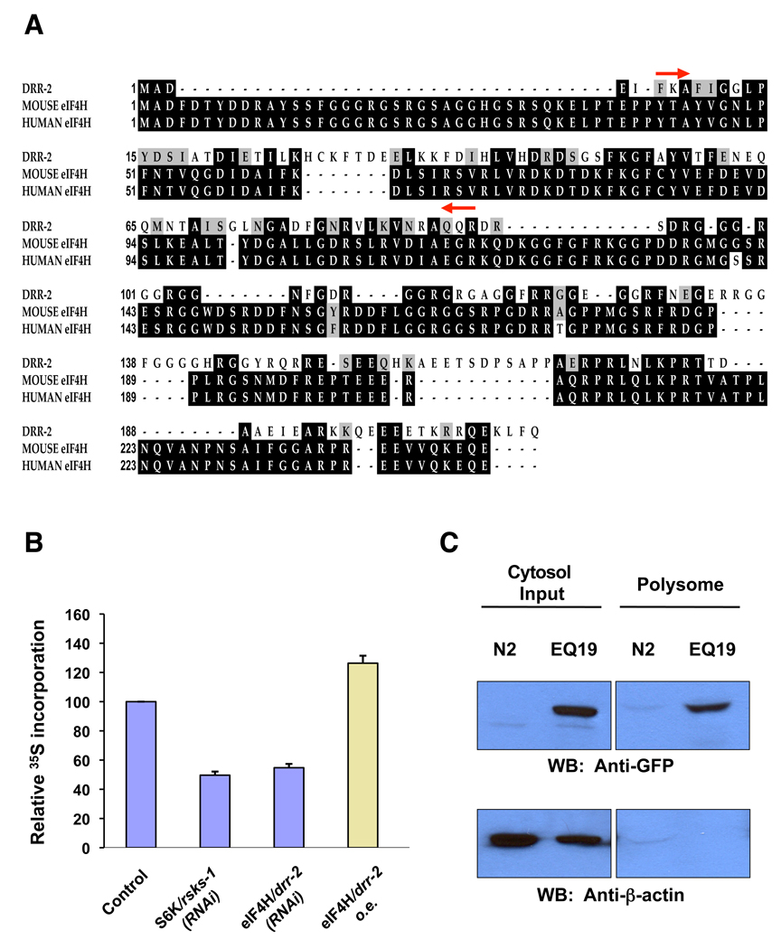
DRR-2 was originally identified as an unknown protein containing an RNA recognition motif (Hansen et al. 2005). Further analysis using the NCBI protein blast (Basic Local Alignment Search Tool) program identified the eukaryotic translation initiation factor 4H (eIF4H) as the closest human ortholog to DRR-2. The predicted DRR-2 protein is 52% similar and 38% identical to the human eIF4H; and 53% similar and 39% identical to the mouse eIF4H (Fig. 1A). The RNA recognition motif (RRM) of DRR-2 is 57% similar and 34% identical to its human counterpart.
Fig. 1. drr-2 encodes an ortholog of human eukaryotic translation initiation factor, eIF4H.
(A) The predicted DRR-2 protein is 52% similar and 38% identical to the human eIF4H; and 53% similar and 39% identical to the mouse eIF4H. Alignment of proteins was performed using VectorNTI. Black shading indicates identical amino acids. Grey shading indicates similar amino acids. The RNA recognition motifs (RRM) are bracketed by red arrows. (B) Alterations of drr-2 expression affect the rate of accumulation of newly synthesized protein. Relative levels of 35S-methionine incorporation in 1-day-old wild-type (N2) adults fed with vector control or RNAi bacteria from hatching were shown in blue columns; and relative level of 35S-methionine incorporation in transgenic animals over-expressing drr-2∷gfp (EQ19) was shown in yellow column. Bar graph represents mean incorporated 35S-methionine of three independent experiments normalized to total protein levels of the indicated RNAi treated animals compared to control animals; error bars represent standard deviation (STD). The p value for rsks-1 RNAi, drr-2 RNAi, and drr-2 o.e., when compared to vector control by two-sided, paired t-test, are 0.0008, 0.0011, and 0.0007, respectively. (C) DRR-2 is present in polysomes. Polysomes of N2 control or EQ19 animals were pelleted by centrifugation as described in method section. The DDR-2∷GFP fusion proteins, detected by anti-GFP antibodies, co-sediments with polysomes. Polysome purity was assessed by western blotting analysis using anti-β-actin antibodies, which serve as a cytosolic marker.
The function of eIF4H in mammals is to enhance the RNA helicase activity of eIF4A that works to unwind the secondary structure in the 5’-untranslated region of mRNA. It is proposed that eIF4H may act via protein-protein interactions to stabilize the conformational changes that occur in eIF4A during RNA binding, ATP hydrolysis, and RNA duplex unwinding (Richter et al. 1999). Reduction in eIF4H expression is expected to slow down the mRNA translation. Therefore, we measured the accumulation of newly synthesized protein in animals subjected to drr-2 RNAi, as well as transgenic animals over-expressing drr-2∷gfp (EQ19). As expected, the global translation rate was reduced to 55% in drr-2 knockdown animals, and increased by 26% in animals over-expressing drr-2 (Fig. 1B). This level of reduction is similar to those observed with rsks-1 (the worm ortholog of S6K) RNAi, which has been previously reported in C. elegans (Hansen et al. 2007; Pan et al. 2007). Translation factors belonging to the eIF4 protein family has been reported to be co-purified with polysomes (Merrick 1979; Bjork et al. 2003). To validate that drr-2 indeed encodes an ortholog of eIF4H that is present in the polysomes to modulate protein translation, we isolated polysomes from N2 wild-type and EQ19 animals. By using anti-GFP antibodies, we found that DRR-2∷GFP fusion proteins co-sediments with polysomes, implicating the association of C. elegans DRR-2 with polysomes (Fig. 1C).
eIF4H/DRR-2 is widely expressed
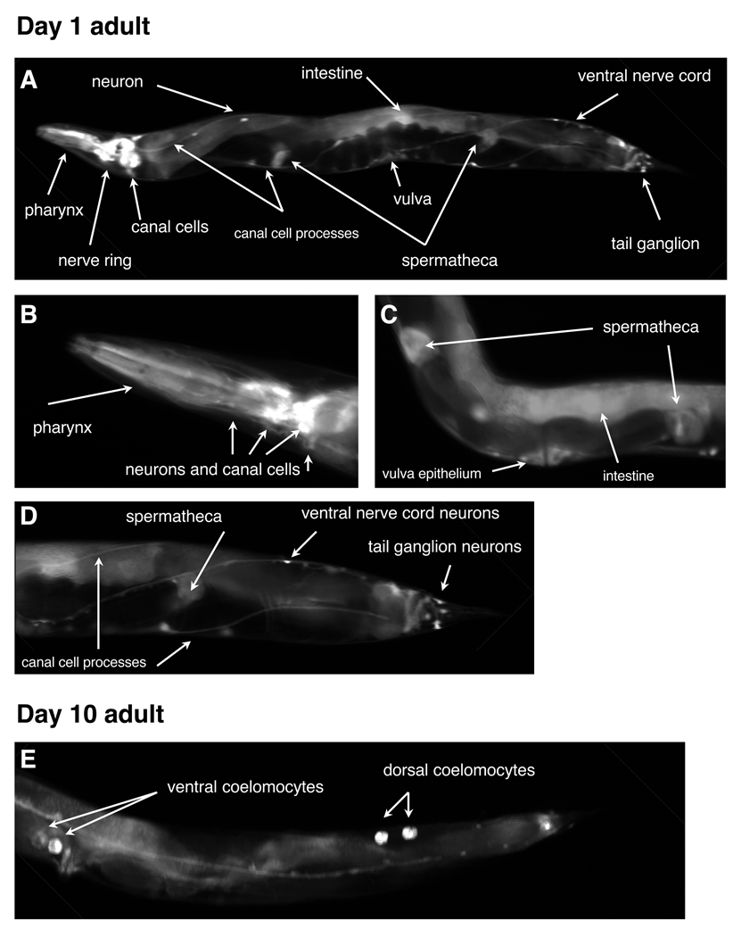
The expression pattern of eIF4H/DRR-2 was examined in transgenic animals containing a translational fusion of the genomic drr-2 locus to GFP. The construct contains the entire genomic coding region from drr-2, including the 5’ upstream regulatory sequences, fused in frame at the carboxyl terminus to GFP. The eIF4H/DRR-2∷GFP expression is observed in all stages of larval development and is maintained throughout the life of the animals. In adults, eIF4H/DRR-2 is expressed in many neurons, including those localized in head, tail, and ventral nerve cord (Fig. 2A–B, D). Expression is also observed in spermatheca and vulva epithelium cells (Fig. 2C). Additional tissues that consistently express eIF4H/DRR-2 include intestine, pharyngeal muscle, and canal cells (Fig. 2A–B). Interestingly, in aged adults, we observed large amount of GFP fusion proteins accumulated in the coelomocytes (Fig. 2E), which are scavenger cells that continuously and nonspecifically take up various molecules from the body cavity fluid. This suggests that eIF4H/DRR-2 may be secreted into the pseudocoelomic space. However, the reason why eIF4H/DRR-2 may be circulating in the body fluid remains unclear. The broad expression pattern of this gene is consistent with its function in the normal regulation of mRNA translation.
Fig. 2. The expression pattern of eIF4H/drr-2 in C. elegans.
Images of (A–D) 1-day-old adult or (E) 10-days-old adult transgenic animals (EQ19) carrying drr-2p∷drr-2∷gfp. Various cells and organs are indicated by white arrows.
Reduction in expression of eIF4H/drr-2 is essential for DR to extend lifespan
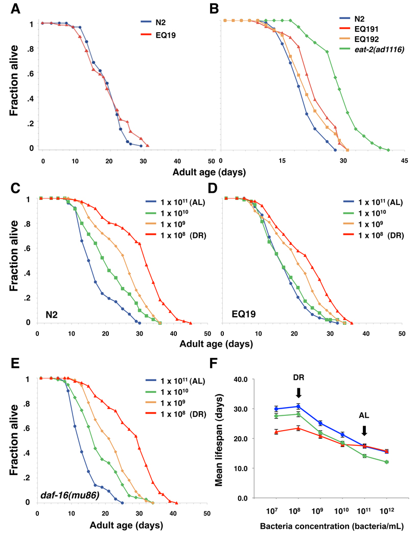
We have previously demonstrated that inhibition of drr-2 by RNAi produces a variety of DR-like phenotypes, and increases the lifespan of wild-type animals but not the lifespan of eat-2 (ad1116) mutants. We have also found that the mRNA levels of drr-2 is significantly lower in eat-2(ad1116) mutants, suggesting that DR might trigger a regulated reduction in drr-2 expression to initiate its longevity response (Hansen et al. 2005). To test this idea, we created transgenic animals carrying additional copies of drr-2 genes and asked whether elevation in drr-2 expression is sufficient to diminish the longevity effect of DR. Indeed, we found that the lifespan extension produced by eat-2 mutations, which is thought to genetically mimic DR, is largely suppressed by the over-expression of drr-2 (Fig. 3A–B, Table S1).
Fig. 3. eIF4H/DRR-2 is partially required for dietary restriction to extend lifespan.
(A) Survival curves of wild-type (N2) animals (blue line) or transgenic animals carrying additional copies of drr-2∷gfp (EQ19) (red line) at 20°C. Transgenic lines over-expressing DRR-2 without GFP tag were also obtained, and were found to have no effects on wild-type lifespan (Table S1). Please see Table S1 for statistical details, and for a repetition of this experiment. (B) Survival curves of wild-type (N2) animals (blue line), eat-2(ad1116) mutants (green line), or transgenic animals carrying additional copies of drr-2∷gfp in eat-2(ad1116) background (EQ191, EQ192) (red line) at 20°C. EQ191 and EQ192 were generated by crossing the drr-2∷gfp array into the eat-2(ad1116) mutant background from EQ19. (C–E) Survival curves of N2, daf-16(mu86) or transgenic animals over-expressing drr-2∷gfp (EQ19) grown on E coli at different concentrations (1 × 10⁷ to 1 × 10¹² bacteria/mL). 1 × 10¹¹ bacteria/ml was considered to be the ad libitum control. DR was initiated at day 1 of adulthood. Details of the solid plate DR protocol we utilized were described in method section. (F) Mean lifespan of N2, daf-16(mu86) or transgenic animals over-expressing drr-2∷gfp (EQ19) fed with E coli at different concentrations (1 × 10⁷ to 1 × 10¹² bacteria/mL). The drr-2 over-expressing animals show diminished lifespan extension under DR, while daf-16 mutants show similar degree of lifespan extension under DR when compared to AL. Please see Table S1 for statistical details, and for a repetition of this experiment.
It has been reported recently that different DR regimens may extend lifespan by both independent and overlapping genetic pathways in C. elegans (Greer & Brunet 2009). Thus, to further confirm this finding, we examined the effect of drr-2 over-expression on direct DR, using an agar plate-based DR protocol modified from a previously described dietary deprivation (DD) method (Kaeberlein et al. 2006). In brief, an overnight culture of OP50 bacteria was diluted into different concentrations (1 × 10¹² to 1 × 10⁷ bacteria/ml; 1 × 10¹¹ bacteria/ml is considered to be the ad libitum control) before being seeded on regular NGM plates. The bacteria were allowed to grow for 1 hr at 37°C before an antibiotics mix or UV light was applied to stop the growth. The DR treatment was initiated at day 1 of adulthood (day 4 of life) for all experiments, and the worms were transferred to fresh DR plates and scored for viability every two days. Remarkably, the lifespan extension induced by direct DR (i.e. solid plate-based DR) is significantly diminished by the over-expression of drr-2 (Fig. 3C–D and F, Table S1).
The majority of DR regimens in C. elegans (axenic growth, liquid DR, dietary deprivation, and eat-2 mutations) extend lifespan independent of the activity of DAF-16/FoxO transcription factor. To examine whether this is also true for the modified solid plate-based DR method we employed, the lifespan of daf-16(mu86) mutant worms grown on the solid plate-based DR conditions described above were assayed. Indeed, we found that solid plate-based DR treatment results in a similar lifespan extension of daf-16(mu86) mutant worms compared to that of wild-type worms (Fig. 3E–F, Table S1), suggesting that DAF-16 is not required for the longevity response to solid plate-based DR we employed, as observed in most of the other DR regimens in worms. This finding, however, is not consistent with a similar study using a different the solid plate-based DR regiment (Greer et al. 2007). The differences in the age of animals at which treatments were initiated or how bacteria were prepared may contribute to this discrepancy.
Lowering the level of eIF4H/DRR-2 is required for TOR but not S6K to extend lifespan
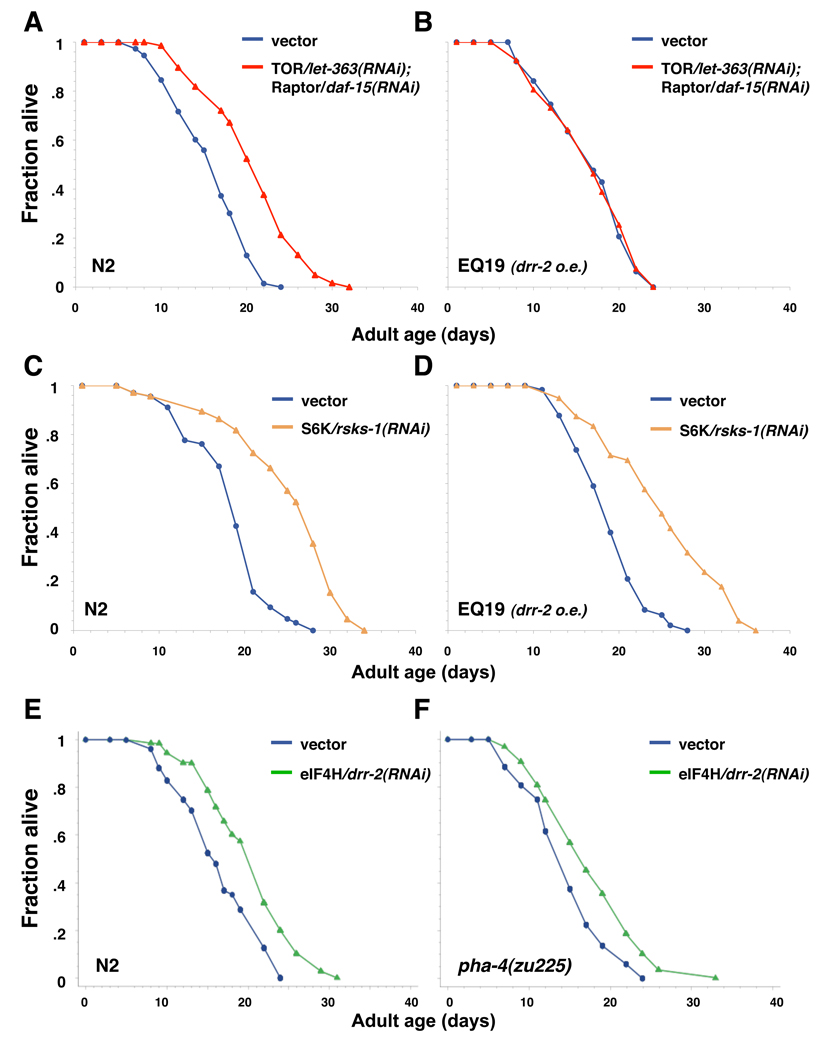
Inhibition of let-363 (worm homolog of TOR) has been shown to extend lifespan in C. elegans (Vellai et al. 2003). In both flies and worms, it has also been reported that reducing TOR activity results in little or no lifespan increase under conditions of DR, suggesting that DR may extend lifespan by lowering TOR activity in these organisms (Kapahi et al. 2004; Hansen et al. 2007). Moreover, TOR is known to influence mRNA translation by regulating several translation initiation factors (eIFs), such as eIF4E, in response to changes in the level of nutrients (Wang & Proud 2006). Given the findings that lowering translation extends worm lifespan, it is plausible that the effects of TOR on lifespan may be at least partially mediated by its ability to influence mRNA translation rate. If so, one might expect the lifespan extension resulting from TOR/let-363 inhibition to be suppressed by over-expression of eIF genes, such as the eIF4H/drr-2. Indeed, we found that inhibition of TOR signaling by knocking down both TOR/let-363 and Raptor/daf-15 (a binding partner of TOR that is necessary for TOR activity) or TOR/let-363 alone in the EQ19 (i.e. the eIF4H/drr-2 over-expressing line) background failed to extend lifespan (Fig. 4A–B, Table S2). This finding strongly implies that a common mechanism mediates the lifespan effects of TOR and eIF4H/drr-2, and that eIF4H/drr-2 might function genetically downstream of TOR to influence lifespan.
Fig. 4. Genetic interaction of eIF4H/drr-2 with let-363, rsks-1, and pha-4.
(A–B) Lowering the level of eIF4H/DRR-2 is required for C. elegans TOR/let-363 to influence longevity. Survival curves of wild-type (N2) animals or transgenic animals over-expressing drr-2∷gfp (EQ19) grown on either vector control (blue lines) or a 1:1 mix of TOR/let-363 and Raptor/daf-15 RNAi bacteria (red lines) at 25°C. The RNAi treatments were initiated at day 1 of adulthood. Please see Table S2 for statistical details, and for a repetition of this experiment. (C–D) Lowering the level of eIF4H/DRR-2 is not required for S6K/rsks-1 inhibition to increase longevity. Survival curves of N2 or EQ19 animals fed with either vector control (blue lines) or S6K/rsks-1 RNAi bacteria (orange lines) at 20°C. Please see Table S2 for statistical details, and for a repetition of this experiment. (E–F) FoxA transcription factor, PHA-4, is not required for eIF4H/drr-2 inhibition to increase longevity. Survival curves of wild-type or pha-4(zu225) animals grown on either vector control (blue lines) or eIF4H/drr-2 RNAi bacteria (green lines) at 20°C. Please see Table S3 for statistical details, and for a repetition of this experiment. All RNAi treatments described in (C–F) were initiated from hatching.
To control mRNA translation and protein synthesis, a number of eIFs or eEFs are regulated by TOR signaling through either an S6K-dependent pathway (e.g. eIF4B) or a 4E-BP-dependent pathway (e.g. eIF4E). While no structural 4E-BP homolog is apparent in the C. elegans genome (Syntichaki et al. 2007), recent studies have shown that reducing expression of S6K/rsks-1 results in a significant lifespan extension in worms (Hansen et al. 2007; Pan et al. 2007) and mammals (Selman et al. 2009). Therefore, to examine whether TOR acts via S6K to affect eIF4H/DRR-2 activity, we carried out lifespan analysis on animals over-expressing eIF4H/drr-2 and grown on S6K/rsks-1 RNAi bacteria. Interestingly, we found that over-expression of eIF4H/drr-2 did not have any effect on the lifespan of S6K/rsks-1 RNAi animals (Fig. 4 C–D, Table S2). Although S6K is known to act downstream of TOR to regulate translation by modulating the activity of several eIFs, our finding suggests that eIF4H/DRR-2 might be regulated by TOR via an S6K-independent pathway.
eIF4H/DRR-2 modulates lifespan independently of PHA-4 transcription factor
It has been previously reported that the FoxA transcription factor PHA-4 controls adult lifespan in response to DR, that is induced by dilutions of liquid food source or by eat-2 mutations in worms (Panowski et al. 2007). Furthermore, recent studies have suggested that pha-4 might act downstream of the S6K signaling to control adult lifespan (Sheaffer et al. 2008). Since we have shown that eIF4H/DRR-2 also functions downstream of TOR signaling in response to DR, it would be intriguing to examine whether drr-2 and pha-4 act in a common pathway to influence lifespan. Interestingly, we found that pha-4 is not required for eIF4H/drr-2 inhibition to extend lifespan, as the RNAi of eIF4H/drr-2 extends lifespan to a similar extent in the pha-4(zu225) mutant background compared to that in wild types (Fig. 4 E–F, Table S3). This finding is consistent with a previous model proposed by Susan Mango’s lab, in which pha-4 acts downstream of S6K, but in parallel to eIF4E, in response to TOR signaling and food limitation (Sheaffer et al. 2008). Thus, eIF4H/drr-2 may also act in a pathway independent of the S6K/PHA-4 pathway to control lifespan in response to TOR signaling and DR.
eIF4H/DRR-2 may act downstream of SAMS-1 and RAB-10 to influence longevity
Previous RNAi longevity screens have identified several drr (dietary restriction response) genes that might mediate the longevity response to DR (Hansen et al. 2005). These genes include the eIF4H/drr-2, rab-10 and sams-1. rab-10 encodes a Rab-like GTPase similar to those that regulate vesicle trafficking, whereas sams-1 encodes an S-adenosyl methionine synthetase, a protein that catalyzes the biosynthesis of S-adenosyl methionine (SAM). SAM is known to function as a universal methyl group donor in the majority of transmethylation reactions. Inhibition of this enzyme can affect methylation of histones, DNA, RNA, proteins, phospholipids and other small molecules (Chiang et al. 1996).
Inhibition of these genes by RNAi extend the lifespan of wild types, but not the dietary restricted animals (Hansen et al. 2005). It also produces several DR-like phenotypes (Hansen et al. 2005). In addition to eIF4H/drr-2 (Fig. 3), over-expression of rab-10 and sams-1 also suppress, at least partially, the lifespan extension of DR animals (Ching & Hsu, unpublished results). As observed for eIF4H/drr-2, the expression of both rab-10 and sams-1 are down-regulated in response to DR as well (Hansen et al. 2005). Taken together, these findings suggest that DR might trigger a regulated reduction in the expression of these drr genes to initiate its longevity response. However, the genetic interactions between eIF4H/drr-2 and other drr genes remain unclear.
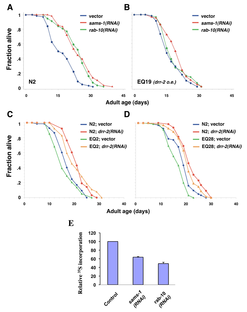
To examine how eIF4H/drr-2 genetically interacts with other drr genes, such as rab-10 and sams-1, we performed genetic epistasis analysis using different transgenic lines over-expressing eIF4H/drr-2, rab-10 or sams-1. We first asked whether over-expression of eIF4H/drr-2 is sufficient to suppress the lifespan extension caused by inhibition of rab-10 and sams-1. We found that inhibition of rab-10 or sams-1 by RNAi does not extend the lifespan of EQ19 (drr-2 over-expression) significantly, while inhibition of rab-10 or sams-1 extends the lifespan of wild-type animals by 20 – 40% (Fig. 5A–B, Table S2), suggesting a common mechanism that mediates the lifespan effects of eIF4H/drr-2, rab-10, and sams-1. We then examined whether it is also true that reducing expression levels of rab-10 or sams-1 is required for eIF4H/drr-2 to influence longevity. Our results show that inhibition of eIF4H/drr-2 consistently extends the lifespan of wild-type animals and animals over-expressing rab-10 (EQ28) or sams-1 (EQ2) (Fig. 5C–D, Table S3). Taken together, these results strongly imply that eIF4H/drr-2 may act downstream of both rab-10 and sams-1 and its activity might be essential for RAB-10 and SAMS-1 to mediate the longevity effects of DR. Consistent with this idea, the global translation rate was also reduced in sams-1 and rab-10 knockdown animals, presumably by at least partly affecting eIF4H/DDR-2 activity (Fig. 5E).
Fig. 5. Genetic interaction of eIF4H/drr 2 with sams-1 and rab-10.
(A–B) Over-expression of eIF4H/DRR-2 significantly suppresses the lifespan extension by sams-1 or rab-10 RNAi. Survival curves of wild-type (N2) animals or transgenic animals over-expressing drr-2∷gfp (EQ19) grown on either vector control bacteria (blue lines), sams-1 RNAi bacteria (red lines), or rab-10 RNAi bacteria (green lines) at 20°C. Please see Table S2 for statistical details, and for a repetition of this experiment. (C–D) Over-expression of sams-1 or rab-10 does not suppress the lifespan extension by drr-2 inhibition. Survival curves of N2 animals, or transgenic animals over-expressing either (C) sams-1∷gfp (EQ2) or (D) rab-10∷gfp (EQ28) grown on either vector control or eIF4H/drr-2 RNAi bacteria at 20°C. Please see Table S3 for statistical details, and for a repetition of this experiment. (E) Relative levels of 35S-methionine incorporation in 1-day-old wild-type (N2) adults fed with vector control, sams-1, or rab-10 RNAi bacteria from hatching. Bar graph represents mean incorporated 35S-methionine of three independent experiments normalized to total protein levels of the indicated RNAi treated animals compared to control animals; error bars represent standard deviation (STD). The p value for sams-1 RNAi, and rab-10 RNAi, when compared to vector control by paired t-test, are 0.036 and 0.004, respectively.
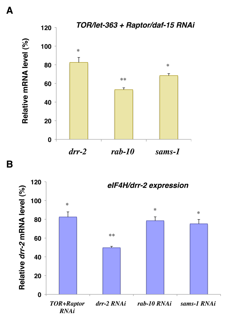
As described above, the mRNA level of drr-2 is significantly down-regulated in DR animals (Hansen et al. 2005). The expression levels of drr-2 genes appear to be tightly regulated in response to DR, and therefore may be critical for its role in the DR longevity response. Moreover, an increase in the drr-2 expression level is sufficient to suppress the lifespan extension induced by DR as well as TOR, rab-10, and sams-1 RNAi knockdown (Fig. 3, 4A–B, 5A–B). For this reason, we examined the expression level of drr-2 in response to different RNAi treatments. First, we found that, as observed in DR animals, the mRNA level of drr-2 is significantly down-regulated in animals fed with a mixture of TOR/let-363 and Raptor/daf-15 RNAi bacteria (Fig. 6A). Not surprisingly, we have also observed a similar down-regulation in the mRNA levels of rab-10 and sams-1 (Fig. 6A). These findings suggest that drr-2, rab-10 and sams-1 may all act downstream of the TOR signaling to mediate the longevity effects of DR. Next, we examined the mRNA level of drr-2 in animals fed with rab-10 or sams-1 RNAi bacteria. Consistent with our findings in longevity epistasis analysis, the mRNA levels of drr-2 are down-regulated in all three RNAi treatments, suggesting that drr-2 may act as a major effecter downstream of TOR signaling, RAB-10 and SAMS-1.
Fig. 6. Effects of TOR/Raptor, rab-10, and sams-1 RNAi on eIF4H/drr-2 expression.
(A) Relative mRNA levels of drr-2, rab-10, and sams-1 in animals fed with 1:1 mixture of TOR/let-363 and Raptor/daf-15 RNAi bacteria compared to those fed with control bacteria were measured by quantitative RT-PCR and the average of three different sample sets are shown. The relative mRNA levels were normalized against act-1 (beta-actin) levels. Error bars: ± STD. (B) Relative levels of drr-2 mRNA in animals fed with various RNAi bacteria compared to those fed with control bacteria. Average of three different sample sets are shown. Error bars: ± STD. *P < 0.05; **P < 0.01.
Discussion
Dietary restriction (DR) results in a robust increase in lifespan while maintaining the physiology of much younger animals in a wide range of species (Masoro 2005). However, the genetic and molecular mechanism by which DR slows aging and extends lifespan remains largely unclear. The nematode Caenorhabditis elegans has provided a good model to study the genetics of longevity response to DR. Both environmental and genetic manipulations have been used to model DR and have shown to extend lifespan in C. elegans [summarized in (Greer & Brunet 2009)]. Over the past few years, only a handful of genes have been identified that might mediate the longevity response to DR in worms, including drr-2.
drr-2 was first identified from a genome-wide RNAi screen for longevity genes. In addition to its longevity phenotype, inhibition of drr-2 also produces several phenotypes that are often observed in DR animals (Hansen et al. 2005). Subsequent genetic epistasis analyses suggest that drr-2 might play an important role in the longevity response to DR, as RNAi knockdown of drr-2 can increase the lifespan of wild-type animals, but fail to further extend the lifespan of eat-2 mutants (Hansen et al. 2005). Finally, we found that the expression level of drr-2 is down-regulated in DR animals (Hansen et al. 2005). Taken together, we hypothesized a model, in which DR may extend lifespan by lowering the expression level of drr-2 and the other drr genes. While the absence of an additive effect between two treatments (i.e. eat-2 mutation and drr-2 RNAi) strongly implies a common mechanism, this experiment does not, however, provide a conclusive answer on its own. Therefore, to further test our hypothesis, we examined the effect of drr-2 over-expression on the longevity response to DR. Indeed, we found that increasing the expression level of drr-2 is sufficient to suppress a majority of the lifespan extensions observed in two different regiments (i.e. eat-2 and solid plate DR). This result strongly suggests an essential role for drr-2 in the DR pathway to influence longevity.
The next question is what is the molecular function of DRR-2 protein? It turns out that drr-2 encodes an ortholog of human eIF4H protein. In mammals, eIF4H is a small protein that stimulates overall protein synthesis, possibly by enhancing the RNA helicase activity of eIF4A, one of the components of a heterotrimeric complex eIF4F. The function of eIF4A is to facilitate the initiation of mRNA translation by unwinding the secondary structure in the 5’-untranslated region of mRNA. Indeed, our results have confirmed that reduction in eIF4H/drr-2 expression slows down protein synthesis (Fig. 1B). We have also shown that DRR-2 protein co-precipitates with polysomes (Fig. 1C). Consistent with our finding, it has been previously reported that the rate of protein synthesis is significantly reduced in DR animals (Hansen et al. 2007). Since we found that reduction in eIF4H/drr-2 expression is essential for DR to extend lifespan (Fig. 3), it is reasonable to hypothesize that DR might initiate its longevity effects by triggering a regulated reduction in protein synthesis via an eIF4H/drr-2-dependent mechanism. So, how might reduction in translation mediate the lifespan effect of DR? One possibility is that, in response to decreases in nutrients (DR), animals need to make an adjustment as to where they spend their limited resources. A potential strategy is to shift their investment toward maintenance and repair but away from reproduction and growth by inhibiting protein synthesis, a very energy costly process. In addition to conserving the energy spent on protein synthesis, the act of inhibiting certain eIFs may also trigger a cellular response that extends lifespan. For instance, some of the stress response programs may be differentially up-regulated in response to eIFs inhibition when global mRNA translations is attenuated. In fact, similar regulations have been observed in unfolded protein response (UPR). In response to ER stress, phosphorylation of eIF2α by PERK kinase is known to attenuate global translation, while select stress-response genes, such as ATF4, are translationally up-regulated (Harding et al. 2003; Fels & Koumenis 2006).
In addition to eIF4H, inhibition of the other two components of the eIF4F complex, eIF4E and eIF4G, have both been previously shown to slow down protein synthesis and extend lifespan in worms (Hansen et al. 2007; Pan et al. 2007; Syntichaki et al. 2007). Contrary to our finding on eIF4H/drr-2, reduction in eIF4E/ife-2 or eIF4G/ifg-1 expression further extends the lifespan of eat-2 mutants. However, this does not necessarily indicate that DR and inhibition of eIF4E or 4G affect longevity in distinct mechanisms, since it is possible that the maximal lifespan extension resulting from DR may have not yet been achieved by eat-2 mutations. In fact, it has been reported that direct DR can further increase the lifespan of eat-2 mutants (Hansen et al. 2007). Most likely, eIF4E/4G and DR may act in both a distinct and shared pathway to influence longevity, while most of the longevity effects observed in eIF4H/drr-2 mutants are DR-related.
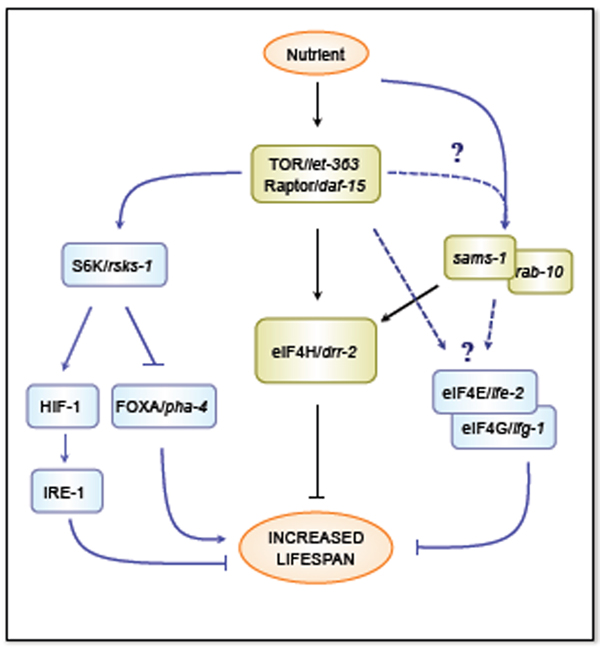
To further understand how eIF4H/DRR-2 mediates the longevity effects of DR, we asked how eIF4H/drr-2 might interact with other known longevity genes that have been implicated in the DR pathway. The TOR signaling pathway has been proposed to mediate the longevity response to DR in several different model organisms (Kapahi et al. 2004; Hansen et al. 2007). In response to nutrient deficiency, TOR is known to inhibit protein synthesis, partly by controlling the initiation steps of mRNA translation (i.e. eIFs activities) (Wang & Proud 2006; Wullschleger et al. 2006). Studies in worms have confirmed that the rate of protein synthesis is indeed decreased in both TOR/let-363 and eat-2 mutants (Hansen et al. 2007). Although inhibition of eIF4E/ife-2 or eIF4G/ifg-1 has been reported to further increase the lifespan of TOR/let-363 mutants (Hansen et al. 2007; Pan et al. 2007; Syntichaki et al. 2007), it is still possible, as discussed above, that the effects of TOR on lifespan are mediated in part by its ability to influence the rate of mRNA translation. Consistent with this idea, we found that the lifespan extension resulting from TOR/let-363 inhibition is largely suppressed by over-expressing eIF4H/drr-2 (Fig. 4A–B). Furthermore, the mRNA level of drr-2 is reduced when both TOR/let-363 and Raptor/daf-15 are inactivated (Fig. 6). These findings strongly support a role of eIF4H/DRR-2 in the TOR signaling pathway to influence longevity (Fig. 7). However, it is not clear whether eIF4H, −4E, and −4G may be regulated by TOR through a different or a common mechanism.
Fig. 7. Model for the role of eIF4H/DRR-2 in the longevity response to DR.
Genetic epistasis suggests that eIF4H/drr-2 acts downstream of TOR/let-363, sams-1 and rab-10 but parallel to the S6K pathway, which controls other DR mediators such as PHA-4 and HIF-1, to influence protein translation and longevity. It is not clear, however, whether other translation initiation factors (eIFs), such as eIF4E/ife-2 and eIF4G/ifg-1, act similarly as eIF4H/drr-2 to influence longevity in worms.
In mammals, mTOR controls activities of several eIFs and the rate of mRNA translation via an S6K-1-dependent pathway (e.g. eIF4A, −4B) or a 4E-BP1-dependent pathway (e.g. eIF4E). Recently, studies have shown that inhibition of S6K/rsks-1 extends lifespan in C. elegans (Hansen et al. 2007; Pan et al. 2007). Conversely, no structural homolog of mammalian 4E-BP1 has been identified in C. elegans (Syntichaki et al. 2007). It is worth noting, however, that Drosophila 4E-BP has been reported to be up-regulated upon DR and is required for the longevity effects of DR in flies (Zid et al. 2009). Therefore, we asked whether TOR signaling functions through S6K to affect eIF4H/DRR-2 activity. We found that, in contrast with our observation on TOR/let-363, over-expression of eIF4H/drr-2 does not prevent S6K/rsks-1 RNAi from extending lifespan (Fig. 4C–D), suggesting that eIF4H/DRR-2 and S6K may act in distinct pathways to influence lifespan. Similarly, additive effects have been observed when inhibiting S6K/rsks-1 in eIF4G/ifg-1 mutants background (Pan et al. 2007). In fact, while our data strongly implicates eIF4H as a downstream effector of TOR, the role of S6K in TOR-mediated DR longevity effects is far from clear. Conflicting results have been reported on whether inhibition of TOR/let-363 further extends the lifespan extension by S6K/rsks-1 mutations (Hansen et al. 2007; Pan et al. 2007). Recently, several genes have been proposed to function downstream of S6K to mediate the longevity effects of DR in worms. These include HIF-1 (hypoxia inducible factor-1), an endoplasmic reticulum (ER) stress regulator IRE-1 (inositol-requiring protein-1), and a FoxA transcription factor PHA-4. Inhibition of hif-1 extends the lifespan of wild types, but fails to extend the lifespan of rsks-1 mutants, while inhibition of egl-9, a HIF-1 enhancer, diminishes the lifespan extension of rsks-1 mutants (Chen et al. 2009). The increased longevity by hif-1 mutations appears to be dependent on IRE-1 activity (Chen et al. 2009). The presence of PHA-4 is required for DR (i.e. eat-2 mutants and liquid DR) to extend lifespan (Panowski et al. 2007). It is also required for TOR/let-363 or S6K/rsks-1 mutations to extend lifespan (Sheaffer et al. 2008). Conversely, inhibition of pha-4 has no effects on the lifespan of eIF4E/ife-2 mutants, suggesting that PHA-4 and eIF4E may act in different pathways downstream of TOR (Sheaffer et al. 2008). This is consistent with our findings that over-expression of eIF4H/drr-2 does not significantly suppress the lifespan extensions resulting from inhibition of rsks-1 or pha-4 (Fig. 4C–F). Therefore, the S6K/PHA-4 pathway and eIFs may act in parallel pathways to mediate the longevity response to DR (Fig. 7).
In addition to eIF4H/drr-2, over-expression of rab-10 and sams-1 also suppress the lifespan extension of DR animals (Ching & Hsu, unpublished results). Both of these genes were identified from the same genetic screen as drr-2 with similar DR-like phenotypes (Hansen et al. 2005). RAB-10 is thought to be involved in vesicle trafficking, whereas SAMS-1 is responsible for the biosynthesis of the universal methyl group donor, SAM (S-adenosylmethionine). However, how these two genes mediate the longevity response to DR remains unclear. Our results suggest that eIF4H/drr-2 might act genetically downstream of both rab-10 and sams-1 to influence lifespan (Fig. 5). How might rab-10 and sams-1 influence eIF4H/DRR-2 activity in response to nutrient changes? Given that the level of eIF4H/drr-2 transcription appears to play an important role in mediating DR longevity responses, and that the mRNA level of eIF4H/drr-2 is also reduced when rab-10 and sams-1 is inhibited by RNAi, one likely scenario is that both RAB-10 and SAMS-1 are somehow involved in the regulation of eIF4H/drr-2 transcription in response to DR. Alternatively, rab-10 and sams-1 mutations may increase lifespan mainly by reducing mRNA translation as eIF4H/drr-2, but via an eIF4H/drr-2-independent mechanism. Therefore, when eIF4H/drr-2 is over-expressed and mRNA translation is accelerated, inhibition of rab-10 or sams-1 can no longer produce significant lifespan effects, assuming that changes in the global translation level is a key determinant of longevity in worms. It is worth noting that the enzymatic product of SAMS-1 (i.e. SAM) is required for most of the transmethylation reactions, which is an important step for the maturation of rRNA and tRNA. Thus, it is very likely that inhibition of sams-1 may also have direct impact on translation. Although our results strongly suggest that eIF4H/drr-2 acts genetically downstream of both rab-10 and sams-1 to influence lifespan, it is still possible that other eIFs may not be regulated by rab-10 and sams-1.
In summary, our analysis of drr-2 has revealed its molecular function as a translation initiation factor and its essential role in the longevity response to DR. Thus, translation regulation appears to be an important component of the lifespan regulation, especially when food resources are changed in the environment. Our findings have also suggested that eIF4H/DRR-2 may function downstream of TOR, SAMS-1, and RAB-10, but not the S6K/PHA-4 pathway to mediate the longevity effect of DR. An intriguing subject for future studies will be to determine how changes in translation might influence longevity and how eIF4H/DRR-2 activity may be regulated in response to TOR signaling.
Experimental Procedures
Strains and generation of transgenic lines
DA1116: eat-2(ad1116)II, CF1037: daf-16(mu86)I, SM190: smg-1(cc546ts)I; pha-4(zu225)V, EQ2: iqEx1[sams-1p∷sams-1∷gfp + rol-6], EQ18: iqEx6[drr-2p∷drr-2 + rol-6], EQ19: iqEx7[drr-2p∷drr-2∷gfp + rol-6], EQ28: iqEx10[rab-10p∷rab-10 + rol-6], EQ49: eat-2(ad1116)II; iqEx19 [drr-2p∷drr-2∷gfp], EQ191: eat-2(ad1116)II; iqEx19 [drr-2p∷drr-2∷gfp], EQ192: eat-2(ad1116)II; iqEx19 [drr-2p∷drr-2∷gfp]. DA1116, CF1037 and wild-type Caenorhabditis elegans (N2) strains were obtained from the Caenorhabditis Genetic Center. SM190 strain was provided by Dr. Susan Mango. All strains were maintained and handled as described previously (Brenner 1974; Riddle et al. 1997). SM190 strain was cultured at the permissive temperature (Gaudet & Mango 2002), while the other strains were cultured at 20°C. For the generation of transgenic animals, a plasmid DNA mix was microinjected into the gonad of young adult hermaphrodite animals, using the standard method (Mello et al. 1991). F1 progeny were selected on the basis of the roller phenotype or RFP expression. Individual F2 progenies were isolated to establish independent lines. For the generation of the EQ2 strain, the plasmid DNA mix consisted of 30ng/µl pAH9(sams-1p∷sams-1∷gfp) and 80ng/µl pRF4 (rol-6). For generation of EQ18, the plasmid DNA mix consisted of 30ng/µl pAH28(drr-2p∷drr-2) and 80ng/µl pRF4 (rol-6). For the generation of EQ19, the plasmid DNA mix consisted of 30ng/µl pAH11(drr-2p∷drr-2∷gfp) and 80ng/µl pRF4 (rol-6). For the generation of EQ28, the plasmid DNA mix consisted of 30ng/µl pAH7(rab-10p∷rab-10∷gfp) and 80ng/µl pRF4 (rol-6). Wild-type (N2) animals were microinjected to generate these strains. Microinjecting N2 animals with 100ng/µl pRF4 (rol-6) alone did not affect the mean lifespan of N2 animals grown on either OP50, HT115, 1% DR, or RNAi bacteria that was examined in this study (data not shown). For the generation of EQ49, 30ng/µl of pAH11(drr-2p∷drr-2∷gfp) plasmid was injected into eat-2(ad1116) mutants. EQ191 and EQ192 were generated by crossing the pAH11(drr-2p∷drr-2∷gfp) array into the eat-2(ad1116) mutant background.
RNA-interference (RNAi) clones
The identity of all RNAi clones was verified by sequencing the inserts using M13-forward primer. The TOR/let-363 RNAi clone was obtained from Dr. Malene Hansen. The S6K/rsks-1 clone was from Marc Vidal’s RNAi library. All other clones were from Julie Ahringer’s RNAi library. HT115 bacteria transformed with RNAi vectors expressing dsRNA of the genes of interest were grown at 37°C in LB with 10 µg/ml tetracycline and 50 µg/ml carbenicillin, then seeded onto NG-carbenicillin plates and supplemented with 100 µl 0.1M IPTG.
Lifespan analysis
Lifespan analysis was conducted at 20°C as described previously (Kenyon et al. 1993; Apfeld & Kenyon 1999; Hsu et al. 2003) unless otherwise stated. Strains were grown at 20°C or 25°C (SM190) for at least two generations before the experiments were initiated. 60–90 animals were tested in each experiment. RNAi treatments were carried out by adding synchronized eggs, unless noted otherwise, to plates seeded with the RNAi bacteria of interest. Worms were moved to fresh RNAi plate every two days until reproduction ceased. Worms were then moved to new plates every 5–7 days for the rest of the lifespan analysis. Viability of the worms was scored every two days. In all experiments, the pre-fertile period of adulthood was used as t = 0 for lifespan analysis. Statview 5.01 (SAS) software was used for statistical analysis to determine the means and percentiles. In all cases, P values were calculated using the log-rank (Mantel-Cox) method.
Solid plate dietary restriction
The solid plate DR protocol used in our lab was adapted and modified based on the method previously described by others (Kaeberlein et al. 2006). OP50 bacteria were grown to near saturation overnight. The concentration of the overnight bacteria culture was measured before diluting into various concentrations (1 × 10⁷ ~ 1 × 10¹² bacteria/ml; 1 × 10¹¹ bacteria/ml was considered to be the ad libitum control). 200 µl of these solutions were seeded on each 35mm plate. The plates were kept in 37°C incubator for 1 hrs before adding 20 µl of an antibiotics mix (1:1 ratio of 100 µg/ml carbenicillin and 50 µg/ml katamycin) on each plate to stop the growth of bacteria. Both control and DR plates were prepared and stored at 4°C for no more than two days before being used. 12–15 adult worms were placed on each plate containing different concentrations of bacteria starting at day 1 of adulthood (day 4 of life). If indicated, 100 µg/ml of FUDR was added to each plate before placing the worms on the plate to stop the progeny production. Otherwise, adults were separated from their progeny by transferring to fresh plates every 20–24 hrs during reproduction period. Worms were transferred to fresh plates and scored for viability every two days.
Quantitative RT-PCR (qRT-PCR) analysis
Total RNA was isolated from approximately 5,000 day 1 adult worms, and cDNA was made from 4 µg of RNA using Superscript III RT (Invitrogen). TaqMan real-time qPCR experiments were performed for each primer and probe sets using the Chromo 4 system (MJ Research). Relative mRNA level of the genes of interest were calculated and normalized against the internal control (act-1, the β-actin). Primers and probes designed specifically for act-1, sme-1, rab-10, drr-1 and drr-2 are listed below:
Primers:
act-1-720F: 5’-CTACGAACTTCCTGACGGACAAG-3’
act-1-821R: 5’-CCGGCGGACTCCATACC-3’
sams-1-209F: 5’-TCCGTCGTGTCATCGAAAAG-3’
sams-1-275R: 5’-TTGCAGGTCTTGTGGTCGAA-3’
rab-10-514F: 5’-GCTAAGATGCCTGATACCACTGA-3’
rab-10-585R: 5’-ACTCTGCCTCTGTGGTTGCA-3’
drr-2-530F: 5’-TGAAGCCCCGTACCACAGA-3’
drr-2-596R: 5’-CTTGGTCTCCTCTTCTTCTTGCT-3’
Probes (All probes listed here were labeled with FAM™ at 5’ end and Black hole Quencher at 3’ end):
act-1-T: 5’-AAACGAACGTTTCCGTTGCCCAGAGGCTAT-3’
sams-1-230T: 5’-TTGGATTCACCGACTCCAGCATTGG-3’
rab-10-541T: 5’-CAATCCCGCGATACGGTGAATCCA-3’
drr-2-551T: 5’-CGCTGAGATCGAGGCTCGCAA-3’
35S-methionine incorporation
To assess the level of global translation, 35S-methionine incorporation assays were performed as previously described (Hansen et al. 2007) with minor modifications. OP50 bacteria were cultured in LB containing 10–15 µCi 35S-methionine (Perkin-Elmer, #NEG 709A001MC) for 12 hr and then concentrated 10-fold. ~2000 Day 1 adult worms treated with 100 µg/ml FUDR were mixed with the bacteria and incubated for 3 hr at room temperature. Negative controls were produced by mixing worms with non-radioactive labeled bacteria. All worm samples were washed twice with M9 buffer and incubated with non-radioactive bacteria for 30 min before being flash frozen twice in liquid nitrogen. The samples were boiled in 100 µl of 1% SDS and centrifuged at 16,000g for 20min. The supernatants were precipitated by adding 100 µl of 10% trichloroacetic acid (TCA) and incubated on ice for 1 hr. Protein concentrations were measured using the BCA protein assay (Pierce #23225). 35S radioactivity levels in protein extracts were measured after filtration with a spin column (Pierce) by liquid scintillation. To determine the levels of newly synthesized proteins, 35S incorporation levels were calculated by normalizing 35S counts per min to total protein levels.
Polysome isolation
Cytoplasmic extracts were prepared from synchronized Day 1 adults. Worms were washed three times with M9 buffer and once with HB buffer ( 20 mM HEPES pH 7.6, 100 mM NaCl, 1.5 mM MgCl2, 10 mM KCl, 0.5 mM EGTA, 0.1 mM EDTA, 44 mM Sucrose, 100 U/mL of RNasin, Roche complete protease inhibitor tablet). Worms were then resuspended in three volumes of HB buffer and lysed by two freeze-and-thaw cycles. The frozen worms were homogenized 30 strokes with pestle B of the Dounce homogenizer. The lysate was collected and spun at 14,000 × g for 20 min at 4°C. The supernatant was centrifuged in an SW41Ti rotor (Beckman) at 100,000 × g for 3 h at 4°C to pellet polysomes. The purity of the polysome fraction was validated by western blotting analysis using anti-β-actin antibodies (cytosolic maker).
Western blotting analysis
The protein samples were subjected to SDS-PAGE and transferred to a PVDF membrane (Millipore). The transblotted membrane was washed three times with TBS containing 0.05% Tween 20 (TBST). After blocking with TBST containing 5% nonfat milk for 60 min, the membrane was incubated with the primary antibody indicated (e.g. anti-GFP; abcam #ab6556) at 4°C for 12 h and washed three times with TBST. The membrane was then probed with HRP-conjugated secondary antibody for 1 h at room temperature and washed with TBST three times. Finally, the immunoblots were detected using a chemiluminescent substrate (Pierce) and visualized by autoradiography.
Supplementary Material
Acknowledgements
We thank Dr. Malene Hansen, The Burnham Institute for Medical Research, for providing the TOR/let-363 RNAi clone. We thank Dr. Susan Mango, University of Utah, for providing the SM190 strain. We thank the Caenorhabditis Genetic Center for providing the DA1116 and CF1037 strains. We also thank Carol Mousigan, Wei-Chun Chiang and Meng-Yin Hsieh for technical assistance. This work was supported in part by grants from the Ellison Medical Foundation and the National Institutes of Health (NIA) to Ao-Lin Hsu.
Contributor Information
Tsui-Ting Ching, Email: ttching@umich.edu.
Ao-Lin Hsu, Email: aolinhsu@umich.edu.
References
- Apfeld J, Kenyon C. Regulation of lifespan by sensory perception in Caenorhabditis elegans. Nature. 1999;402:804–809. doi: 10.1038/45544. [DOI] [PubMed] [Google Scholar]
- Bjork P, Bauren G, Gelius B, Wrange O, Wieslander L. The Chironomus tentans translation initiation factor eIF4H is present in the nucleus but does not bind to mRNA until the mRNA reaches the cytoplasmic perinuclear region. J Cell Sci. 2003;116:4521–4532. doi: 10.1242/jcs.00766. [DOI] [PubMed] [Google Scholar]
- Brenner S. The genetics of Caenorhabditis elegans. Genetics. 1974;77:71–94. doi: 10.1093/genetics/77.1.71. [DOI] [PMC free article] [PubMed] [Google Scholar]
- Chen D, Thomas EL, Kapahi P. HIF-1 modulates dietary restriction-mediated lifespan extension via IRE-1 in Caenorhabditis elegans. PLoS Genet. 2009;5:e1000486. doi: 10.1371/journal.pgen.1000486. [DOI] [PMC free article] [PubMed] [Google Scholar]
- Chiang PK, Gordon RK, Tal J, Zeng GC, Doctor BP, Pardhasaradhi K, McCann PP. S-Adenosylmethionine and methylation. FASEB J. 1996;10:471–480. [PubMed] [Google Scholar]
- Dorrello NV, Peschiaroli A, Guardavaccaro D, Colburn NH, Sherman NE, Pagano M. S6K1- and betaTRCP-mediated degradation of PDCD4 promotes protein translation and cell growth. Science. 2006;314:467–471. doi: 10.1126/science.1130276. [DOI] [PubMed] [Google Scholar]
- Fels DR, Koumenis C. The PERK/eIF2alpha/ATF4 module of the UPR in hypoxia resistance and tumor growth. Cancer Biol Ther. 2006;5:723–728. doi: 10.4161/cbt.5.7.2967. [DOI] [PubMed] [Google Scholar]
- Gaudet J, Mango SE. Regulation of organogenesis by the Caenorhabditis elegans FoxA protein PHA-4. Science. 2002;295:821–825. doi: 10.1126/science.1065175. [DOI] [PubMed] [Google Scholar]
- Greer EL, Brunet A. Different dietary restriction regimens extend lifespan by both independent and overlapping genetic pathways in C. elegans. Aging Cell. 2009;8:113–127. doi: 10.1111/j.1474-9726.2009.00459.x. [DOI] [PMC free article] [PubMed] [Google Scholar]
- Greer EL, Dowlatshahi D, Banko MR, Villen J, Hoang K, Blanchard D, Gygi SP, Brunet A. An AMPK-FOXO pathway mediates longevity induced by a novel method of dietary restriction in C. elegans. Curr Biol. 2007;17:1646–1656. doi: 10.1016/j.cub.2007.08.047. [DOI] [PMC free article] [PubMed] [Google Scholar]
- Hamilton B, Dong Y, Shindo M, Liu W, Odell I, Ruvkun G, Lee SS. A systematic RNAi screen for longevity genes in C. elegans. Genes Dev. 2005;19:1544–1555. doi: 10.1101/gad.1308205. [DOI] [PMC free article] [PubMed] [Google Scholar]
- Hansen M, Hsu AL, Dillin A, Kenyon C. New genes tied to endocrine, metabolic, and dietary regulation of lifespan from a Caenorhabditis elegans genomic RNAi screen. PLoS Genet. 2005;1:119–128. doi: 10.1371/journal.pgen.0010017. [DOI] [PMC free article] [PubMed] [Google Scholar]
- Hansen M, Taubert S, Crawford D, Libina N, Lee SJ, Kenyon C. Lifespan extension by conditions that inhibit translation in Caenorhabditis elegans. Aging Cell. 2007;6:95–110. doi: 10.1111/j.1474-9726.2006.00267.x. [DOI] [PubMed] [Google Scholar]
- Harding HP, Zhang Y, Zeng H, Novoa I, Lu PD, Calfon M, Sadri N, Yun C, Popko B, Paules R, Stojdl DF, Bell JC, Hettmann T, Leiden JM, Ron D. An integrated stress response regulates amino acid metabolism and resistance to oxidative stress. Mol Cell. 2003;11:619–633. doi: 10.1016/s1097-2765(03)00105-9. [DOI] [PubMed] [Google Scholar]
- Holz MK, Ballif BA, Gygi SP, Blenis J. mTOR and S6K1 mediate assembly of the translation preinitiation complex through dynamic protein interchange and ordered phosphorylation events. Cell. 2005;123:569–580. doi: 10.1016/j.cell.2005.10.024. [DOI] [PubMed] [Google Scholar]
- Houthoofd K, Braeckman BP, Johnson TE, Vanfleteren JR. Life extension via dietary restriction is independent of the Ins/IGF-1 signalling pathway in Caenorhabditis elegans. Exp Gerontol. 2003;38:947–954. doi: 10.1016/s0531-5565(03)00161-x. [DOI] [PubMed] [Google Scholar]
- Houthoofd K, Vanfleteren JR. The longevity effect of dietary restriction in Caenorhabditis elegans. Exp Gerontol. 2006;41:1026–1031. doi: 10.1016/j.exger.2006.05.007. [DOI] [PubMed] [Google Scholar]
- Hsu AL, Murphy CT, Kenyon C. Regulation of aging and age-related disease by DAF-16 and heat-shock factor. Science. 2003;300:1142–1145. doi: 10.1126/science.1083701. [DOI] [PubMed] [Google Scholar]
- Jia K, Chen D, Riddle DL. The TOR pathway interacts with the insulin signaling pathway to regulate C. elegans larval development, metabolism and life span. Development. 2004;131:3897–3906. doi: 10.1242/dev.01255. [DOI] [PubMed] [Google Scholar]
- Kaeberlein M, Powers RW, 3rd, Steffen KK, Westman EA, Hu D, Dang N, Kerr EO, Kirkland KT, Fields S, Kennedy BK. Regulation of yeast replicative life span by TOR and Sch9 in response to nutrients. Science. 2005;310:1193–1196. doi: 10.1126/science.1115535. [DOI] [PubMed] [Google Scholar]
- Kaeberlein TL, Smith ED, Tsuchiya M, Welton KL, Thomas JH, Fields S, Kennedy BK, Kaeberlein M. Lifespan extension in Caenorhabditis elegans by complete removal of food. Aging Cell. 2006;5:487–494. doi: 10.1111/j.1474-9726.2006.00238.x. [DOI] [PubMed] [Google Scholar]
- Kapahi P, Zid BM, Harper T, Koslover D, Sapin V, Benzer S. Regulation of lifespan in Drosophila by modulation of genes in the TOR signaling pathway. Curr Biol. 2004;14:885–890. doi: 10.1016/j.cub.2004.03.059. [DOI] [PMC free article] [PubMed] [Google Scholar]
- Kenyon C. The plasticity of aging: insights from long-lived mutants. Cell. 2005;120:449–460. doi: 10.1016/j.cell.2005.02.002. [DOI] [PubMed] [Google Scholar]
- Kenyon C, Chang J, Gensch E, Rudner A, Tabtiang R. A C. elegans mutant that lives twice as long as wild type. Nature. 1993;366:461–464. doi: 10.1038/366461a0. [DOI] [PubMed] [Google Scholar]
- Lakowski B, Hekimi S. The genetics of caloric restriction in Caenorhabditis elegans. Proc Natl Acad Sci U S A. 1998;95:13091–13096. doi: 10.1073/pnas.95.22.13091. [DOI] [PMC free article] [PubMed] [Google Scholar]
- Masoro EJ. Subfield history: caloric restriction, slowing aging, and extending life. Sci Aging Knowledge Environ. 2003;2003:RE2. doi: 10.1126/sageke.2003.8.re2. [DOI] [PubMed] [Google Scholar]
- Masoro EJ. Overview of caloric restriction and ageing. Mech Ageing Dev. 2005;126:913–922. doi: 10.1016/j.mad.2005.03.012. [DOI] [PubMed] [Google Scholar]
- Mello CC, Kramer JM, Stinchcomb D, Ambros V. Efficient gene transfer in C.elegans: extrachromosomal maintenance and integration of transforming sequences. EMBO J. 1991;10:3959–3970. doi: 10.1002/j.1460-2075.1991.tb04966.x. [DOI] [PMC free article] [PubMed] [Google Scholar]
- Merrick WC. Purification of protein synthesis initiation factors from rabbit reticulocytes. Methods Enzymol. 1979;60:101–108. doi: 10.1016/s0076-6879(79)60010-1. [DOI] [PubMed] [Google Scholar]
- Oldham S, Hafen E. Insulin/IGF and target of rapamycin signaling: a TOR de force in growth control. Trends Cell Biol. 2003;13:79–85. doi: 10.1016/s0962-8924(02)00042-9. [DOI] [PubMed] [Google Scholar]
- Pan KZ, Palter JE, Rogers AN, Olsen A, Chen D, Lithgow GJ, Kapahi P. Inhibition of mRNA translation extends lifespan in Caenorhabditis elegans. Aging Cell. 2007;6:111–119. doi: 10.1111/j.1474-9726.2006.00266.x. [DOI] [PMC free article] [PubMed] [Google Scholar]
- Panowski SH, Wolff S, Aguilaniu H, Durieux J, Dillin A. PHA-4/Foxa mediates diet-restriction-induced longevity of C. elegans. Nature. 2007;447:550–555. doi: 10.1038/nature05837. [DOI] [PubMed] [Google Scholar]
- Raught B, Peiretti F, Gingras AC, Livingstone M, Shahbazian D, Mayeur GL, Polakiewicz RD, Sonenberg N, Hershey JW. Phosphorylation of eucaryotic translation initiation factor 4B Ser422 is modulated by S6 kinases. Embo J. 2004;23:1761–1769. doi: 10.1038/sj.emboj.7600193. [DOI] [PMC free article] [PubMed] [Google Scholar]
- Richter NJ, Rogers GW, Jr, Hensold JO, Merrick WC. Further biochemical and kinetic characterization of human eukaryotic initiation factor 4H. J Biol Chem. 1999;274:35415–35424. doi: 10.1074/jbc.274.50.35415. [DOI] [PubMed] [Google Scholar]
- Riddle DL, Blumenthal T, Meyer BJ, Priess JR. Cold Spring Harbor, NY: Cold Spring Harbor Laboratory Press; 1997. [PubMed] [Google Scholar]
- Rogers GW, Jr, Richter NJ, Lima WF, Merrick WC. Modulation of the helicase activity of eIF4A by eIF4B, eIF4H, and eIF4F. J Biol Chem. 2001;276:30914–30922. doi: 10.1074/jbc.M100157200. [DOI] [PubMed] [Google Scholar]
- Selman C, Tullet JM, Wieser D, Irvine E, Lingard SJ, Choudhury AI, Claret M, Al-Qassab H, Carmignac D, Ramadani F, Woods A, Robinson IC, Schuster E, Batterham RL, Kozma SC, Thomas G, Carling D, Okkenhaug K, Thornton JM, Partridge L, Gems D, Withers DJ. Ribosomal protein S6 kinase 1 signaling regulates mammalian life span. Science. 2009;326:140–144. doi: 10.1126/science.1177221. [DOI] [PMC free article] [PubMed] [Google Scholar]
- Sheaffer KL, Updike DL, Mango SE. The Target of Rapamycin pathway antagonizes pha-4/FoxA to control development and aging. Curr Biol. 2008;18:1355–1364. doi: 10.1016/j.cub.2008.07.097. [DOI] [PMC free article] [PubMed] [Google Scholar]
- Syntichaki P, Troulinaki K, Tavernarakis N. eIF4E function in somatic cells modulates ageing in Caenorhabditis elegans. Nature. 2007;445:922–926. doi: 10.1038/nature05603. [DOI] [PubMed] [Google Scholar]
- Vellai T, Takacs-Vellai K, Zhang Y, Kovacs AL, Orosz L, Muller F. Genetics: influence of TOR kinase on lifespan in C. elegans. Nature. 2003;426:620. doi: 10.1038/426620a. [DOI] [PubMed] [Google Scholar]
- Walker G, Houthoofd K, Vanfleteren JR, Gems D. Dietary restriction in C. elegans: from rate-of-living effects to nutrient sensing pathways. Mech Ageing Dev. 2005;126:929–937. doi: 10.1016/j.mad.2005.03.014. [DOI] [PubMed] [Google Scholar]
- Wang X, Li W, Williams M, Terada N, Alessi DR, Proud CG. Regulation of elongation factor 2 kinase by p90(RSK1) and p70 S6 kinase. EMBO J. 2001;20:4370–4379. doi: 10.1093/emboj/20.16.4370. [DOI] [PMC free article] [PubMed] [Google Scholar]
- Wang X, Proud CG. The mTOR pathway in the control of protein synthesis. Physiology (Bethesda) 2006;21:362–369. doi: 10.1152/physiol.00024.2006. [DOI] [PubMed] [Google Scholar]
- Weindruch R, Walford R. The Retardation of Aging and Disease by Dietary Restriction. Springfield, IL: Thomas; 1988. [Google Scholar]
- Wullschleger S, Loewith R, Hall MN. TOR signaling in growth and metabolism. Cell. 2006;124:471–484. doi: 10.1016/j.cell.2006.01.016. [DOI] [PubMed] [Google Scholar]
- Zid BM, Rogers AN, Katewa SD, Vargas MA, Kolipinski MC, Lu TA, Benzer S, Kapahi P. 4E-BP extends lifespan upon dietary restriction by enhancing mitochondrial activity in Drosophila. Cell. 2009;139:149–160. doi: 10.1016/j.cell.2009.07.034. [DOI] [PMC free article] [PubMed] [Google Scholar]
Associated Data
This section collects any data citations, data availability statements, or supplementary materials included in this article.


