Abstract
A/J mice were found to have amino acid differences in Naip5, one of the NOD-like receptors (NLRs) involved in the cytosolic recognition of pathogen-associated molecular patterns and one of the adaptor proteins for caspase-1 activation. This defect was associated with a susceptibility to Legionella infection, suggesting an important role for Naip5 in the immune response also to other intracellular pathogens, such as Mycobacterium leprae. In this study, the immune responses of macrophages from A/J mice against M. leprae were compared to those of macrophages from C57BL/6 mice. Infection with M. leprae induced high levels of TNF-α production and NF-κB activation in A/J and C57BL/6 macrophages. Caspase-1 activation and IL-1β secretion were also induced in both macrophages. However, macrophages from A/J mice exhibited reduced caspase-1 activation and IL-1β secretion compared to C57BL/6 macrophages. These results suggest that NLR family proteins may have a role in the innate immune response to M. leprae.
1. Introduction
Mycobacterium leprae is an intracellular pathogen that often resides within specialized compartments and replicates in macrophages. This pathogen can induce macrophages to release inflammatory cytokines, such as IFN-γ, TNF-α, and IL-12, which are involved in the innate control of bacterial replication and the coordination of adaptive immune responses [1, 2].
There are two major classes of pattern recognition receptor (PRR) in the innate immune system. Toll-like receptors (TLRs) detect conserved microbial components at the cell surface and within endosomes, whereas NOD-like receptors (NLRs) recognize a variety of bacterial products in the cytosol [3, 4]. Activation of NLRs, like that of TLRs, by bacterial products can stimulate the nuclear transcription factor (NF)-κB pathway, a key regulator of the proinflammatory response, activating genes that are involved in immune responses. NLRs can also activate caspase-1 which cleaves proIL-1β to active IL-1β.
Recent evidence has shown that a deficiency in NLR proteins associated with caspase-1 could alter IL-1β expression. Naip5 is a member of the NLR protein family, and A/J mice, which have a defect in the naip5 protein, exhibit a reduced ability to kill Legionella pneumophila compared to wild-type mice [5]. Previously, it was revealed that TLR2 recognizes the antigens from Mycobacterium leprae and is involved in the immune responses and intracellular signaling in macrophages [1, 2]. However, the association between M. leprae and NLRs in the immune response is not well defined. We therefore assessed the ability of M. leprae to induce caspase-1 activity and IL-1β secretion in macrophages from C57BL/6 and A/J mice, representing mouse strain with restrictive and permissive Naip5 alleles, respectively.
2. Materials and Methods
2.1. Mycobacterium leprae
M. leprae was obtained from infected nude mouse footpads as described by Kang et al. [6]. Footpads from M. leprae-infected nude mice were dissected, soaked in 1% iodine solution, and chopped finely with no. 10 and 15 disposable scalpels. These samples were then homogenized in 2 mL of DPBS with 25–30 glass beads in a Mickle homogenizer (Mickle Laboratory Engineering Co., Surrey, UK). An aliquot of the supernatant was stained with Ziehl-Neelsen's stain for AFB, which was quantified by the procedure of Shepard and McRae [7].
2.2. Cell Lysate Preparation of M. leprae
M. leprae lysate (MLL) was used as a stimulant for activation of NLRs in murine macrophages. M. leprae lysate was isolated as previously described [6]. In brief, M. leprae from mouse footpads were suspended in sonication buffer (50 mM Tris-HCl, 10 mM MgCl2, sodium azide 0.02%, pH 7.4) and treated ultrasonically for 45 min at 75 W with a Sonifier 250 (Branson Ultrasonic, USA) in an ice-water bath. The sonicated material was centrigued at 12,000 × g for 30 min and supernatants were stored at −20°C as cell lysate.
2.3. Mouse and Macrophage
A/J and C57BL/6J mice were obtained from (Central Lab. Animal, Inc. Seoul, Korea). Murine peritoneal cells were obtained as described previously [8]. Primary peritoneal macrophages were obtained from mice 4 days after intraperitoneal inoculation of 3 mL of 3% thioglycolate. Peritoneal fluid was drawn through the abdominal wall with a 23-gauge needle. Fluid from mice was pooled and washed, total cell counts were determined using a hemocytometer, and the remaining fluid was centrifuged at 380 × g for 10 min at 4°C. Washed cell suspensions were adjusted to 106 macrophages per ml in culture medium containing RPMI 1640 with 10% fetal bovine serum and antibiotics. Animal treatment and maintenance were carried out in accordance with the Principle of Laboratory Animal Care (NIH publication No. 85–23 revised 1985) and the Animal Care and Use Guidelines of Sahmyook University, Korea.
2.4. Macrophage Infection
Peritoneal macrophages were cultured and infected with M. leprae in a multiplicity-of-infection (MOI)-dependent manner. In some experiment the cells were treated with M. leprae lysates. Macrophages were also stimulated with LPS (derived from E. coli O111:B4, Sigma). Culture supernatants were assayed for mouse IL-12, TNF-α, and IL-1β by ELISA (DuoSet, R & D).
2.5. Caspase-1 Assay
Caspase-1 activity assays were performed in vitro as previously described [9] using the caspase-1 assay kit (Calbiochem). Cell lysates were centrifuged at 10,000 × g for 5 min at 4°C, and caspase-1 activity was measured. The total increase in the optical density at 405 nm versus that of the sample alone was then calculated. Caspase-1 activity was expressed as follows: (maximum OD405/microgram protein) × 10,000.
2.6. NF-κB Activity
Cytosolic and nuclear extracts were isolated and assayed for NF-κB activity by the colorimetric method (NF-κB EZ-TFA Transcription Factor Assay, Upstate) according to the manufacturer's instructions.
2.7. Statistical Analysis
All data were expressed as mean ± SD. Student's t test was used to analyze the data for statistical significance (GraphPad Prism), and significance was accepted at P < .05.
3. Results
3.1. Caspase-1 Activity and IL-1β Secretion in Response to M. leprae Was Reduced in Macrophages from A/J Mice
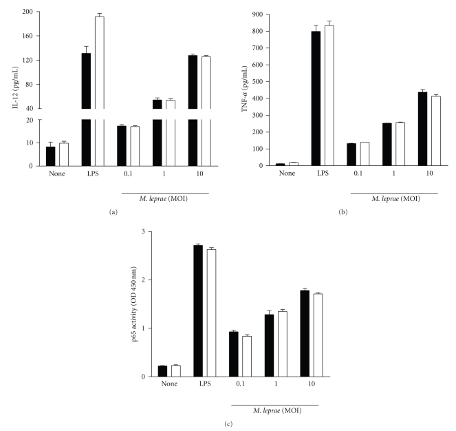
Macrophages from C57BL/6 and A/J mice were infected with M. leprae and the levels of IL-12 and TNF-α produced by macrophages were measured by ELISA. The production of two cytokines was similar in macrophages from both mice (Figures 1(a) and 1(b)). NF-κB activity levels measured with the p65 activity kit were also similar in both macrophages following infection with M. leprae (Figure 1(c)).
Figure 1.
TNF-α and IL-12 production and NF-κB activation in response to M. leprae infection in macrophages from A/J and C57BL/6 mice. Macrophages (106) from C57BL/6 and A/J mice were treated with LPS (100 ng/ml) and M. leprae (MOI of 0.1, 1.0, and 10.0) for 18 h, and supernatants and cell extracts were assayed for cytokines (IL-12 and TNF-α) and NF-κB, respectively. Open bar and closed bar represent macrophages from C57BL/6 and A/J mice, respectively. Data are representative of at least three independent experiments, each performed in triplicate.
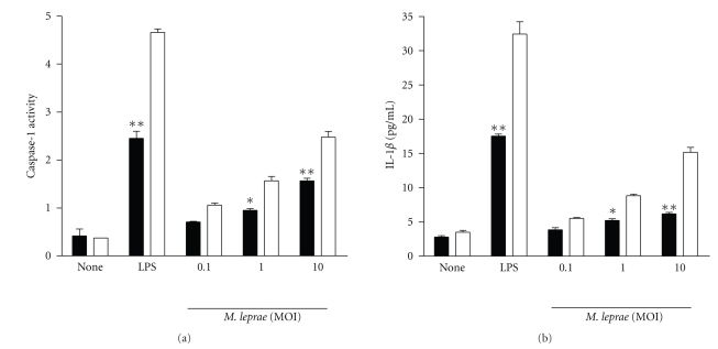
A previous report showed the differences in Legionella-induced IL-1β levels between A/J and C57BL/6 macrophages [5]. We measured the activation of caspase-1 after infection of macrophages with M. leprae because the maturation and secretion of IL-1β is dependent on the activation of caspase-1. Caspase-1 activity was lower in macrophages from A/J mice than in those from C57BL/6 mice (Figure 2(a)). We next studied the production of IL-1β during infection with M. leprae. As expected, IL-1β production was rather low in A/J macrophages whereas significant levels of mature IL-1β were measured in the culture supernatants of C57BL/6 macrophages (Figure 2(b)). The reduction in caspase-1 activation and in IL-1β production was however only partial in cells from the A/J mice stimulated with either LPS or M. leprae.
Figure 2.
Caspase-1 activity and IL-1β production in response to M. leprae infection in macrophages from A/J and C57BL/6 mice. Macrophages (106) from C57BL/6 and A/J mice were treated with LPS (100 ng/ml) and M. leprae (MOI of 0.1, 1.0, and 10.0) for 18 h, and supernatants and cell lysates were assayed for IL-1β concentrations and for caspase-1 activity, respectively. Open bar and closed bar represent macrophages from C57BL/6 and A/J mice, respectively. Data are representative of at least three independent experiments, each performed in triplicate. *P < .05; **P < .01.
3.2. Caspase-1 Activity and IL-1β Secretion by MLL Was Also Reduced in A/J Macrophages
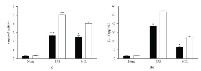
Our previous data showed that upon exposure to MLL, macrophages produced the proinflammatory cytokine IL-12 and TNF-α [1, 2]. In addition, we also investigated caspase-1 activity and IL-1β production in response to M. leprae lysate (MLL) in macrophages from C57BL/6 and A/J mice. The results show that caspase-1 activity and IL-1β secretion were lower in macrophages from A/J mice than in those from C57BL/6 mice (Figure 3).
Figure 3.
Caspase-1 activity and IL-1β production in response to M. leprae lysate (MLL) in macrophages from A/J and C57BL/6 mice. Macrophages (106) from C57BL/6 and A/J mice were treated with LPS (100 ng/ml) and the cell lysates of M. leprae (1 μg/ml) for 18 h, and supernatants and cell lysates were assayed for IL-1β concentrations and for caspase-1 activity, respectively. Open bar and closed bar represent macrophages from C57BL/6 and A/J mice, respectively. Data are representative of at least three independent experiments, each performed in triplicate. *P < .05; **P < .01.
4. Discussion
NLR proteins play an important role in the surveillance of mammalian cytosol, providing a crucial interface between invading bacterial pathogens and the host immune system. Intracellular detection of the bacterium itself and of bacterially derived molecules might signal a danger to the host cell that is amplified and synergized with signals from cell surface receptors, such as the TLRs. The ultimate outcome of cytosolic NLR signaling is to trigger a proinflammatory response by activation and secretion of cytokines via the NF-κB pathway and the inflammasome [10, 11].
IL-1β is one of proinflammatory cytokines and is expressed following an inflammatory stimulus. In the inflammasome, which induces caspase-1-mediated generation of IL-1β, IL-1β has a critical role in the prevention of intracellular pathogens, including Shigella, Salmonella, Listeria, Legionella, Francisella, as well as Bacillus anthracis [10, 12, 13].
Our data suggest that allelic difference in Naip5 found between A/J and C57BL/6 mice [5] partially controls caspase-1 activity and IL-1β secretion by macrophages in response to M. leprae infection.
Previous studies concluded that infection with Legionella activates Naip5 by delivering flagellin through its type IV secretion system, which then induces Ipaf-mediated caspase-1 activation and cell death to restrict Legionella replication [5, 14–16]. M. leprae is an intracellular pathogen and may also translocate into the cytosol [17] in which is able to interact with some NLRs. We also used MLL as another stimulant, and the results were similar to the data shown in M. leprae strain. Although flagellin is responsible for naip5-mediated caspase-1 activation in response to legionella, we suggest that presumably another component of M. leprae is responsible for its interaction with Naip5. Future study should include the analysis of cell wall components of mycobacteria and how they correlate with immune modulation via Naip 5.
The previously described susceptibility of Legionella was shown at low MOIs of 1 or 0.5 [5]. Our study used at MOIs 0.1, 1.0, and 10 of M. leprae per macrophage and the differences in response to M. leprae in macrophage from the two mouse strains were not significant unless MOIs of 1 or 10 were used. Because M. leprae is slow-growing bacteria (doubling time is about 21 days) in contrast to rapid-growing E. coli and legionella, it is likely that the response of macrophages to M. leprae is induced at high MOI.
In our future studies, we will investigate whether susceptibility to mycobacterial infection, such as tuberculosis and leprosy, is increased in the absence of caspase-1 or IL-1β, as would indicate the importance of the inflammasome in host defense against mycobacterial infection. In addition, a downstream molecular target of Naip5 will be identified.
5. Conclusions
Although we did not find a component of M. leprae, which is responsible for the interaction with Naip5, it is likely that Naip5 is partially required for caspase-1 activation and IL-1β secretion by macrophages in response to M. leprae infection in our study using A/J mice, suggesting the possibility that NLRs may have a role in the innate immune response to M. leprae.
Acknowledgment
This paper was supported by the Sahmyook University Research Fund in 2008.
References
- 1.Kang TJ, Lee S-B, Chae G-T. A polymorphism in the toll-like receptor 2 is associated with IL-12 production from monocyte in lepromatous leprosy. Cytokine. 2002;20(2):56–62. doi: 10.1006/cyto.2002.1982. [DOI] [PubMed] [Google Scholar]
- 2.Kang TJ, Yeum CE, Kim BC, You E-Y, Chae G-T. Differential production of interleukin-10 and interleukin-12 in mononuclear cells from leprosy patients with a Toll-like receptor 2 mutation. Immunology. 2004;112(4):674–680. doi: 10.1111/j.1365-2567.2004.01926.x. [DOI] [PMC free article] [PubMed] [Google Scholar]
- 3.Kawai T, Akira S. TLR signaling. Cell Death and Differentiation. 2006;13(5):816–825. doi: 10.1038/sj.cdd.4401850. [DOI] [PubMed] [Google Scholar]
- 4.Philpott DJ, Girardin SE. The role of Toll-like receptors and Nod proteins in bacterial infection. Molecular Immunology. 2004;41(11):1099–1108. doi: 10.1016/j.molimm.2004.06.012. [DOI] [PubMed] [Google Scholar]
- 5.Lamkanfi M, Amer A, Kanneganti T-D, et al. The Nod-like receptor family member Naip5/Birc1e restricts Legionella pneumophila growth independently of caspase-1 activation. Journal of Immunology. 2007;178(12):8022–8027. doi: 10.4049/jimmunol.178.12.8022. [DOI] [PubMed] [Google Scholar]
- 6.Kang T-J, You J-C, Chae G-T. Identification of catalase-like activity from Mycobacterium leprae and the relationship between catalase and isonicotinic acid hydrazide (INH) Journal of Medical Microbiology. 2001;50(8):675–681. doi: 10.1099/0022-1317-50-8-675. [DOI] [PubMed] [Google Scholar]
- 7.Shepard CC, McRae DH. A method for counting acid-fast bacteria. International Journal of Leprosy and Other Mycobacterial Diseases. 1968;36(1):78–82. [PubMed] [Google Scholar]
- 8.Kang TJ, Fenton MJ, Weiner MA, et al. Murine macrophages kill the vegetative form of Bacillus anthracis . Infection and Immunity. 2005;73(11):7495–7501. doi: 10.1128/IAI.73.11.7495-7501.2005. [DOI] [PMC free article] [PubMed] [Google Scholar]
- 9.Joshi VD, Kalvakolanu DV, Hebel JR, Hasday JD, Cross AS. Role of caspase 1 in murine antibacterial host defenses and lethal endotoxemia. Infection and Immunity. 2002;70(12):6896–6903. doi: 10.1128/IAI.70.12.6896-6903.2002. [DOI] [PMC free article] [PubMed] [Google Scholar]
- 10.Lamkanfi M, Kanneganti T-D, Franchi L, Núñez G. Caspase-1 inflammasomes in infection and inflammation. Journal of Leukocyte Biology. 2007;82(2):220–225. doi: 10.1189/jlb.1206756. [DOI] [PubMed] [Google Scholar]
- 11.Martinon F, Burns K, Tschopp J. The Inflammasome: a molecular platform triggering activation of inflammatory caspases and processing of proIL-β . Molecular Cell. 2002;10(2):417–426. doi: 10.1016/s1097-2765(02)00599-3. [DOI] [PubMed] [Google Scholar]
- 12.Henry T, Brotcke A, Weiss DS, Thompson LJ, Monack DM. Type I interferon signaling is required for activation of the inflammasome during Francisella infection. Journal of Experimental Medicine. 2007;204(5):987–994. doi: 10.1084/jem.20062665. [DOI] [PMC free article] [PubMed] [Google Scholar]
- 13.Kang TJ, Basu S, Zhang L, et al. Bacillus anthracis spores and lethal toxin induce IL-1β via functionally distinct signaling pathways. European Journal of Immunology. 2008;38(6):1574–1584. doi: 10.1002/eji.200838141. [DOI] [PMC free article] [PubMed] [Google Scholar]
- 14.Zamboni DS, Kobayashi KS, Kohlsdorf T, et al. The Birc1e cytosolic pattern-recognition receptor contributes to the detection and control of Legionella pneumophila infection. Nature Immunology. 2006;7(3):318–325. doi: 10.1038/ni1305. [DOI] [PubMed] [Google Scholar]
- 15.Lightfield KL, Persson J, Brubaker SW, et al. Critical function for Naip5 in inflammasome activation by a conserved carboxy-terminal domain of flagellin. Nature Immunology. 2008;9(10):1171–1178. doi: 10.1038/ni.1646. [DOI] [PMC free article] [PubMed] [Google Scholar]
- 16.Losick VP, Stephan K, Smirnova II, Isberg RR, Poltorak A. A hemidominant Naip5 allele in mouse strain MOLF/Ei-derived macrophages restricts Legionella pneumophila intracellular growth. Infection and Immunity. 2009;77(1):196–204. doi: 10.1128/IAI.01011-08. [DOI] [PMC free article] [PubMed] [Google Scholar]
- 17.van der Wel N, Hava D, Houben D, et al. M. tuberculosis and M. leprae translocate from the phagolysosome to the cytosol in myeloid cells. Cell. 2007;129(7):1287–1298. doi: 10.1016/j.cell.2007.05.059. [DOI] [PubMed] [Google Scholar]


