Figure 1.

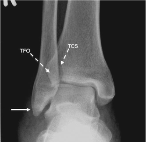
Anterior-posterior view of the ankle
Standard radiographs for suspected ankle injury include anterior-posterior (AP), lateral, and mortise views.1,2 On this AP radiograph, the solid white arrow demonstrates a subtle fracture of the distal fibula; the ankle mortise is intact. On AP ankle films, cortical disruption or talar tilt should be identified. If tibiofibular overlap (TFO)—the distance between the lateral border of the tibia and the medial border of the fibula—is less than 10 mm, or the tibiofibular clear space (TCS)—the distance between the medial border of the fibula and the lateral aspect of the posterior tibial malleolus—is greater than 5 mm, associated syndesmotic injury is likely. Greater than 2 mm difference between the lateral and medial joint space above the talus indicates talar tilt suggestive of medial or lateral disruption of this joint.1,2
Figure 2.

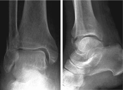
Bimalleolar Ankle Fracture
Anterior-posterior (AP) view (left) of the ankle demonstrates fracture of the fibula visualized as cortical disruption along the lateral border and a subtle distal tibia fracture seen approximately 2 mm above the distal tip, with preservation of the posterior border of the tibia (seen on lateral view [right]). In addition, the AP view reveals widening of the medial aspect of the superior talar joint space compared with the lateral space, suggesting talar tilt. This pattern of distal fibula fracture with medial malleolus involvement is often due to supination-external rotation injury and is likely associated with significant joint instability if the deltoid ligament is disrupted.1 A small avulsion of the talar neck is also seen along the medial border, opposite the site of the distal tibia fracture.
Figure 3.

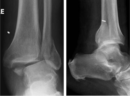
Trimalleolar Ankle Fracture
Anterior-posterior (AP) (left) and lateral (right) views demonstrate fracture of the distal fibula, medial malleolus, and posterior tibial malleolus with associated shortening. Note the decreased tibiofibular overlap (TFO) and significant talar tilt on the AP radiograph. Fractures that can't be reduced or which involve widening of the ankle mortise require urgent orthopedic consultation for possible open reduction internal fixation (ORIF) to prevent complications of avascular necrosis, malunion, or nonunion. Subtle nondisplaced fractures or displaced ankle fractures that have been anatomically reduced can be treated with a posterior splint and stirrup, crutches and non-weight-bearing, with close orthopedic follow-up.1
Figure 4.

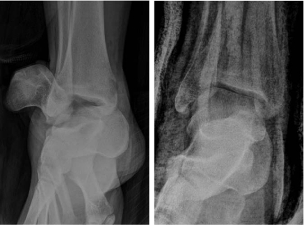
Talar neck fracture-dislocation
Slightly oblique anterior-posterior radiographs show a talar neck fracture-dislocation with associated subluxation of the subtalar joint pre- and postreduction. The talus was reduced into better anatomic position, but talar tilt and joint instability are still evident postreduction. Open ankle fractures (such as this case) are surgical (orthopedic) emergencies, requiring immediate reduction, irrigation and antibiotics, and tetanus vaccination if indicated. This Hawkins Type IV fracture has a near 100% likelihood of avascular necrosis due to the extreme level of displacement.3
References
- Koval KJ, Zuckerman JD, editors. Handbook of fractures. 3rd ed. Philadelphia, PA: Lippincott Williams & Wilkins; 2006. Ankle fractures; pp. 398–423. p. [Google Scholar]
- del Castillo J. Foot and ankle injuries. In: Adams JG, editor. Emergency medicine. 1st ed. Philadelphia, PA: Saunders Elsevier; 2008. pp. 897–909. p. [Google Scholar]
- Koval KJ, Zuckerman JD, editors. Handbook of fractures. 3rd ed. Philadelphia, PA: Lippincott Williams & Wilkins; 2006. Talus; pp. 435–42. p. [Google Scholar]


