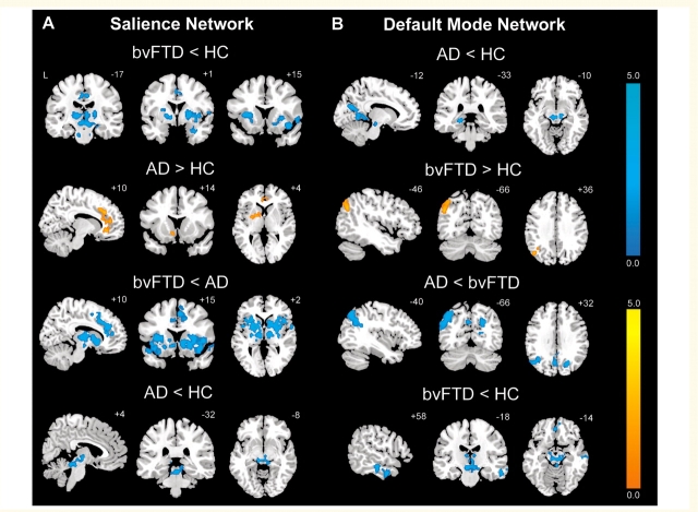
Figure 2.
BvFTD and Alzheimer’s disease feature divergent Salience Network and DMN dynamics. Group difference maps illustrate clusters of significantly reduced or increased connectivity for each ICN. In the Salience Network (A), patients with bvFTD showed distributed connectivity reductions compared to healthy controls (HC) and patients with Alzheimer’s disease (AD), whereas patients with Alzheimer’s disease showed increased connectivity in anterior cingulate cortex and ventral striatum compared to healthy controls. In the DMN (B), patients with Alzheimer’s disease showed several connectivity impairments compared to healthy controls and patients with bvFTD, whereas patients with bvFTD showed increased left angular gyrus connectivity. Patients with bvFTD and Alzheimer’s disease further showed focal brainstem connectivity disruptions within their ‘released’ network (DMN for bvFTD, Salience Network for Alzheimer’s disease). Results are displayed at a joint height and extent probability threshold of P < 0.05, corrected at the whole brain level. Colour bars represent t-scores, and statistical maps are superimposed on the Montreal Neurological Institute template brain.

