

Abstract
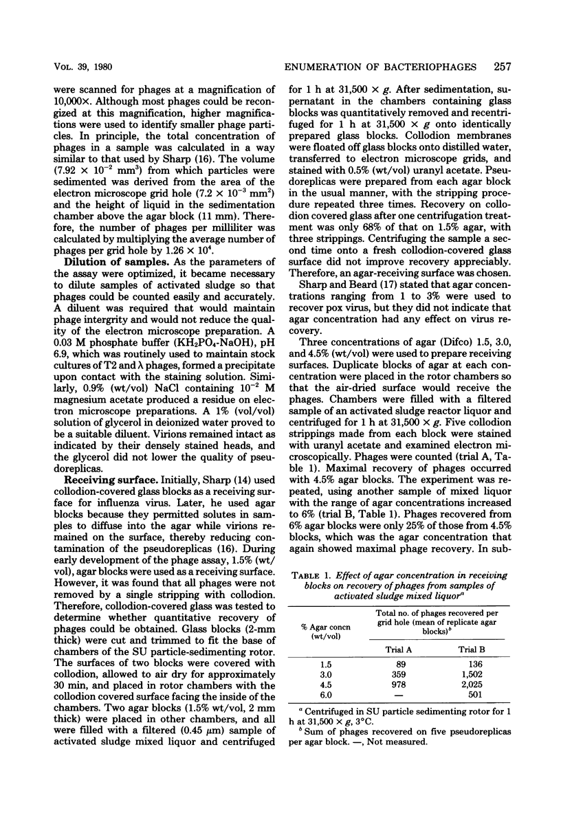
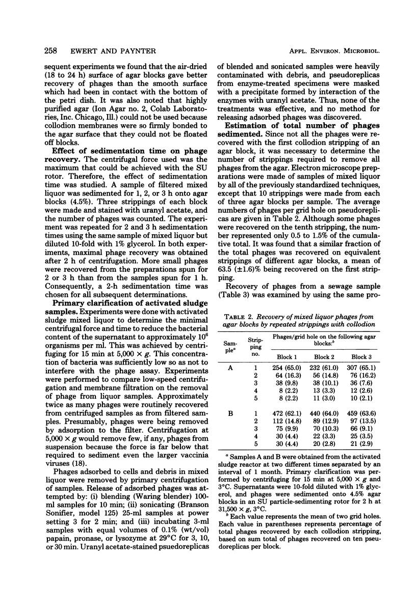
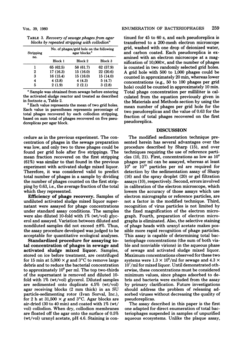
A direct method is described for measuring bacteriophage concentrations in complex aqueous systems. Conditions for sample clarification, phage recognition, and recovery were optimized. In contrast to the plaque assay, this procedure permits quantitation of total numbers of phages independent of bacterial host. Also, the modifications increase the sensitivity of the sedimentation assay, permitting detection of particles at a minimum concentration of 10(4) per ml. Maximal total phage concentrations in the aqueous phase of sewage and activated sludge mixed liquor were 1.3 x 10(6) and 4.3 x 10(7) per ml, respectively.
Full text
PDF
Images in this article
Selected References
These references are in PubMed. This may not be the complete list of references from this article.
- Anderson T. F. The Activation of the Bacterial Virus T4 by l-Tryptophan. J Bacteriol. 1948 May;55(5):637–649. doi: 10.1128/jb.55.5.637-649.1948. [DOI] [PMC free article] [PubMed] [Google Scholar]
- BRENNER S., HORNE R. W. A negative staining method for high resolution electron microscopy of viruses. Biochim Biophys Acta. 1959 Jul;34:103–110. doi: 10.1016/0006-3002(59)90237-9. [DOI] [PubMed] [Google Scholar]
- Bradley D. E. Ultrastructure of bacteriophage and bacteriocins. Bacteriol Rev. 1967 Dec;31(4):230–314. doi: 10.1128/br.31.4.230-314.1967. [DOI] [PMC free article] [PubMed] [Google Scholar]
- HUXLEY H. E., ZUBAY G. Preferential staining of nucleic acid-containing structures for electron microscopy. J Biophys Biochem Cytol. 1961 Nov;11:273–296. doi: 10.1083/jcb.11.2.273. [DOI] [PMC free article] [PubMed] [Google Scholar]
- KELLENBERGER E., ARBER W. Electron microscopical studies of phage multiplication. I. A method for quantitative analysis of particle suspensions. Virology. 1957 Apr;3(2):245–255. doi: 10.1016/0042-6822(57)90091-0. [DOI] [PubMed] [Google Scholar]
- LURIA S. E., WILLIAMS R. C., BACKUS R. C. Electron micrographic counts of bacteriophage particles. J Bacteriol. 1951 Feb;61(2):179–188. doi: 10.1128/jb.61.2.179-188.1951. [DOI] [PMC free article] [PubMed] [Google Scholar]
- Mayor H. D., Jamison R. M., Jordan L. E., Melnick J. L. Structure and Composition of a Small Particle Prepared from a Simian Adenovirus. J Bacteriol. 1965 Jul;90(1):235–242. doi: 10.1128/jb.90.1.235-242.1965. [DOI] [PMC free article] [PubMed] [Google Scholar]
- SHARP D. G., BEARD J. W. Counts of virus particles by sedimentation on agar and electron micrography. Proc Soc Exp Biol Med. 1952 Oct;81(1):75–79. doi: 10.3181/00379727-81-19782. [DOI] [PubMed] [Google Scholar]
- SHARP D. G., OVERMAN J. R. Enumeration of vaccinia virus particles in crude extracts of infected tissues by electron microscopy. Proc Soc Exp Biol Med. 1958 Nov;99(2):409–413. doi: 10.3181/00379727-99-24366. [DOI] [PubMed] [Google Scholar]
- SMITH K. O., MELNICK J. L. A method for staining virus particles and identifying their nucleic acid type in the electron microscope. Virology. 1962 Jul;17:480–490. doi: 10.1016/0042-6822(62)90143-5. [DOI] [PubMed] [Google Scholar]
- Sturdza S. A., Russu-Pandelesco M. Recherches écologiques phago-bactériennes dans le milieu extérieur. (Un bilan) Arch Roum Pathol Exp Microbiol. 1967 Mar;26(1):125–154. [PubMed] [Google Scholar]