Abstract

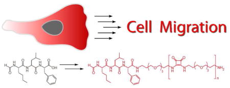
Polyethylene glycol (PEG) is used widely, and many biologically active molecules are modified with oligoethylene glycol substituents to enhance their half-life in circulation. The pervasive use of PEG substituents is partly due to their presumed inertness. Our investigation of formyl peptide receptor (FPR)-mediated chemotaxis reveals that oligoethylene glycol substitution can enhance the ability of the peptide chemoattractant fMLF to activate signal transduction through FPR, a transmembrane G-protein coupled receptor.
Polyethylene glycol (PEG) often is used to enhance key properties of biologically active molecules.1, 2 PEG substituents can augment the efficacy of protein and peptide therapeutic agents, by protecting them from proteolytic degradation3 or decreasing their rate of clearance from plasma.2 The widespread use of PEG for these purposes stems from its low toxicity, excellent aqueous solubility, and low antigenicity. These properties appear to be shared by ethylene glycol oligomers. Indeed, oligoethylene glycol groups have been employed to tether biological recognition elements (e.g. to form dimers4 or higher order oligomers5). Because their persistence length can be estimated,6 oligoethylene glycol moieties are attractive linkers in building potent multivalent ligands.4,5 Moreover, oligoethylene glycol moieties on surfaces or in biomaterials resist non-specific protein binding.7 Thus, the widespread use of oligoethylene substituents also stems from their presumed inertness. Here, we present results demonstrating that an oligoethylene glycol substituent can enhance the potency of a ligand for a transmembrane G-protein coupled receptor (GPCR).
Our studies originated from our interest in chemotactic signaling. A key initiator of neutrophil chemotaxis is the formyl peptide receptor (FPR). FPR belongs to the largest and the most diverse family of integral membrane signaling receptors, the GPCR family.8 FPR, which is present at high levels on the surface of neutrophils and monocytes, mediates chemotactic responses to N-formylated peptides, including the canonical chemoattractant N-formyl-methionine-leucine-phenylalanine (fMLF). Formylated peptides are produced from sources that include the mitochondrial proteins of ruptured host cell and the proteins of invading pathogens.9 The molecular details of FPR–ligand complexes have not yet been elucidated; however, modeling of the seven transmembrane α-helices10 suggests that FPR binding site can accommodate four to five amino acids.11 Structure-activity relationship data indicate that formyl peptide derivatives with C-terminal substituents can retain the activity of the parent compound.12,4a Because we were interested in generating formyl peptide probes of chemotactic signaling, we tested the consequences of adding C-terminal linker substituents.
Precedent suggested that a tether based on oligoethylene glycol would have little effect on signaling. To test this assumption, we appended a series of ethylene glycol oligomers to the C-terminus of a formyl peptide. The FPR ligand we employed, N-formyl-norleucine-leucine-phenylalanine (fNleLF), is a chemoattractant.13 Though less potent than fMLF, its chemical stability is superior. Specifically, the methionine residue in fMLF can undergo oxidation, thereby complicating the synthesis and handling of its derivatives. In contrast, fNleLF-based compounds are stable. To assemble the target compounds, oligoethylene glycol building blocks 2 and 4 were synthesized.5a These precursors could be conjugated to the peptidic chemoattractant to yield a series of C-terminal modified fNleLF derivatives.
We used squarate-derived building block 4 and the free peptide (1) to assemble a series of derivatives possessing C-terminal substituents with six (5), nine (6), or twelve (7) ethylene glycol units. The resulting compounds were evaluated for their abilities to activate signaling in FPR-transfected U937 cells, a monocytic cell line.14 Like neutrophils, these cells can respond to even a shallow gradient of chemoattractant.15 To assay chemotactic responses, we employed a simplified multi-well Boyden chamber assay, and the number of migrating cells was determined by using a cell proliferation assay.16,17 All of the fNleLF derivatives promote cell migration and therefore serve as attractants. Their differential effects on chemotaxis, however, were surprising. Specifically, the more hydrophilic ethylene glycol unit might be expected to decrease the ability of fNleLF to bind to its transmembrane receptor and thereby mitigate attractant activity. Unexpectedly, these substituents had a dramatic positive effect on chemotaxis (Figure 1A). Compound 5, with six ethylene glycol units, is a more powerful attractant than the free N-formyl peptide. Compound 6 with nine ethylene glycol units is even more potent. Indeed, compared to the non-derivatized formyl peptide 1, compound 6 is >20-fold more active. The trend, however, did not continue beyond nine units. Compound 7, which possesses an oligoethylene glycol substituent of twelve units, is less active than compound 6. These results suggest that the oligoethylene glycol substituent is not inert—it increases chemotactic activity, and the extent of that increase depends upon its length.
Figure 1.
Effects of the formyl peptide derivatives. (A) Chemotactic responses of FPR-transfected U937 cells to formyl peptides. Data shown are from three separate experiments conducted in triplicate. The standard error is depicted. (B) Change in intracellular Ca2+ concentration induced by formyl peptides. Cells were loaded with ratiometric dye Indo-1,18 and emission ratios were measured using a Photon Technology International fluorimeter. The results shown are from a representative experiment using formyl peptides 1 and 5–7 (10 nM). Experiments also were performed at different peptide concentrations (see supporting information).
To test whether the differences in the cell migration assay depend on FPR signaling, we evaluated the ability of the fNleLF derivatives to elevate a key indicator of chemotactic signaling—intracellular calcium ion concentration ([Ca2+]i). As shown in Figure 1B, formyl peptide 1 induced an increase in [Ca2+]i,and oliogethuylene glycol derivatives 5–7 likewise activated signaling. The most active fNleLF derivatives in this assay were also most potent attractants. Specifically, compound 6, a powerful chemoattractant, caused the greatest increase in [Ca2+]i; it was >10-fold more potent than formyl peptide 1. These findings link the observed changes in chemotactic activity to increases in intracellular signaling.
We conducted several additional experiments to determine whether the responses observed were mediated through the FPR. To test whether the oligoethylene glycol unit alone can elicit chemotaxis or FPR-mediated signaling, we synthesized compound 8 (see supporting information). This compound neither promoted chemotaxis nor signaling (see supporting information). We also assessed whether the effects of 1 and 5–7 depend upon FPR by evaluating their ability to elicit chemotaxis of FPR-negative U937 cells. None of the compounds induce chemotactic activity in this cell line. These results indicate that the observed activity arises from formyl peptide recognition by FPR.
The observed enhancements in chemotactic activity might stem solely from C-terminal substitution and not from the nature of the substituent. To test for this possibility, we synthesized formyl peptide derivative 9 (see supporting information), which possesses an alkyl linker with a length comparable to that of the ethylene glycol substituent in 5. Though 5 is a more potent attractant than the unsubstituted 1, alkyl derivative 9 has no enhanced activity in either assay. These data indicate that the unexpected increase in the activity of the formyl peptide derivatives 5–7 is due to the oligoethylene glycol substituent.
PEG substituents are known to exhibit a long-range protein repellent effect. At short range, however, the interaction between PEG and protein can become attractive and therefore facilitate binding.19 Thus, oligoethylene glycol substituents might contribute to the binding affinity of the ligands to the receptor. To test for this possibility, we performed a competitive binding assay of oligoethylene glycol substituented fNleLF derivatives using commercially available fNleLFNleYK-FITC. Our data indicate that oligoethylene glycol substitution could contribute to binding affinity as the dissociation constants (Kd) of oligoethylene glycol substituents are slightly lower (4-7-fold) than that of the non-derivatized fNleLF (see supporting information). Still, the affinity differences for these formyl peptide derivatives are subtle, suggesting that other factors contribute to the differences in the cellular responses they elicit. For example, the oligoethylene glycol substituent may stabilize an active signaling conformation, increase the conformational flexibility of FPR or alter its oligomerization state. Regarding the latter, several studies show that agonists can disrupt GPCR oligomerization.20 An ethylene glycol group could serve in that capacity. It is also interesting to note that the activity of the compound with the largest oligoethylene glycol substituent, 7, is less active than compound 6. As the ethylene glycol substituent becomes more sterically demanding, it may impede binding.
In summary, ethylene glycol-substituted chemoattractants show enhanced activities. The magnitude of the increase depends upon the length of the oligoethylene glycol substituent. The physiological importance of GPCRs is underscored by the many drugs that target them. Our finding that an oligoethylene glycol unit can enhance the activity of a chemotactic agonist provides a blueprint for generating highly potent GPCR agonists.
Supplementary Material
Scheme 1.
Route to formyl peptides designed to bind to FPR.
Acknowledgments
This research was supported by the NIH (R01 GM055984). We thank Dr. E. R. Prossnitz for the FPR-transfected U937 cell line and Dr. A. C. Lamanna for helpful scientific discussions.
Footnotes
Supporting Information Available: Synthetic methods and experimental details (PDF). This material is available free of charge via the internet at http://pubs.acs.org.
References
- 1.(a) Delgado C, Francis GE, Fisher D. Crit Rev Ther Drug. 1992;9:249–304. [PubMed] [Google Scholar]; (b) Roberts MJ, Bentley MD, Harris JM. Adv Drug Deliver Rev. 2002;54:459–476. doi: 10.1016/s0169-409x(02)00022-4. [DOI] [PubMed] [Google Scholar]
- 2.Yamaoka T, Tabata Y, Ikada Y. J Pharm Sci. 1994;83:601–606. doi: 10.1002/jps.2600830432. [DOI] [PubMed] [Google Scholar]
- 3.Harris JM, Chess RB. Nat Rev Drug Discovery. 2003;2:214–221. doi: 10.1038/nrd1033. [DOI] [PubMed] [Google Scholar]
- 4.(a) Miyazaki M, Kodama H, Fujita I, Hamasaki Y, Miyazaki S, Kondo M. J Biochem. 1995;117:489–494. doi: 10.1093/oxfordjournals.jbchem.a124734. [DOI] [PubMed] [Google Scholar]; (b) Kramer RH, Karpen JW. Nature. 1998;395:710–713. doi: 10.1038/27227. [DOI] [PubMed] [Google Scholar]; (c) Karellas P, McNaughton M, Baker SP, Scammells PJ. J Med Chem. 2008;51:6128–6137. doi: 10.1021/jm800613s. [DOI] [PubMed] [Google Scholar]
- 5.(a) Fan E, Zhang Z, Minke WE, Hou Z, Verlinde CLMJ, Hol WGJ. J Am Chem Soc. 2000;122:2663–2664. [Google Scholar]; (b) Solomon D, Kitov PI, Paszkiewicz E, Grant GA, Sadowska JM, Bundle DR. Org Lett. 2005;7:4369–4372. doi: 10.1021/ol051529+. [DOI] [PubMed] [Google Scholar]; (c) Gestwicki JE, Strong LE, Borchardt SL, Cairo CW, Schnoes AM, Kiessling LL. Bioorg Med Chem. 2001;9:2387–2393. doi: 10.1016/s0968-0896(01)00162-6. [DOI] [PubMed] [Google Scholar]; (d) Kiessling LL, Gestwicki JE, Strong LE. Annu Rep Med Chem. 2000;35:321–330. [Google Scholar]; (e) Kim Y, Hechler B, Gao ZG, Gachet C, Jacobson KA. Bioconjugate Chem. 2009;20:1888–1898. doi: 10.1021/bc9001689. [DOI] [PMC free article] [PubMed] [Google Scholar]
- 6.Knoll D, Hermans J. J Biol Chem. 1983;258:5710–5715. [PubMed] [Google Scholar]
- 7.(a) Pale-Grosdemange C, Simon ES, Prime KL, Whitesides GM. J Chem Soc. 1991;113:12–20. [Google Scholar]; (b) Prime KL, Whitesides GM. Science. 1991;252:1164–1167. doi: 10.1126/science.252.5009.1164. [DOI] [PubMed] [Google Scholar]; (c) Elbert DI, Hubbell JA. Annu Rev Mater Sci. 1996;26:365–394. [Google Scholar]
- 8.Murphy PM. Annu Rev Immunol. 1994;12:593–633. doi: 10.1146/annurev.iy.12.040194.003113. [DOI] [PubMed] [Google Scholar]
- 9.(a) Schiffmann E, Corcoran BA, Wahl SM. Proc Natl Acad Sci. 1975;72:1059–1062. doi: 10.1073/pnas.72.3.1059. [DOI] [PMC free article] [PubMed] [Google Scholar]; (b) Prossnitz ER, Ye RD. Pharmacol Ther. 1997;74:73–102. doi: 10.1016/s0163-7258(96)00203-3. [DOI] [PubMed] [Google Scholar]
- 10.Miettinen HM, Mills JS, Gripentrog JM, Dratz EA, Granger BL, Jesaitis AJ. J Immunol. 1997;159:4045–4054. [PubMed] [Google Scholar]
- 11.Sklar LA, Fay SP, Selismann BE, Freer RJ, Muthukumaraswamy N, Mueller H. Biochemistry. 1990;29:313–6. doi: 10.1021/bi00454a002. [DOI] [PubMed] [Google Scholar]
- 12.Freer RJ, Day AR, Muthukumaraswamy N, Pinon D, Wu A, Showell HJ, Becker EL. Biochemistry. 1982;21:257–263. doi: 10.1021/bi00531a009. [DOI] [PubMed] [Google Scholar]
- 13.Freer RJ, Day AR, Becker EL, Showell HJ, Schiffmann E, Gross E. Pept Struct Biol Funct, Proc Am Pept Symp, 13th. 1979:749–51. [Google Scholar]
- 14.Kew RR, Peng T, Dimartino SJ, Madhavan D, Weinman SJ, Cheng D, Prossnitz ER. J Leukocyte Biol. 1997;61:329–337. doi: 10.1002/jlb.61.3.329. [DOI] [PubMed] [Google Scholar]
- 15.Servant G, Weiner OD, Herzmark P, Bella T, Sedat JW, Bourne HR. Science. 2000;287:1037–1040. doi: 10.1126/science.287.5455.1037. [DOI] [PMC free article] [PubMed] [Google Scholar]
- 16.Boyden SV. J Exp Med. 1962;115:453–466. doi: 10.1084/jem.115.3.453. [DOI] [PMC free article] [PubMed] [Google Scholar]
- 17.Jones LJ, Gray M, Yue ST, Haugland RP, Singer VL. J Immunol Methods. 2001;254:85–98. doi: 10.1016/s0022-1759(01)00404-5. [DOI] [PubMed] [Google Scholar]
- 18.Grynkiewicz G, Poenie M, Tsien RY. J Biol Chem. 1985;260:3440–50. [PubMed] [Google Scholar]
- 19.(a) Sheth SR, Leckband D. Proc Natl Acad Sci USA. 1997;94:8399–8404. doi: 10.1073/pnas.94.16.8399. [DOI] [PMC free article] [PubMed] [Google Scholar]; (b) Israelachvili J. Proc Natl Acad Sci USA. 1997;94:8378–8379. doi: 10.1073/pnas.94.16.8378. [DOI] [PMC free article] [PubMed] [Google Scholar]
- 20.(a) Cheng ZJ, Miller LJ. J Biol Chem. 2001;276:48040–48047. doi: 10.1074/jbc.M105668200. [DOI] [PubMed] [Google Scholar]; (b) Latif R, Graves P, Davies TF. J Biol Chem. 2002;277:45059–45067. doi: 10.1074/jbc.M206693200. [DOI] [PubMed] [Google Scholar]
Associated Data
This section collects any data citations, data availability statements, or supplementary materials included in this article.


