Summary
A sensitive and specific FRET biosensor was developed by Mizutani et al. and applied to detect the activity of BCR-ABL kinase in live cells. This biosensor allowed the detection of cancerous and drug-resistant cells, and the evaluation of kinase inhibitor efficacy. Future biosensor development and imaging can increasingly contribute to cancer diagnosis and therapeutics.
Keywords: FRET, Biosensor, Live Cell Imaging, Cancer, Drug Screening
In this issue of Clinical Cancer Research, Mizutani and colleagues reported the development and application of a sensitive and specific biosensor based on fluorescence resonance energy transfer (FRET) for the quantification of BCR-ABL activity and its response to drugs in living cells(1). This work pioneers the application of FRET-based biosensors to evaluate the efficacy of the kinase inhibitors with a clinically relevant experimental design. FRET between two chromophores occurs when the donor chromophore in excited state, through non-radiative dipole coupling, transfers energy to a correctly oriented acceptor at close proximity. Because FRET signals are independent of the chromophore concentration and excitation light intensity, the FRET mechanism has been widely applied to develop fluorescent biosensors for visualizing molecular activities in live cells with high spatiotemporal resolution.
Recently progress in molecular therapy highlights the targeting of certain malfunctioning molecules and pathways in cancer (2). For example, the small molecule kinase inhibitor imatinib has achieved great success in treating chronic myeloid leukemia (CML) and gastrointestinal stromal tumors whose growth is acutely dependent on the expression of specific kinase mutants (3). Ideally, the efficiency of a kinase inhibitor needs to be evaluated at the level of protein interactions, the living cell, and in animal models (4). At the protein level, the potential of kinase inhibitors to inhibit the phosphorylation of a substrate protein or peptide can be evaluated quite conveniently, even via commercially available services. However, there is a lack of technology to accurately evaluate the effect of kinase inhibitors in live cells. The genetically-encoded FRET biosensors based on fluorescent proteins (FPs) can be ideal candidates for this purpose (5). The feasibility of applying FRET biosensors for screening kinase inhibitors is underscored by the recent development of numerous FRET-based biosensors, such as those for the detection of oncogene-related kinase activities including Src, FAK, PKA, EGFR, and Abl (6, 7), and of other molecules important for migration and cancer invasion (5).
In this issue of Clinical Cancer Research, the authors utilized CrkL, a major substrate of the BCR-ABL kinase containing both tyrosine and SH2 domain, to be sandwiched between Venus (a variant of yellow FP) and enhanced cyan FP (ECFP). When the BCR-ABL kinase is active, the phosphorylated tyrosine site on CrkL can bind its intramolecular SH2 domain to cause a conformational change and subsequently FRET increase. The sensitivity of this biosensor was further improved by truncating the C-terminus of the substrate CrkL sequence, circularly permutating the donor ECFP, and monomerizing the acceptor Venus. While it is possible to further improve the biosensor sensitivity by replacing Venus with a bright yellow FP (YPet) which can form weak dimers with ECFP and hence increase the FRET efficiency of the activated biosensor (8), the final version of this FRET biosensor, termed Pickles, displayed a remarkable 80% increase of FRET ratio in vitro upon stimulation by BCR-Abl. The authors then carefully verified the specificity of the Pickles biosensor toward BCR-ABL by screening against an array of kinases. Mutations on the tyrosine site Y207 and the SH2 domains further confirmed that the FRET change of Pickles was indeed due to the designed intramolecular binding between the SH2 domain and phosphorylated tyrosine site within the CrkL sequence.
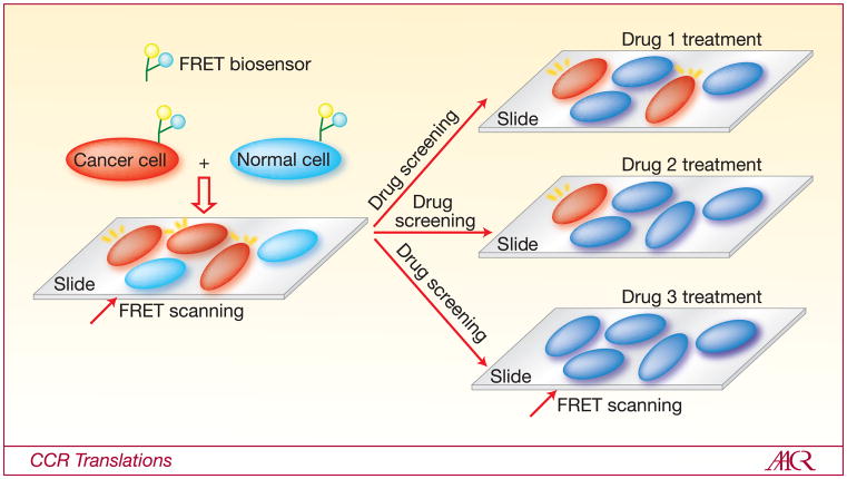
With the BCR-ABL FRET biosensor optimized, the authors further examined the efficacy of several established drugs for the treatment of CML, utilizing cell lines and primary cells expressing this biosensor (Figure 1). The results revealed that the FRET biosensor can detect the inhibitory effect of imatinib at the concentration as low as 0.1 μM. In contrast, established approaches based on western blotting or antibody staining coupled with flow cytometry can only detect the imatinib effect when it reaches the concentration level of 0.5 or 1 μM, respectively. Although it would be a fairer comparison if the biosensor signals were also assayed using flow cytometry when comparing to antibody staining, these quantification results demonstrated that the sensitivity of the FRET biosensor is outstanding and clearly superior over that of western blotting which is widely used for inhibitor screening (9). Further results revealed and verified that the two second-generation BCR-ABL inhibitors, nilotinib and dasatinib, were more potent than imatinib.
Figure 1. The application of FRET biosensor for drug screening.
Cancer (red) and normal (blue) cells in biopsy samples can be introduced with FRET biosensors to detect cancerous molecular activities, e.g. BCR-ABL kinase activity. The FRET scanning can identify the cancer cells and quantify their cancerous activities based on the FRET signals. The biopsy samples and cells expressing the biosensors can be subjected to different drug treatments to assess the efficacy of different drugs in inhibiting the target molecular activities
More importantly, the authors showed that the FRET biosensor in combination with flow cytometry can be used to detect a small percentage of drug-resistant cancer cells mixed in a large cell population. This is exciting since these drug-resistant cells may likely constitute the main reason for CML relapse and therapeutic failures. Therefore, the FRET biosensor developed here can provide a powerful tool to assess the biopsy samples from a particular patient and predict the future probability of his/her resistance to specific drugs. This should provide invaluable information for clinicians/physicians to identify and design alternative therapeutic approaches with a better efficiency and higher successful chance.
With the power demonstrated by the current paper, it is envisioned that numerous new FRET biosensors will be developed for cancer medicine. While the future potential is tremendous, the broad application of the FRET technology for cancer detection and drug screening is in need of further improvement on several aspects. First, the FRET biosensors at the current stage are generally introduced into live cells with methods involving liposome-based delivery, electroporation, or virus infection. The typical efficiency of these methods is around 20–30%. This low efficiency of delivering biosensors into cells, particularly primary cells, may constitute a major obstacle for the detection of rare cancer cells such as those detected in circulation, e.g. circulating tumor cells (CTCs) (10). Hence, new ways of delivering biosensors into cells with high efficiency are needed to promote the field of FRET application in cancer detection. Second, advanced flow cytometry with high-resolution imaging capability will be helpful to overcome the problem of cell heterogeneity and allow the high throughput screening of drugs with high accuracy and subcellular resolutions. Third, the majority of the biosensors were developed semi-rationally, mostly in a trial-and-error fashion based on personal experience and published literature. Systematic and high throughput screening approaches are hence in great need to automate the biosensor development and optimization. Finally, FRET biosensors with distinctive colors for the simultaneous visualization of multiple cancer-related molecular events, such as those previously published to visualize Src and MT1-MMP activities in the same single cell, can be very useful to provide precise assessment and prediction of cancer characteristics and drug efficacy (11).
In summary, FRET biosensors are ideal to screen the efficacy of the kinase inhibitors or drugs in live cells. In addition to drug screening, specific and sensitive biosensors can be developed for biomarkers with applications in early cancer detection, cancer prognosis, and monitoring of therapeutic efficacy. More importantly, FRET biosensors can be applied to visualize subcellular molecular signaling events in real time for the identification of novel targeting molecules and pathways, which may lead to a new era in clinical cancer research.
Abbreviations
- FRET
fluorescence resonance energy transfer
- CML
chronic myeloid leukemia
- FP
fluorescent proteins
- ECFP
enhanced cyan FP
- CTCs
circulating tumor cells
References
- 1.Mizutani T, Kondo T, Darmanin S, et al. A novel FRET-based biosensor for the measurement of BCR–ABL activity and its response to drugs in living cells. Clin Cancer Res. 2010:16. doi: 10.1158/1078-0432.CCR-10-0548. [DOI] [PubMed] [Google Scholar]
- 2.Sebolt-Leopold JS, English JM. Mechanisms of drug inhibition of signalling molecules. Nature. 2006;441:457–62. doi: 10.1038/nature04874. [DOI] [PubMed] [Google Scholar]
- 3.Sawyers C. Targeted cancer therapy. Nature. 2004;432:294–7. doi: 10.1038/nature03095. [DOI] [PubMed] [Google Scholar]
- 4.Zhang J, Yang PL, Gray NS. Targeting cancer with small molecule kinase inhibitors. Nat Rev Cancer. 2009;9:28–39. doi: 10.1038/nrc2559. [DOI] [PMC free article] [PubMed] [Google Scholar]
- 5.Wang Y, Shyy JY, Chien S. Fluorescence proteins, live-cell imaging, and mechanobiology: seeing is believing. Annu Rev Biomed Eng. 2008;10:1–38. doi: 10.1146/annurev.bioeng.010308.161731. [DOI] [PubMed] [Google Scholar]
- 6.Wang Y, Botvinick EL, Zhao Y, et al. Visualizing the mechanical activation of Src. Nature. 2005;434:1040–5. doi: 10.1038/nature03469. [DOI] [PubMed] [Google Scholar]
- 7.Zhang J, Allen MD. FRET-based biosensors for protein kinases: illuminating the kinome. Mol Biosyst. 2007;3:759–65. doi: 10.1039/b706628g. [DOI] [PubMed] [Google Scholar]
- 8.Ouyang M, Sun J, Chien S, Wang Y. Determination of hierarchical relationship of Src and Rac at subcellular locations with FRET biosensors. Proc Natl Acad Sci U S A. 2008;105:14353–8. doi: 10.1073/pnas.0807537105. [DOI] [PMC free article] [PubMed] [Google Scholar]
- 9.Bain J, Plater L, Elliott M, et al. The selectivity of protein kinase inhibitors: a further update. Biochem J. 2007;408:297–315. doi: 10.1042/BJ20070797. [DOI] [PMC free article] [PubMed] [Google Scholar]
- 10.Cristofanilli M, Budd GT, Ellis MJ, et al. Circulating tumor cells, disease progression, and survival in metastatic breast cancer. N Engl J Med. 2004;351:781–91. doi: 10.1056/NEJMoa040766. [DOI] [PubMed] [Google Scholar]
- 11.Ouyang M, Huang H, Shaner NC, et al. Simultaneous visualization of protumorigenic Src and MT1-MMP activities with fluorescence resonance energy transfer. Cancer Res. 70:2204–12. doi: 10.1158/0008-5472.CAN-09-3698. [DOI] [PMC free article] [PubMed] [Google Scholar]


