



Abstract
The potential usefulness of artificially selected peptides as probes to detect specific proteins has been proposed because of the ease and low cost of syntheses, manipulation and genetic expression. However, the affinities of these peptides to their target proteins are generally too low to be practical as diagnostic or bio-analytical reagents. One approach to this problem is to incorporate a redox-active amino acid 3,4-dihydroxy-L-phenylalanine (L-DOPA) which selectively forms a covalent linkage to the target protein. Such peptide-based probes can also be fused to tailored reporter proteins and easily expressed in bacterial cultures. As a demonstration, a candidate peptide TOP1 that weakly binds to the target protein, the SH3 domain of human Abl kinase, was fused to green fluorescent protein (GFP) and L-DOPA was site-specifically incorporated into the peptide region (TOP1-DOPA-GFP). TOP1-DOPA-GFP produced from E. coli was used in a Western blot-type experiment to show that the Abl SH3 domain can be detected in one step by observing the fluorescence. The molecular design presented in this work is significant in that the same approach could be used to transform many other protein-binding peptides with insufficient affinities into protein detection probes with a variety of fused reporter or therapeutic proteins.
Keywords: Peptides, Genetic Expression, Protein Detection, Cross-Linking, L-DOPA
Introductory Statement
Detection of specific proteins and protein-protein interactions is of a paramount importance in current biochemical and clinical laboratories. To supplement and improve the existing array of techniques in protein chemistry, the use of artificially selected peptides and peptide mimetic as probes to detect specific proteins has long been proposed [1; 2]. This is an attractive idea since synthetic peptides can be easily manipulated and/or prepared in large quantities at reasonable costs. Peptide ligands to certain proteins have been routinely isolated by various techniques such as phage display and in vitro one-bead-one-compound (OBOC) libraries [3; 4]. Significant advances have also been made recently in computational biology to simulate protein-protein and other protein-ligand interactions [5; 6]. Nonetheless, affinities of these laboratory-isolated peptides to their target proteins are often too low to be practically useful in any bio-analytical application. Whereas various In vitro chemical methods such as “click chemistry” [7] and other combinatorial chemistry [8] have been used to enhance the affinity of these peptides to the target proteins, the other approaches include the chemical coupling of the peptide to the target protein. Chemical cross-linking is relatively easy and inexpensive with a variety of commercially available cross-linking agents. However, the disadvantage of these small molecule-based coupling agents is that most of them are limited with low efficiency and/or selectivity and can form heterogeneous mixture of protein/peptide complexes, which could complicate the detection steps [9].
One solution to this problem has been suggested using a synthetic peptide containing a redox-active amino acid 3,4-dihydroxy-L-phenylalanine (L-DOPA, Figure 1) [10]. L-DOPA forms a reactive ortho-quinone intermediate upon selective oxidation by sodium periodate (NaIO4) and reacts with a nucleophile in its close proximity, thereby forming a stable covalent linkage between the DOPA-containing peptide and the interacting protein [10; 11; 12]. L-DOPA is a naturally occurring derivative of tyrosine and serves as a synthetic precursor for chatecholamine neurotransmitters in mammals [13]. L-DOPA is also found in some natural proteins such as mussel-adhesive proteins and related species [14; 15]. In these cases, L-DOPA is not encoded by the 64 codons of mRNA’s, but rather post- or co-translationally incorporated into the primary sequence of proteins through enzymatic modification of tyrosine [16; 17]. More recently, it has been demonstrated that L-DOPA can be genetically and site-specifically incorporated into recombinant proteins in Escherichia coli by suppressing an amber stop codon (TAG) [18]. L-DOPA can be a very powerful protein cross-linking agent based on its reactivity, chemical orthogonality to other conventional amino acids and the site-specific labeling of recombinant proteins. In fact, genetically incorporated L-DOPA has been shown to mediate a highly efficient and selective cross-linking of a protein-protein complex with moderate to weak interaction [19]. We hypothesize that peptides with low affinities could be transformed into practical molecular probes by utilizing the genetically encoded L-DOPA as a cross-linker and with various fused reporter proteins and other functional proteins. Moreover, such peptide-based probes can be easily produced in large quantities from bacterial cultures with a slight modification to the routine protein expression and purification procedures. As a proof-of-concept example, herein we present the design of a peptide-based molecular probe using a peptide with a (GFP) as a reporter protein. We then demonstrate that this fusion protein can be used in a Western blot-type experiment to detect the model target protein (Figure 1).
Figure 1.

Design and application of peptide-based molecular probe expressed in E. coli. Genetically tailored L-DOPA forms covalent linkage to the target protein to alleviate the low affinity of the peptide.
Materials and Methods
Vector Constructions
Plasmids pTre-Tight-AcGFP and pTetOff were purchased from Clontech. Restriction enzyme sites AgeI and KpnI were introduced into pTetOff previously to afford pTOAgeIKpnI [20]. The coding sequences for TOP1 and BOT1 were purchased as complementary oligonucleotides (Invitrogen) and annealed. Resulting double-stranded DNA fragments were digested sequentially with AgeI and KpnI and ligated to the vector pTOAgeIKpnI also digested with AgeI and KpnI to afford pTetOff -TOP1 and pTetOff-BOT1. The coding sequence of the SH3 domain of Abl tyrosine kinase was amplified by polymerase chain reaction (PCR) from cDNA BC117451.1:pCR4-TOPO (Open Biosystems). The PCR product was digested with AgeI and KpnI and ligated to pTOAgeIKpnI also digested with AgeI and KpnI to afford the vector pTetOff-SH3. To construct the expression vectors for E. coli, the coding sequences for TOP1 and BOT1 were purchased as complementary oligonucleotides (Invitrogen), annealed, and double-digested with NcoI and XhoI. Resulting digested DNA fragments were ligated to the vector pET41b (Novagen) also double-digested with NcoI and XhoI to afford pET41-GST-TOP1 and pET41-GST-BOT1. The coding sequence for Abl SH3 domain was amplified by PCR from the cDNA mentioned above, sequentially digested with KpnI and BamHI, and ligated to the vector pMAL-c4E (New England Biolabs) also digested with KpnI and BamHI to afford pMAL-MBP-SH3. To obtain pET28-SH3-His, the coding sequence of Abl SH3 domain was amplified by PCR, double-digested with NcoI and XhoI, and ligated to the vector pET28b (Novagen) also double-digested with NcoI and XhoI. The coding sequence of GFP was amplified by PCR from the plasmid pGFPuv (Clontech), double-digested with EcoRI and HindIII, and ligated to pET28b also double-digested with EcoRI and HindIII to afford pET28-GFP. Coding sequence of TOP1 which contains part of the vector sequence from pET28b and an amber stop codon (TAG) immediately after the last codon of TOP1 sequence was purchased as two half-complementary oligonucleotides (Invitrogen), annealed and extended at the 5′-end with DNA polymerase Klenow fragment. Resulting double-stranded DNA fragment was double-digested with XbaI and EcoRI and ligated to pET28-GFP also double-digested with XbaI and EcoRI to afford pET28-TOP1(+1)TAG-GFP. To afford the plasmid pET28-wtTOP1-GFP, the amber stop codon was mutated to a codon for alanine (GCG) using the QuickChange Site-Directed Mutagenesis Kit (Stratagene) and pET28-TOP1(+1)TAG-GFP as a template according to the manufacturer’s instructions. All vector sequences were confirmed by DNA sequencing (The University of Texas at Austin ICMB DNA Core Facility). The vector pAC-DHPheRS-6TRN which carries one copy of mutant Methanococcus jannaschii Tyr-tRNA synthetase and six copies of mutant M. jannaschii tRNATyr was a gift from Dr. Peter G. Schulz (The Scripps Research Institute, La Jolla, CA).
Protein Purification
To obtain SH3-His and wtTOP1-GFP proteins, E. coli BL21 (Novagen) was individually transformed with the plasmid pET28-SH3-His or pET28-wtTOP1-GFP and grown in Luria-Bertani broth supplemented with 50 μg/mL kanamycin. Over-expression of the protein was induced with 1 mM IPTG for 5 hours at 30 °C. L-DOPA was incorporated into TOP1-DOPA-GFP as previously described [19]. Briefly, E. coli BL21 was co-transformed with the plasmid pET28-TOP1(+1)TAG-GFP and pAC-DHPheRS-6TRN. A stock solution of L-DOPA (100 mM) was freshly prepared each time by dissolving solid L-DOPA (Acros Organics) in double-distilled water and adjusting the pH to 0.9 using conc. HCl. The double-transformed culture was grown at 37°C in glucose minimal media supplemented with 50 μg/mL kanamycin and 12.5 μg/mL tetracycline until OD600 reached about 0.6. The concentration of tetracycline was increased to 18.75 μg/mL and L-DOPA was added to the media to the final concentration of 1 mM. The culture was incubated at 30°C for 40 minutes before the protein expression was induced with 1 mM IPTG for 6 hours at 30 °C. SH3-His, wtTOP1-GFP, and TOP1-DOPA-GFP were purified using Ni-NTA agarose beads (Qiagen). After the overexpression of proteins, cells were harvested and frozen at −80°C. The cells were then resuspended in lysis buffer (50 mM NaH2PO4, 300 mM NaCl, 10 mM imidazole, 1 mM DTT or 10 mM β-mercaptoethanol, pH 8.0) and incubated on ice for 1 hour in the presence of 1 mg/mL lysozyme. The samples were sonicated and centrifuged at 15,000 rpm for 40 minutes at 4°C. The cell lysates were collected and applied to the resin pre-equilibrated with the lysis buffer. Unbound proteins were washed off with wash buffer (50 mM NaH2PO4, 300 mM NaCl, 30 mM imidazole, 1 mM DTT or 10 mM β-mercaptoethanol, pH 8.0) and the His6-tagged proteins were eluted with elution buffer (50 mM NaH2PO4, 300 mM NaCl, 500 mM imidazole, 1 mM DTT or 10 mM β-mercaptoethanol, pH 8.0). Purified proteins were then dialyzed against phosphate-buffered saline (PBS, 5 mM NaH2PO4, 140 mM NaCl, 1 mM DTT, pH 7.3 for SH3-His and 50 mM NaH2PO4, 150 mM NaCl, 1 mM DTT, pH 8.0 for TOP1-DOPA-GFP). In some experiments, the His6-tagged proteins were further purified by size-exclusion gel chromatography using HighLoad 16/60 Superdex 75 preparative column (GE Healthcare) equipped with BioLogic DuoFlow system (Bio-Rad) eluted with PBS (100 mM NaH2PO4, 150 mM NaCl, 1 mM DTT, pH 7.4). Insoluble precipitate in SH3-His was removed by centrifuging the dialyzed sample twice at 14,000 rpm for 15 minutes. Final protein concentrations were calculated by measuring the absorbance at 280 nm using NanoDrop spectrophotometer (Thermo Scientific). Prepared protein samples were resolved by SDS-PAGE and visualized by Coomassie Brilliant Blue for total proteins. Incorporated L-DOPA was detected by redox-cycling staining as described by Paz et al [21] as well as Arnow assay [22] as described by Waite and Benedict [23].
Tetracycline Repressor-Based Mammalian Two-Hybrid Assay
Human embryonic kidney (HEK) 293 cells were maintained in high glucose DMEM with 10% v/v heat-inactivated Fetal Bovine Serum (FBS) Premium Select (Atlanta Biologicals) in a humidified incubator at 37°C with 5% CO2. All transfections were performed at 60–80% confluency in a tissue-culture treated 24-well plate (Corning). Plasmids pTRE-Tight-AcGFP, pTetOff, pTetOff-TOP1, pTetOff-BOT1, and pTetOff-SH3 were transiently transfected into HEK-293 cells using FuGene6 transfection reagent (Roche) according to the manufacturer’s instructions. GFP expression was observed 48 hours post-transfection with a Nikon Eclipse TE2000-S microscope equipped with a FITC HyQ filter (Chroma, Rockingham). The excitation and the emission wavelengths were set to 460–500 nm and 510–560 nm, respectively.
GST Affinity Chromatography
E. coli BL21 was individually transformed with plasmids pMAL-c4E, pMAL-MBP-SH3, pET41b, pET41-GST-TOP1 and pET41-GST-BOT1 and the corresponding proteins MBP, MBP-SH3, GST, GST-TOP1 and GST-BOT1 were expressed in 1 L each Luria-Bertani broth supplemented with appropriate antibiotics. Cell lysates expressing GST, GST-TOPI, and GST-BOT1 were prepared and incubated for 1 hour at 4°C with 1 mL of pre-washed glutathione-sepharose 4 Fast Flow beads (GE Healthcare) in GST binding buffer (140 mM NaCl, 2.7 nM KCl, 10 nM Na2HPO4, 1.8 mM KH2PO4, pH 7.3), supplemented with 1 mM PMSF. The resin was packed into individual columns after washing off unbound proteins with 10 times the bed volume of GST binding buffer. Cell lysates expressing MBP and MBP-SH3 were applied to the columns with the gravity flow. Unbound proteins were washed off with 10 times the bed volume of GST binding buffer and eluted with 10 mL of GST elution buffer (50 mM Tris-HCl, 10mM reduced glutathione, pH 8.0). Eluents were resolved by SDS-PAGE and analyzed by both Western blot probed with anti-MBP antibody conjugated with horse-radish peroxidase (New England Biolabs) and Coomassie Brilliant Blue staining.
Fluorescence Anisotropy
Binding solutions (60 μL) containing 215 nM TOP1-FITC (Celtek Peptides) and various concentrations (4–426 μM) of SH3-His in PBS were prepared and incubated on ice for 2 hours before taking the measurements. All fluorescence measurements were taken on a Fluorolog Model FL3-11 (HORIBA Jobin Yvon) at 25°C using a fluorescence grade quartz cuvette with a 1.0 cm path length. The experimental samples were excited at 495 nm and fluorescence intensity was measured at 520 nm. The excitation and emission slit widths were set to 5 nm and 15 nm, respectively. The integration time for each measurement was 300 milliseconds. Measurements were taken every 15 seconds for a total of 60 seconds for each sample. Multiple data sets were fitted to the equation using the program GraFit (Erithacus Software), where r is the total anisotropy, rf and rb are the anisotropies of free and bound TOP1-FITC, respectively, Et is the total concentration of SH3-His, and Kd is the equilibrium dissociation constant.
Abl SH3 domain Western blot probed with TOP1-DOPA-GFP
SH3-His (4.2–70 μg to be probed with Top1-DOPA-GFP and 25 μg to be probed with wtTOP1-GFP) and Sortase A (SrtA, 75 μg to be probed with Top1-DOPA-GFP) were resolved by non-denaturing PAGE using 14 % Tris-glycine polyacrylamide gel (Invitrogen) with native running buffer (2.5 mM Tris-base, 19.2 mM glycine, pH 8.5). Proteins were then transferred to a nitrocellulose membrane for 3 hours at 50 V using native transfer buffer (2.5 mM Tris-base, 19.2 mM glycine, 20 % methanol, pH 8.5). The membrane was blocked with 5 % dry milk in TBST (200 mM Tris base, 150 mM NaCl, 0.01 % Tween-20, pH 7.5) for more than 1 hour at 4°C on a rotary shaker. The membrane was then rinsed three times with PBS (50 mM NaH2PO4, 150 mM NaCl, pH 8.0) and incubated with 3–6 mL of purified TOP1-DOPA-GFP solution (0.35 mg/mL in PBS) or wtTop1-GFP (0.35 mg/mL) overnight at 4°C on the shaker. NaIO4 was added to both membranes to the final concentration of 2 mM and the membrane was incubated for additional 2 hours at 4°C. The membrane was then washed on the shaker with PBS three times for 10–20 minutes each time and visualized under ultra violet light at 365 nm.
Results and Discussion
We chose the Src Homology 3 (SH3) domain of Human Abelson tyrosine kinase (Abl) as a model target protein. SH3 domain is normally 50–70 amino acids long and is the most abundant modular domain in the human proteome. SH3 domain functions in a wide variety of regulatory mechanisms involving both intra- and intermolecular interactions with the peptide regions in the binding partners [24; 25; 26]. SH3 domains generally bind to the proline-rich peptides that forms a left-handed poly-Pro type II helix with the minimal consensus PXXP (X represents any residue) motif. Abl has a wide range of functions including signal transduction, cytoskeletal and cell cycle regulation, neural development, and reaction to oxidative stress [27]. Mutational studies have shown that the Abl SH3 domain is implicated in negative regulation of the Abl kinase by mediating protein-protein interactions [28; 29]. A number of peptides that would bind to the Abl SH3 domain have been computationally predicted from the human proteome [30; 31] (Personal communication). One binding peptide (APAFPPPSPP, termed TOP1) and one non-binding peptide (AAAMQKPSLP, termed BOT1) were selected for further biochemical analyses before application in designing the molecular probe against the Abl SH3 domain.
We first tested the interaction of TOP1 and BOT1 to the Abl SH3 domain in vivo using the established tetracycline repressor-based mammalian two hybrid system (trM2H) that is sensitive to weak interactions [20]. This system takes advantages of the dimeric TetR transcription regulator which binds to the Tet-responsive element (TRE) sequence at upstream of reporter gene (AcGFP) to induce its expression. The dimerization domain of the TetR was replaced with “bait” and “prey”, in this case the Abl SH3 domain and TOP1 or BOT1. Positive expression of AcGFP indicates the interaction of bait and prey. Human embryonic kidney (HEK) 293 cells were co-transfected with the combination of the plasmids pTre-Tight-AcGFP, pTetOff, pTetOff-SH3, pTetOff-TOP1 and pTetOff-BOT1, which carry the genes AcGFP, TetR, TetR-SH3, TetR-TOP1 and TetR-BOT1, respectively. The cells were visualized using a fluorescence-equipped microscope 48 hours post transfection. Figure 2 indicates the positive interaction of TOP1 and the Abl SH3 domain (Panel vi). The signal obtained from the interaction of TetR-TOP1 and TetR-SH3 is estimated to be 4 to 5-fold higher than the negligible amount of background GFP expression resulted from the homodimerization of TetR-TOP1 (Panel iii). Roughly equal amount of background signal is observed from TetR-BOT1 homodimerization (Panel ii) and TetR-BOT1/TetR-SH3 interaction (Panel v) as TetR-TOP1 homodimerization (Panel iii), indicating that TetR-BOT1 does not bind to TetR-SH3. Enlarged photographs of Figure 1 and corresponding bright field images are available in the Supplementary Material (Figure S1). In conclusion, these genetic results support that TOP1 peptide binds to the Abl SH3 domain in vivo.
Figure 2.
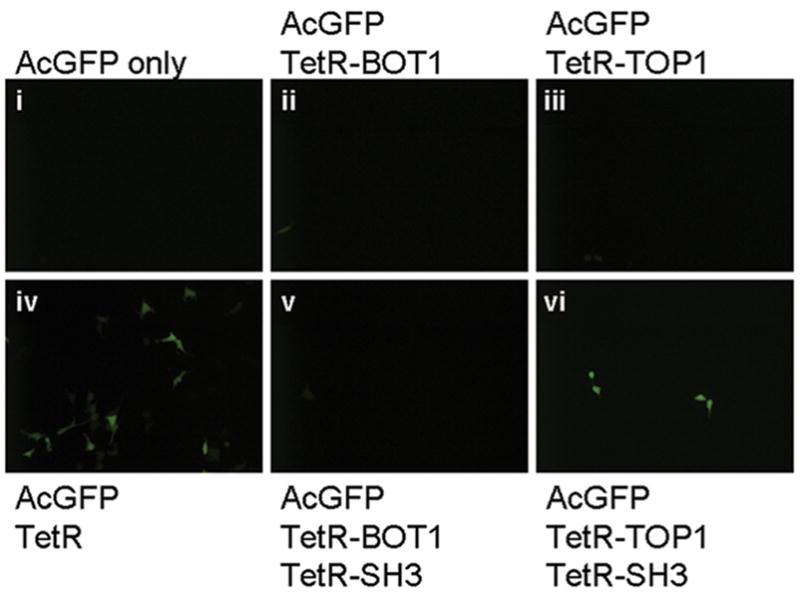
In vivo binding assay using trM2H to evaluate the interaction of TOP1 and BOT1 to the Abl SH3 domain. TetR transcription regulator binds to the Tet-responsive element (TRE) sequence upon dimerization. The binding of dimeric TetR to TRE which is at upstream of the reporter gene (AcGFP) results in the reporter gene expression (Panel iv, positive control). In this system, the dimerization domain of the TetR is replaced with “bait” and “prey”, which are the Abl SH3 domain and TOP1 or BOT1. Positive expression of GFP indicates the interaction of bait and prey.
We further characterized the interaction of TOP1 peptide to the Abl SH3 domain with various in vitro assays. In an affinity chromatography assay, TOP1 and BOT1 peptides were fused to glutathione-S-transferase (GST), purified and immobilized on glutathione sepharose resin. The Abl SH3 domain was also fused to maltose binding protein (MBP) and expressed in E. coli. The resin carrying GST-TOP1 and GST-BOT1 were incubated with the cell lysate expressing MBP-SH3 fusion protein. Unbound proteins were washed off and GST fusion proteins were eluted from the column. Co-purified MBP-SH3 was detected by Western blot probed with anti-MBP antibody. Figure 3A, lane 4 indicates that MBP-SH3 interacts with GST-TOP1. No association is observed between MBP and GST (lane 1) nor MBP-SH3 and either GST-BOT1 (lane 2) or GST (lane 3). A small amount of MBP was co-purified with GST-TOP1 (lane 5) indicating a weak interaction between TOP1 and MBP. However, in comparison to the interaction of GST-TOP1 and MBP-SH3 (lane 4), the background signal observed in lane 5 is negligible. Total protein staining confirmed that the different amounts of co-purified MBP fusion proteins are not due to the varying amounts of GST fusions eluted (Figure S2). We also determined the equilibrium dissociation constant (Kd) between the TOP1 peptide and the Abl SH3 domain without fusion proteins by measuring fluorescent anisotropy (Figure 3B) using the TOP1 peptide labeled with fluorescein isothiocyanate (TOP1-FITC). C-terminal His6-tagged Abl SH3 domain can be readily expressed in E. coli (Figure S3). The obtained value of Kd = 16 ± 1 μM (Figure 3B) is consistent with the dissociation constants reported for other SH3 domain binding partners, which are typically in the μM range [32]. Taken together, these data support that the TOP1 peptide weakly but specifically binds to the Abl SH3 domain both as fusion protein and as a fluorescein-labeled peptide.
Figure 3.

In vitro characterization of TOP1 and BOT1 interacting with the Abl SH3 domain. (A) Affinity chromatography assay. Fusion proteins GST-TOP1 and GST-BOT1 were immobilized on glutathione sepharose resin and incubated with the E. coli cell lysate expressing MBP-SH3. Unbound proteins were washed off and GST fusion proteins were eluted from the column. Co-purified MBP-SH3 was detected by Western blot probed with anti-MBP antibody. (B) Determination of equilibrium dissociation constant (Kd) between the Abl SH3 domain and TOP1-FITC peptide by measuring fluorescence anisotropy, where max is the anisotropy at the saturation and R is the offset factor.
We designed a molecular probe against the Abl SH3 domain using the low-affinity TOP1 peptide. TOP1 peptide was fused to the N-terminus of GFP and an amber stop codon (TAG) was introduced in the linker region between the TOP1 and the GFP, with a His6 tag at the C-terminus (Figure 4A). The resulting plasmid pET28-TOP1(+1)TAG-GFP encodes the corresponding amino acid sequence MGAGAPAFPPPSPP@EF-(GFP)-His6 (where bold face is the TOP1 sequence and @ represents the amber codon). Plasmid pAC-DHPheRS-6TRN carries the orthogonal mutant tRNATyr and mutant tyrosyl-tRNA synthetase which as a pair recognize the amber stop codon and incorporate L-DOPA into the newly synthesized polypeptide chain [18]. E. coli BL21 was co-transformed with pET28-TOP1(+1)TAG-GFP and pAC-DHPheRS-6TRN and the protein expression was induced in the presence of L-DOPA (1 mM) as described previously [19]. Resulting DOPA-containing protein TOP1-DOPA-GFP was purified using Ni-NTA resin and resolved on SDS-PAGE. Figure 4B, lane 2 shows the high yield of TOP1-DOPA-GFP based on the total protein staining. A small amount of endogenous amino acid(s) were incorporated when L-DOPA was omitted from the growth media (lane 1), but compared to lane 2, the level of such background expression is insignificant. A similar gel was also blotted to a nitrocellulose membrane and stained with the redox-cycling method which detects quino-proteins [21] (Figure 4C). This detection method utilizes the reaction of quinone groups on proteins which oxidize free glycine at a basic pH to produce a superoxide in a cyclic reaction, which in turn reduces nitroblue tetrazolium (NBT) to form a purple formazan. Redox-cycling staining detects incorporated L-DOPA in both diphenolic and dopaquinone forms. We routinely use this method to detect L-DOPA in proteins because of its superb sensitivity. Unlike other naturally found DOPA-bearing proteins in which multiple L-DOPA residues are present in each protein molecule (3–40 mol% L-DOPA) [14; 33; 34], TOP1-DOPA-GFP presumably contains only one L-DOPA residue per polypeptide chain (0.4 mol% L-DOPA). Moreover, since the subsequent cross-linking reaction (see below) is based on the dopaquinone, indifferential detection of diphenolic L-DOPA and dopaquinone should not pose a significant problem for the purpose of this study. The strong band in Figure 4C, lane 2 indicates the presence of L-DOPA/dopaquinone within TOP1-DOPA-GFP, supporting the successful incorporation. There are a few endogenous proteins from E. coli that are co-purified with TOP1-DOPA-GFP (Figure 4B). These proteins are most likely metal-binding proteins and typical in the preparation of DOPA-containing proteins by the method described here. All of these endogenous proteins are NBT-negative, suggesting that none of them contains L-DOPA (Figure 4C). Based on our previous study, these contaminating proteins do not affect the efficiency of L-DOPA-mediated protein cross-linking [19]. Successful incorporation of L-DOPA into TOP1-DOPA-GFP was also confirmed by more sensitive Arnow assay [22] which only detects diphenolic L-DOPA, according to Waite and Benedict [23] (Figure S4). We concluded that the efficiency of L-DOPA incorporation into TOP1-DOPA-GFP is sufficient for the following application.
Figure 4.

Expression of the molecular probe TOP1-DOPA-GFP. (A) A schematic diagram of 30.0 kDa TOP1-DOPA-GFP. (B) Total protein analyses of L-DOPA incorporation into TOP1-DOPA-GFP. TOP1-DOPA-GFP was expressed in the presence and absence of L-DOPA, purified with Ni-NTA resin, and resolved by SDS-PAGE. The gel was stained with Coomassie Brilliant Blue. (C) Redox cycling staining of TOP1-DOPA-GFP. Proteins from a similar gel as (B) were blotted to a nitrocellulose membrane and stained with NBT reagent (2 M sodium glycinate, 0.24 mM NBT, pH 10). This method detects quino-proteins and confirmed the presence of L-DOPA/dopaquinone in TOP1-DOPA-GFP.
A larger quantity of TOP1-DOPA-GFP was prepared to demonstrate the ability of TOP1-DOPA-GFP to detect the Abl SH3 domain in a subsequent Western blot-type experiment. The contaminating proteins shown in Figure 4B were reduced to the minimum by extensive washing of the Ni-NTA resin and/or size exclusion chromatography. As a control, a “wild type” counterpart of TOP1-DOPA-GFP, in which the L-DOPA residue was replaced with alanine (wtTOP1-GFP), was also prepared. A typical preparation of purified TOP1-DOPA-GFP and wtTOP1-GFP resolved by SDS-PAGE and detected with Coomassie Brilliant Blue as well as redox-cycling staining are shown in Figure 5A and 5B, respectively. Purified SH3-His was resolved by non-denaturing PAGE and blotted to nitrocellulose membranes. The membranes were blocked with 5 % dry milk in TBST, and incubated with TOP1-DOPA-GFP or wtTOP1-GFP in phosphate-buffered saline (PBS). NaIO4 (2 mM) was added to both membranes probed with TOP1-DOPA-GFP and wtTOP1-GFP. Up to 4 mM of NaIO4 was confirmed not to affect the fluorescence of GFP in a separate experiment (data not shown). The membranes were washed with PBS and visualized under ultra-violet light at 365 nm (Figure 5C). Similar gels were also analyzed by Coomassie Brilliant Blue staining (Figure 5D). The SH3-His is readily detected by TOP1-DOPA-GFP with the signals roughly proportional to the amounts of loaded protein (lanes 2–7, Figure 5C). The two observed protein bands were subjected to in-gel trypsin digestion followed by MALDI-TOF mass-spectrometry to confirm both of their identities as the Abl SH3 domain (Figure S5). No sign of interaction between TOP1-DOPA-GFP and the blocking agent or the non-interacting protein Sortase A (SrtA, Figure 5C, lane 1, negative control) was observed, supporting the specificity of TOP1-DOPA-GFP towards the Abl SH3 domain. The lowest limit of detection of SH3-His using TOP1-DOPA-GFP was estimated to be about 0.5 μg in our current laboratory setting (data not shown). On the other hand, wtTOP1-GFP failed to detect SH3-His (Figure 5C, lane 8) even though the amount of loaded Abl SH3 was well above the detection limit if probed with TOP1-DOPA-GFP. This data supports the critical role of incorporated L-DOPA in enhancing the interaction between the low-affinity TOP1 and the Abl SH3 domain. The concentrations of TOP1-DOPA-GFP and wtTOP1-GFP used in this experiment to probe the membrane were both about 0.35 mg/ml (11 μM) which is greatly less than that required by a previously reported similar experiment using a non-covalent peptide probe (5 mg/ml) [1]. The detection limit and the amount of required probe described above could be even lowered by further optimizing the purification condition of TOP1-DOPA-GFP according to the chemical reactivity of L-DOPA [35]. Taken together, these data demonstrates that TOP1-DOPA-GFP can be employed as a molecular probe to detect the Abl SH3 domain, allowing the instant detection and analysis with enhanced affinity originated from L-DOPA moiety.
Figure 5.

TOP1-DOPA-GFP in Western blot-type protein analysis. Typical preparation of purified TOP1-DOPA-GFP and wtTOP1-GFP resolved by SDS-PAGE and detected with (A) Coomassie Brilliant Blue staining and (B) redox-cycling staining. (C) SH3-His was resolved by non-denaturing PAGE, blotted to nitrocellulose membranes and probed with TOP1-DOPA-GFP (lanes 1–7) or wtTOP1-GFP (lane 8). NaIO4 was added to the membranes to oxidize L-DOPA and induce cross-linking between SH3-His and TOP1-DOPA-GFP. (D) Similar gels containing the same amount of SH3-His as (C) were visualized by Coomassie Brilliant Blue staining.
In summary, we have employed a 10-amino acid peptide TOP1 and a built-in chemical cross-linker L-DOPA to design a peptide-based molecular probe TOP1-DOPA-GFP that can be expressed and purified from E. coli. TOP1 interacts with the Abl SH3 domain weakly but specifically, and the presence of L-DOPA stabilizes the interaction without affecting its specificity to the substrate. We have demonstrated the use of TOP1-DOPA-GFP in Western blot-type experiment with one-step fluorescence-based detection using GFP as a reporter protein. Artificially selected protein-binding peptides are potentially very useful as diagnostic tools and therapeutic reagents [36]; however, majority of laboratory-isolated peptides suffer from low affinity. The molecular design presented here could be applied to many of these peptides, turning the “useless” low-affinity peptides into effective probes and drugs with genetically tailored functional proteins. This technique would add a great flexibility and applicability to the series of peptide based methods currently employed in biomedical and clinical research laboratories.
Supplementary Material
Acknowledgments
Authors thank Prof. Wei Wang and Dr. Tingjun Hou (University of California San Diego) for providing us with the peptide sequences and for helpful discussions. We also thank Prof. Peter G. Schultz (The Scripps Research Institute) for the plasmid pAC-DHPheRS-6TRN. Finally, we appreciate the assistance of YungAh Lee (University of Texas) and Sarah Chun (University of Texas) throughout the conduct of this study.
Footnotes
This work is supported by The Welch Foundation (F1618) and The National Institute of Health (RO1 CA120 168).
Publisher's Disclaimer: This is a PDF file of an unedited manuscript that has been accepted for publication. As a service to our customers we are providing this early version of the manuscript. The manuscript will undergo copyediting, typesetting, and review of the resulting proof before it is published in its final citable form. Please note that during the production process errors may be discovered which could affect the content, and all legal disclaimers that apply to the journal pertain.
References
- 1.Zhang Z, Zhu W, Kodadek T. Selection and application of peptide-binding peptides. Nat Biotechnol. 2000;18:71–4. doi: 10.1038/71951. [DOI] [PubMed] [Google Scholar]
- 2.Kodadek T, Reddy MM, Olivos HJ, Bachhawat-Sikder K, Alluri PG. Synthetic molecules as antibody replacements. Acc Chem Res. 2004;37:711–8. doi: 10.1021/ar030145l. [DOI] [PubMed] [Google Scholar]
- 3.Smith GP, Petrenko VA. Phage Display. Chem Rev. 1997;97:391–410. doi: 10.1021/cr960065d. [DOI] [PubMed] [Google Scholar]
- 4.Lam KS, Lebl M, Krchnak V. The “One-Bead-One-Compound” Combinatorial Library Method. Chem Rev. 1997;97:411–448. doi: 10.1021/cr9600114. [DOI] [PubMed] [Google Scholar]
- 5.Franzosa E, Linghu B, Xia Y. Computational reconstruction of protein-protein interaction networks: algorithms and issues. Methods Mol Biol. 2009;541:89–100. doi: 10.1007/978-1-59745-243-4_5. [DOI] [PubMed] [Google Scholar]
- 6.Villoutreix BO, Renault N, Lagorce D, Sperandio O, Montes M, Miteva MA. Free resources to assist structure-based virtual ligand screening experiments. Curr Protein Pept Sci. 2007;8:381–411. doi: 10.2174/138920307781369391. [DOI] [PubMed] [Google Scholar]
- 7.Agnew HD, Rohde RD, Millward SW, Nag A, Yeo WS, Hein JE, Pitram SM, Tariq AA, Burns VM, Krom RJ, Fokin VV, Sharpless KB, Heath JR. Iterative in situ click chemistry creates antibody-like protein-capture agents. Angew Chem Int Ed Engl. 2009;48:4944–8. doi: 10.1002/anie.200900488. [DOI] [PMC free article] [PubMed] [Google Scholar]
- 8.Reddy MM, Bachhawat-Sikder K, Kodadek T. Transformation of low-affinity lead compounds into high-affinity protein capture agents. Chem Biol. 2004;11:1127–37. doi: 10.1016/j.chembiol.2004.05.013. [DOI] [PubMed] [Google Scholar]
- 9.Denison C, Kodadek T. Toward a general chemical method for rapidly mapping multi-protein complexes. J Proteome Res. 2004;3:417–25. doi: 10.1021/pr034071j. [DOI] [PubMed] [Google Scholar]
- 10.Burzio LA, Waite JH. Cross-linking in adhesive quinoproteins: studies with model decapeptides. Biochemistry. 2000;39:11147–53. doi: 10.1021/bi0002434. [DOI] [PubMed] [Google Scholar]
- 11.Burdine L, Gillette TG, Lin HJ, Kodadek T. Periodate-triggered cross-linking of DOPA-containing peptide-protein complexes. J Am Chem Soc. 2004;126:11442–3. doi: 10.1021/ja045982c. [DOI] [PubMed] [Google Scholar]
- 12.Liu B, Burdine L, Kodadek T. Chemistry of periodate-mediated cross-linking of 3,4-dihydroxylphenylalanine-containing molecules to proteins. J Am Chem Soc. 2006;128:15228–35. doi: 10.1021/ja065794h. [DOI] [PMC free article] [PubMed] [Google Scholar]
- 13.Siegel GJ, Agranoff BW, Albers RW, Fisher SK, Uhler MD. Basic Neurochemistry: Molecular, Cellular, and Medical Aspects. Lippincott, Williams & Wilkins; Philadelphia: 1999. [Google Scholar]
- 14.Waite JH, Tanzer ML. Polyphenolic Substance of Mytilus edulis: Novel Adhesive Containing L-Dopa and Hydroxyproline. Science. 1981;212:1038–1040. doi: 10.1126/science.212.4498.1038. [DOI] [PubMed] [Google Scholar]
- 15.Waite JH. The phylogeny and chemical diversity of quinone-tanned glues and varnishes. Comp Biochem Physiol B. 1990;97:19–29. doi: 10.1016/0305-0491(90)90172-p. [DOI] [PubMed] [Google Scholar]
- 16.Waite JH. Evidence for a repeating 3,4-dihydroxyphenylalanine- and hydroxyproline-containing decapeptide in the adhesive protein of the mussel, Mytilus edulis L. J Biol Chem. 1983;258:2911–5. [PubMed] [Google Scholar]
- 17.Benedict CV, Waite JH. Location and analysis of byssal structural proteins of Mytilus edulis. J Morphol. 1986;189:171–81. doi: 10.1002/jmor.1051890207. [DOI] [PubMed] [Google Scholar]
- 18.Alfonta L, Zhang Z, Uryu S, Loo JA, Schultz PG. Site-specific incorporation of a redox-active amino acid into proteins. J Am Chem Soc. 2003;125:14662–3. doi: 10.1021/ja038242x. [DOI] [PubMed] [Google Scholar]
- 19.Umeda A, Thibodeaux GN, Zhu J, Lee Y, Zhang ZJ. Site-specific protein cross-linking with genetically incorporated 3,4-dihydroxy-L-phenylalanine. Chembiochem. 2009;10:1302–4. doi: 10.1002/cbic.200900127. [DOI] [PMC free article] [PubMed] [Google Scholar]
- 20.Thibodeaux GN, Cowmeadow R, Umeda A, Zhang Z. A tetracycline repressor-based mammalian two-hybrid system to detect protein-protein interactions in vivo. Anal Biochem. 2009;386:129–31. doi: 10.1016/j.ab.2008.11.042. [DOI] [PMC free article] [PubMed] [Google Scholar]
- 21.Paz MA, Gallop PM, Torrelio BM, Fluckiger R. The amplified detection of free and bound methoxatin (PQQ) with nitroblue tetrazolium redox reactions: insights into the PQQ-locus. Biochem Biophys Res Commun. 1988;154:1330–7. doi: 10.1016/0006-291x(88)90285-9. [DOI] [PubMed] [Google Scholar]
- 22.Arnow LE. Colorimetric Determination of the Components of 3,4-Dihydroxyphenylalanine-Tyrosine Mixtures. J Biol Chem. 1937;118:531–537. [Google Scholar]
- 23.Waite JH, Benedict CV. Assay of dihydroxyphenylalanine (dopa) in invertebrate structural proteins. Methods Enzymol. 1984;107:397–413. doi: 10.1016/0076-6879(84)07028-2. [DOI] [PubMed] [Google Scholar]
- 24.Gmeiner WH, Horita DA. Implications of SH3 domain structure and dynamics for protein regulation and drug design. Cell Biochemistry and Biophysics. 2001;35:127–140. doi: 10.1385/CBB:35:2:127. [DOI] [PubMed] [Google Scholar]
- 25.Lim WA, Richards FM, Fox RO. Structural Determinants of Peptide-Binding Orientation and of Sequence Specificity in Sh3 Domains. Nature. 1994;372:375–379. doi: 10.1038/372375a0. [DOI] [PubMed] [Google Scholar]
- 26.Kuriyan J, Cowburn D. Modular peptide recognition domains in eukaryotic signaling. Annual Review of Biophysics and Biomolecular Structure. 1997;26:259–288. doi: 10.1146/annurev.biophys.26.1.259. [DOI] [PubMed] [Google Scholar]
- 27.Van Etten RA. Cycling, stressed-out and nervous: cellular functions of c-Abl. Trends in Cell Biology. 1999;9:179–186. doi: 10.1016/s0962-8924(99)01549-4. [DOI] [PubMed] [Google Scholar]
- 28.Jackson P, Baltimore D. N-Terminal Mutations Activate the Leukemogenic Potential of the Myristoylated Form of C-Abl. Embo Journal. 1989;8:449–456. doi: 10.1002/j.1460-2075.1989.tb03397.x. [DOI] [PMC free article] [PubMed] [Google Scholar]
- 29.Cohen GB, Ren RB, Baltimore D. Modular Binding Domains in Signal-Transduction Proteins. Cell. 1995;80:237–248. doi: 10.1016/0092-8674(95)90406-9. [DOI] [PubMed] [Google Scholar]
- 30.Hou TJ, Chen K, McLaughlin WA, Lu BZ, Wang W. Computational analysis and prediction of the binding motif and protein interacting partners of the Abl SH3 domain. Plos Computational Biology. 2006;2:46–55. doi: 10.1371/journal.pcbi.0020001. [DOI] [PMC free article] [PubMed] [Google Scholar]
- 31.Hou T, Xu Z, Zhang W, McLaughlin WA, Case DA, Xu Y, Wang W. Characterization of domain-peptide interaction interface: a generic structure-based model to decipher the binding specificity of SH3 domains. Mol Cell Proteomics. 2009;8:639–49. doi: 10.1074/mcp.M800450-MCP200. [DOI] [PMC free article] [PubMed] [Google Scholar]
- 32.Sparks AB, Quilliam LA, Thorn JM, Der CJ, Kay BK. Identification and characterization of Src SH3 ligands from phage-displayed random peptide libraries. J Biol Chem. 1994;269:23853–6. [PubMed] [Google Scholar]
- 33.Rzepecki LM, Hansen KM, Waite JH. Characterization of a Cystine-Rich Polyphenolic Protein Family from the Blue Mussel Mytilus edulis L. Biol Bull. 1992;183:123–137. doi: 10.2307/1542413. [DOI] [PubMed] [Google Scholar]
- 34.Taylor SW, Ross MM, Waite JH. Novel 3,4-di- and 3,4,5-trihydroxyphenylalanine-containing polypeptides from the blood cells of the ascidians Ascidia ceratodes and Molgula manhattensis. Arch Biochem Biophys. 1995;324:228–40. doi: 10.1006/abbi.1995.0035. [DOI] [PubMed] [Google Scholar]
- 35.Waite JH. Precursors of quinone tanning: dopa-containing proteins. Methods Enzymol. 1995;258:1–20. doi: 10.1016/0076-6879(95)58033-6. [DOI] [PubMed] [Google Scholar]
- 36.Newton J, Deutscher SL. Phage peptide display. Handb Exp Pharmacol. 2008:145–63. doi: 10.1007/978-3-540-77496-9_7. [DOI] [PubMed] [Google Scholar]
Associated Data
This section collects any data citations, data availability statements, or supplementary materials included in this article.