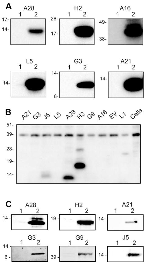
Fig. 1.
Western blots showing expression and immunogenicity of EFC proteins. (A) Expression of VACV EFC genes from plasmids. BS-C-1 cells were transfected with the empty vector plasmid or a plasmid expressing A28, H2, A16, L5, G3 or A21, harvested after 24 h and analyzed by SDS-PAGE under reducing conditions. Following membrane transfer, strips were incubated with the corresponding polyclonal antibody and bound proteins were detected by chemiluminescence. Numbers: 1, empty plasmid vector; 2, plasmid encoding indicated EFC gene. Positions and masses in kDa of protein markers are at the left of each panel. (B) Reactivity of serum from a rabbit immunized with VACV. Western blotting was performed as in panel A except that the membrane was incubated with antiserum from a rabbit that had been immunized with live VACV (Wyatt et al., 2008). The blot was developed with chemiluminescence. The proteins encoded by the plasmids are indicated above each lane. EV refers to empty vector. (C) Antibody responses of mice following DNA immunizations. Pooled mouse sera (n=10) obtained two weeks following the 3rd immunization of A28, H2, A21, G3, G9 or J5 genes were used to probe Western blots containing the corresponding protein from transfected BS-C-1 cell lysates using infrared fluorescence detection (LI-COR Biosciences, Lincoln, NE).

