Abstract
The highly pathogenic avian influenza (HPAI) virus phenotype is restricted to influenza A viruses of the H5 and H7 hemagglutinin (HA) subtypes. To obtain more information on the apparent subtype-specific nature of the HPAI virus phenotype, a low-pathogenic avian influenza (LPAI) H6N1 virus was generated, containing an HPAI H5 RRRKKR↓G multibasic cleavage site (MBCS) motif in HA (the downward arrow indicates the site of cleavage). This insertion converted the LPAI virus phenotype into an HPAI virus phenotype in vitro and in vivo. The H6N1 virus with an MBCS displayed in vitro characteristics similar to those of HPAI H5 viruses, such as cleavage of HA0 (the HA protein of influenza A virus initially synthesized as a single polypeptide precursor) and virus replication in the absence of exogenous trypsin. Studies of chickens confirmed the HPAI phenotype of the H6N1 virus with an MBCS, with an intravenous pathogenicity index of 1.4 and systemic virus replication upon intranasal inoculation, the hallmarks of HPAI viruses. This study provides evidence that the subtype-specific nature of the emergence of HPAI viruses is not at the molecular, structural, or functional level, since the introduction of an MBCS resulted in a fully functional virus with an HPAI virus genotype and phenotype.
Wild birds represent the natural reservoir of avian influenza A viruses in nature (43). Influenza A viruses are classified on the basis of the hemagglutinin (HA) and neuraminidase (NA) surface glycoproteins. In wild birds throughout the world, influenza A viruses representing 16 HA and 9 NA antigenic subtypes have been found in numerous combinations (also called subtypes, e.g., H1N1, H6N1) (12). Besides classification based on the antigenic properties of HA and NA, avian influenza A viruses can also be classified based on their pathogenic phenotype in chickens. Highly pathogenic avian influenza (HPAI) virus, an acute generalized disease of poultry in which mortality may be as high as 100%, is restricted to subtypes H5 and H7. Other avian influenza A virus subtypes are generally low-pathogenic avian influenza (LPAI) viruses that cause much milder, primarily respiratory disease in poultry, sometimes with loss of egg production (6).
The HA protein of influenza A virus is initially synthesized as a single polypeptide precursor (HA0), which is cleaved into HA1 and HA2 subunits by host cell proteases. The mature HA protein mediates binding of the virus to host cells, followed by endocytosis and HA-mediated fusion with endosomal membranes (43). Influenza viruses of subtypes H5 and H7 may become highly pathogenic after introduction into poultry and cause outbreaks of HPAI. The switch from an LPAI phenotype to the HPAI phenotype of these H5 and H7 influenza A viruses is achieved by the introduction of basic amino acid residues into the HA0 cleavage site by substitution or insertion, resulting in the so-called multibasic cleavage site (MBCS), which facilitates systemic virus replication (4, 5, 14, 44). The cleavage of the HA0 of LPAI viruses is restricted to trypsin-like proteases which recognize the XXX(R/K)↓G cleavage motif, where the downward arrow indicates the site of cleavage. Replication of these LPAI viruses is therefore restricted to sites in the host where these enzymes are expressed, i.e., the respiratory and intestinal tract (32, 38). The introduction of an RX(R/K)R↓G or R(R/K)XR↓G minimal MBCS motif into the H5 and H7 subtype viruses facilitates the recognition and cleavage of the HA0 by ubiquitous proprotein convertases, such as furin (20, 32, 41, 45). H5 influenza A viruses with a minimal MBCS motif only have the highly pathogenic phenotype if the masking glycosylation site at position 11 in the HA is replaced by a nonglycosylation site. Otherwise, at least one additional basic amino acid has to be inserted to allow the shift from an LPAI virus phenotype to an HPAI virus phenotype to occur (15, 18, 21, 22, 28). No information is available on the minimal prerequisites of H7 influenza A viruses to become highly pathogenic, but all HPAI H7 viruses have at least 2 basic amino acid insertions in the HA0 cleavage site (22). HA0 with the MBCS is activated in a broad range of different host cells and therefore enables HPAI viruses to replicate systemically in poultry (46). To date, little is known about the apparent subtype-specific nature of the introduction of the MBCS into LPAI viruses and the evolutionary processes involved in the emergence of HPAI viruses. When an MBCS was introduced in a laboratory-adapted strain of influenza virus, A/Duck/Ukraine/1/1963 (H3N8), it did not result in a dramatic change in pathogenic phenotype (35). Here, the effect of the introduction of an MBCS into a primary LPAI H6N1 virus, A/Mallard/Sweden/81/2002, is described. The introduction of an MBCS resulted in trypsin-independent replication in vitro and enhanced pathogenesis in a chicken model. Understanding the basis of the HA subtype specificity of the introduction of an MBCS into avian influenza viruses will lead to a better understanding of potential molecular restrictions involved in emergence of HPAI outbreaks.
MATERIALS AND METHODS
Cells.
Madin-Darby canine kidney (MDCK) cells were cultured in Eagle's minimum essential medium (EMEM; Lonza, Breda, Netherlands) supplemented with 10% fetal calf serum (FCS), 100 IU/ml penicillin, 100 μg/ml streptomycin, 2 mM glutamine, 1.5 mg/ml sodium bicarbonate, 10 mM HEPES, and nonessential amino acids. 293T cells were cultured in Dulbecco's modified Eagle's medium (DMEM; Lonza) supplemented with 10% FCS, 100 IU/ml penicillin, 100 μg/ml streptomycin, 2 mM glutamine, 1 mM sodium pyruvate, and nonessential amino acids.
Viruses.
Influenza virus A/Mallard/Sweden/81/02 (H6N1) was isolated from a migratory mallard (Anas platyrrhynchos) within the framework of our ongoing avian influenza surveillance program (25) and passaged twice in embryonated chicken eggs. Influenza virus A/Hong Kong/156/97 (H5N1) was isolated from the human index case during the 1997 H5N1 outbreak in Hong Kong (9). The wild-type H6N1 (H6N1WT) virus, H5N1WT virus, H6N1 virus with the MBCS in HA (H6N1MBCS), and the H5N1 virus lacking the MBCS (H5N1ΔMBCS) were generated by reverse genetics as described previously (10). The supernatant of the transfected cells was harvested 48 h after transfection and was used to inoculate MDCK cells. The genotypes of all recombinant viruses were confirmed by sequencing. The risk potential of the H6N1MBCS virus was assessed prior to start of the experiments, and it was determined that the anticipated risk of generating this virus would be equivalent to that of generating an HPAI virus. Therefore, all in vivo and in vitro experiments were performed under animal biosafety level 3+ (ABSL3+) containment conditions.
Plasmids.
The 8 gene segments of A/Mallard/Sweden/81/2002 (H6N1) and A/Hong Kong/156/97 (H5N1) were amplified by reverse transcriptase PCR (RT-PCR) and cloned in the BsmBI site of a modified version of plasmid pHW2000 (10). For the construction of the plasmid containing the MBCS in HA of H6N1, a QuikChange multisite-directed mutagenesis kit (Qiagen, Venlo, Netherlands) was used, according to instructions of the manufacturer. The following primers were used for the introduction of nucleotides encoding the RRRKKR↓ GMBCS in the HA gene: GAGGCTTGCAACTGGACTAAGAAATGTTCCACAGAGAAAAAAAAGAGGACTTTTCGGAGCC and TGGACTAAGAAATGTTCCACAGAGAAGAAGAAAAAAAAGAGGACTTTTCG. For the generation of H5N1ΔMBCS, 15 nucleotides encoding 5 amino acids (underlined), PQRERRRKKR↓G, were modulated into PQIETR↓G of the MBCS in HA of the H5N1 virus, as previously described (42).
Replication kinetics and virus titrations.
Multistep replication kinetics were determined by inoculating MDCK cells in the presence and absence of 1 μg/ml trypsin (Lonza), with a multiplicity of infection (MOI) of 0.01 50% tissue culture infective dose (TCID50) per cell. Supernatants were sampled at 6, 12, 24, and 48 h after inoculation. Virus titers in MDCK cells were determined by endpoint titration as described previously (10). MDCK cells were inoculated with a 10-fold serial dilution of culture supernatants or tissue homogenates. One hour after inoculation, cells were washed once with phosphate-buffered saline (PBS) and grown in 200 μl of infection media, consisting of EMEM (Lonza) supplemented with 100 U/ml penicillin, 100 μg/ml streptomycin, 2 mM glutamine, 1.5 mg/ml sodium bicarbonate (Lonza), 10 mM HEPES (Lonza), nonessential amino acids (MP Biomedicals) and 20 μg/ml trypsin (Lonza). Three days after inoculation, the supernatants of infected cell cultures were tested for agglutinating activity, using turkey erythrocytes as indicators of infection of the cells. Infectious titers were calculated from 5 replicates by the method of Spearman-Karber. For the multistep replication kinetics, geometric mean titers were calculated using the infectious titers obtained from two independent experiments.
Western blotting.
293T cells were transfected with plasmids expressing the HA genes of H6N1WT, H6N1MBCS, H5N1WT, and H5N1ΔMBCS. Cells were harvested 48 h after transfection and were treated with either PBS or 2.5 μg/ml trypsin (Lonza) for 1 h at 37°C. Cells were lysed in hot lysis buffer (1% sodium dodecyl sulfate [SDS], 100 mM NaCl, 10 mM EDTA, 10 mM Tris-HCl at pH 7.5) and treated with 3× dissociation loading buffer (2% SDS, 0.01 dithiothreitol, 0.02 M Tris-HCl at pH 6.8) for 5 min at 96°C, and proteins were separated in 10% SDS-polyacrylamide gels. A molecular magic marker (Invitrogen, Leek, Netherlands) was run alongside to determine protein sizes. Proteins were transferred onto nitrocellulose membranes by electroblotting for 1 h in blotting buffer (25 mM Tris, 192 mM glycine, and 20% methanol). The blots were incubated overnight at 4°C in blocking buffer (PBS with 5% [wt/vol] nonfat dried milk and 0.05% Tween-10) and incubated with a 1:2,000 dilution of rabbit antisera (1:1 dilution mixture of rabbit anti-A/Turkey/Massachusetts/65 H6N1 and rabbit anti-A/Shearwater/Australia/1/72 H6N1 for H6 and rabbit anti-A/Hong Kong/156/97 H5N1 for H5) in blocking buffer for 2 h at room temperature. Blots were washed with PBS containing 0.05% Tween and incubated for 1 h with swine anti-rabbit horseradish peroxidase (Dako, Denmark) at a dilution of 1:3,000 in blocking buffer, washed again, and developed with ECL Western blotting detection reagents (GE Healthcare, United Kingdom).
Intravenous pathogenicity index.
The intravenous pathogenicity index (IVPI) of recombinant viruses was determined using 10 six-week-old specific-pathogen-free (SPF) White Leghorn chickens (GDL, Deventer, Netherlands), according to the OIE standards (27). In short, chickens were injected intravenously in the ulnar vein with 0.1 ml of the H6N1WT or H6N1MBCS virus at a dose of 106 TCID50. The development of clinical signs was monitored for 10 days for each individual chicken. Chickens were classified sick if they displayed one clinical sign, such as depression, cyanosis of the comb or wattles, respiratory involvement, diarrhea, edema of the face/head, and nervous signs, and severely sick if they displayed two or more clinical signs. The IVPI was calculated as the mean score per bird per observation. All animal studies and procedures were reviewed and approved by the Institutional Animal Ethics Committee of Erasmus Medical Center and have been conducted according to the national guidelines of the Netherlands.
Intranasal infection of chickens.
Two groups of 10 six-week-old specific-pathogen-free (SPF) White Leghorn chickens (GDL) were inoculated intranasally with 5 × 10⁶ TCID50 of the H6N1WT or H6N1MBCS virus. Oropharyngeal and cloacal swabs were collected and stored in 1 ml transport media; virus titers of MDCK cells in the swabs were determined by endpoint titration. At day 3 and day 6 after inoculation, 5 animals from each group were euthanized, and virus titers in the nasal turbinate, trachea, lung, spleen, liver, heart, intestine, and brain were determined for 3 out of 5 animals; remaining chickens were used for immunohistochemistry. Tissues were homogenized in 3 ml transport medium, consisting of Hank's balanced salt solution containing 10% glycerol, 200 U/ml penicillin, 200 mg/ml streptomycin, 100 U/ml polymyxin B sulfate, and 250 mg/ml gentamicin (ICN, Netherlands), using the FastPrep system (MP Biomedicals) with 2 one-quarter-inch ceramic sphere balls (MP Biomedicals) and centrifuged briefly.
Immunohistochemistry.
Immunohistochemistry was performed with the tissues of chickens inoculated with the H6N1WT and H6N1MBCS viruses. For each virus, 2 chickens were euthanized 3 and 6 days after inoculation by exsanguination. Necropsies and tissue sampling were performed according to a standard protocol (protocol available on request). After fixation in 10% neutral-buffered formalin and embedding in paraffin, tissue sections were stained with an immunohistochemical method, using a monoclonal antibody against the nucleoprotein of influenza A virus as a primary antibody for detection of influenza virus antigen (clone Hb65; ATCC, United Kingdom). The following types of tissue were examined: nasal turbinate, trachea, lung, spleen, liver, heart, intestine, brain, and comb.
Nucleotide sequence accession numbers.
The nucleotide sequences of the 8 gene segments of A/Mallard/Sweden/81/02 (H6N1) are available from GenBank under accession numbers CY060379 to CY060386.
RESULTS
In vitro characteristics of the H6N1MBCS virus.
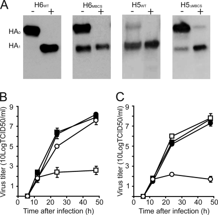
In order to investigate the subtype specificity of the insertion of an MBCS into the HA of LPAI viruses, an MBCS was inserted into the HA0 of A/Mallard/Sweden/81/02 (H6N1WT), resulting in H6N1MBCS. The functionality of the MBCS insertion in the H6 HA protein was first studied by expression of HA in 293T cells upon transfection and determination of the cleavage pattern in the presence and absence of trypsin. The cleavage patterns of H6WT and H6MBCS were compared to the cleavage patterns of HA in HPAI A/HongKong/156/97 H5N1 (H5WT) and HA in this virus from which the MBCS was removed (H5ΔMBCS). H5WT and H6MBCS displayed similar cleavage patterns upon Western blot analysis, with cleavage of the HA0 both with and without supplemental addition of trypsin. H5ΔMBCS displayed a cleavage pattern similar to that of H6WT, with efficient HA0 cleavage only upon the addition of trypsin (Fig. 1 A). Next, the effect of the introduction of the MBCS on the replication kinetics in MDCK cells was determined. Using reverse genetics, 2 wild-type viruses (H6N1WT and H5N1WT) and 2 mutant viruses (H6N1MBCS and H5N1ΔMBCS) were produced. The H6N1WT and H6N1MBCS viruses contained 7 gene segments of A/Mallard/Sweden/81/02 H6N1 and wild-type H6 or H6 HA with MBCS, respectively, and the H5N1WT and H5N1ΔMBCS viruses contained 7 gene segments of A/HongKong/156/97 H5N1 and wild-type H5 HA or H5 HA from which the MBCS was deleted, respectively. The H6N1WT virus was able to replicate to high titers in the presence of trypsin; in the absence of trypsin, the H6N1WT virus failed to replicate efficiently. The H6N1MBCS virus was able to replicate to comparable titers in the presence and absence of trypsin, with only modest differences in maximum virus titers after 48 h (Fig. 1B). The comparison of the replication kinetics of the H6N1WT and H6N1MBCS viruses and those of the HPAI H5N1WT and H5N1ΔMBCS viruses (Fig. 1C) was in agreement with observations from Western blot analysis. The H6N1MBCS and H5N1WT viruses displayed replication kinetics in the absence of trypsin similar to those of the H6N1WT and H5N1ΔMBCS viruses in the presence of trypsin. The H6N1WT and H5N1ΔMBCS viruses did not replicate efficiently in the absence of trypsin, in agreement with an LPAI virus phenotype. Thus, the insertion of an MBCS in an LPAI H6N1 virus conferred an in vitro phenotype comparable to that of an HPAI H5N1 virus.
FIG. 1.
In vitro phenotype of H6N1MBCS in comparison with those of H6N1WT, H5N1WT, and H5N1ΔMBSC influenza A viruses. (A) Western blots of transfected 293T cells with the H6N1WT, H6N1MBCS, H5N1WT, or H5N1ΔMBCS HA open reading frame, treated with (+) or without (−) trypsin. (B, C) Replication kinetics of the H6N1WT (squares) and H6N1MBCS (circles) viruses in MDCK cells in the presence (filled symbols) or absence (open symbols) of trypsin (B) or the H5N1WT (squares) and H5N1ΔMBCS (circles) viruses in MDCK cells in the presence (filled symbols) or absence (open symbols) of trypsin (C). Geometric mean titers were calculated from two independent experiments; error bars indicate standard deviations.
Intravenous pathogenicity index of the H6N1WT and H6N1MBCS virus.
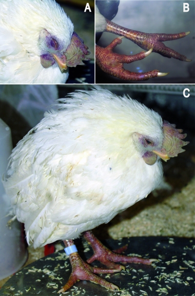
To determine whether the in vitro HPAI phenotype of the H6N1MBCS virus also resulted in an HPAI phenotype in vivo, the intravenous pathogenicity index (IVPI) was determined. The IVPI is the gold standard for the assessment of the pathogenic phenotype of avian influenza viruses in poultry. Avian influenza viruses with an IVPI of <1.2 are considered LPAI viruses, and avian influenza viruses with an IVPI of ≥1.2 are considered HPAI viruses. Ten 6-week-old White Leghorn chickens were inoculated intravenously with 0.1 ml 1 × 10⁶ TCID50/ml of the H6N1WT or H6N1MBCS virus and monitored closely for 10 days. The 10 chickens inoculated with the H6N1WT virus did not show clinical signs of disease over the 10-day period, resulting in an IVPI of 0.0, confirming the LPAI virus phenotype in vivo (Table 1) (27). The chickens inoculated with the H6N1MBCS virus displayed a rapid progressive disease, with 8/10 animals being severely sick at 3 days postinoculation (d.p.i.) and 1 deceased animal at 5 and 6 d.p.i. A variety of clinical symptoms was observed in these birds, including respiratory involvement (2/10 animals), depression (10/10) (Fig. 2), diarrhea (2/10), cyanosis of the exposed skin or wattles (6/10) (Fig. 2), edema of the face and/or head (6/10), and nervous signs (6/10) (Table 1). The overall IVPI score of the H6N1MBCS virus was 1.4, in concordance with an HPAI virus phenotype.
TABLE 1.
Overview of IVPI results for the H6N1WT and H6N1MBCS viruses
| Birds infected with virusa | No. of birds at indicated d.p.i. |
Total no. of birds over 10-day period × scoreb | IVPIc | |||||||||
|---|---|---|---|---|---|---|---|---|---|---|---|---|
| 1 | 2 | 3 | 4 | 5 | 6 | 7 | 8 | 9 | 10 | |||
| H6N1WT | ||||||||||||
| Healthy | 10 | 10 | 10 | 10 | 10 | 10 | 10 | 10 | 10 | 10 | 100 × 0 = 0 | |
| Sick | 0 | 0 | 0 | 0 | 0 | 0 | 0 | 0 | 0 | 0 | 0 × 1 = 0 | |
| Severely sick | 0 | 0 | 0 | 0 | 0 | 0 | 0 | 0 | 0 | 0 | 0 × 2 = 0 | |
| Dead | 0 | 0 | 0 | 0 | 0 | 0 | 0 | 0 | 0 | 0 | 0 × 3 = 0 | |
| Total | 0 | 0 | ||||||||||
| H6N1MBCS | ||||||||||||
| Healthy | 3 | 1 | 1 | 0 | 0 | 0 | 0 | 2 | 3 | 3 | 16 × 0 = 0 | |
| Sick | 7 | 5 | 1 | 2 | 1 | 1 | 6 | 5 | 5 | 5 | 38 × 1 = 38 | |
| Severely sick | 0 | 4 | 8 | 8 | 8 | 7 | 0 | 0 | 0 | 0 | 35 × 2 = 70 | |
| Dead | 0 | 0 | 0 | 0 | 1 | 2 | 2 | 2 | 2 | 2 | 11 × 3 = 33 | |
| Total | 141 | 1.41 | ||||||||||
Sick birds show one of the following signs: respiratory involvement, depression, diarrhea, cyanosis of the exposed skin or wattles, edema of the face and/or head, and nervous signs. Severely sick birds show two or more of the signs mentioned above.
Two groups of 10 chickens were each inoculated intravenously with 0.1 ml 1 × 10⁶ TCID50 of either of the viruses and observed for clinical signs of disease for 10 days. At each observation, each bird is scored 0 if healthy, 1 if sick, 2 if severely sick, and 3 if dead.
The intravenous pathogenicity index (IVPI) is the mean score per bird per observation over the 10-day period.
FIG. 2.
Clinical symptoms of chickens infected with H6N1MBCS virus, consistent with those of chickens infected with HPAI virus, as follows: cyanosis of the comb and wattles (A); subcutaneous hemorrhage of the shanks (B); and severe depression, ruffled feathers with edema of head and neck, and cyanosis of the comb, wattles, and feet (C).
Infection of chickens via the intranasal route.
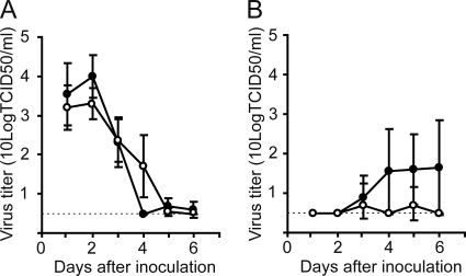
Next, clinical signs, virus shedding, and tissue distribution in chickens were studied upon intranasal inoculation, considered more representative of a natural infection than the intravenous inoculation described above. To this end, 10 chickens were inoculated intranasally with 5 × 10⁶ TCID50/ml of either the H6N1WT or H6N1MBCS virus. The chickens were observed for clinical signs of disease, and cloacal and oropharyngeal swab samples were obtained daily over a 6-day period. None of the chickens inoculated with the H6N1WT virus displayed clinical signs during the course of the experiment, whereas all H6N1MBCS virus-inoculated chickens were lethargic (10/10), and some developed cyanosis of the comb (2/10). Virus shedding from the respiratory tract was observed to start at 1 d.p.i. for both viruses and continued until 4 to 5 d.p.i. The amounts of virus shedding were comparable between the 2 viruses. Virus shedding from the intestinal tract started at 3 d.p.i. for both viruses and continued during the course of the experiment for the H6N1MBCS virus, whereas cloacal shedding of the H6N1WT virus was minimal and below the detection limit after 6 days (Fig. 3). At 3 and 6 d.p.i., 5 chickens from each group were euthanized, and virus titers in the nasal turbinate, trachea, lung, spleen, liver, heart, intestine, and brain were determined for 3 of these animals; the remaining chickens were used for immunohistochemistry. The H6N1WT and H6N1MBCS viruses were detected with comparable titers in the upper respiratory tracts of inoculated chickens at 3 d.p.i. In the lung, the H6N1MBCS virus replicated to higher titers and was detected in all animals, whereas the H6N1WT virus was detected only in 1 animal. The H6N1WT virus was also detected in the spleen (1/3 animals) and intestine (3/3), whereas the H6N1MBCS virus was detected in the brain (2/3) (Table 2). Immunohistochemistry was performed in order to confirm influenza A virus replication in tissues obtained at 3 d.p.i. Using an anti-nucleoprotein (NP) monoclonal antibody, expression of influenza virus antigen was not detected in any of the tissues of the group inoculated with H6N1WT. For the group inoculated with H6N1MBCS, expression of influenza virus antigen was detected in all tissue samples, including tissue of the comb (Fig. 4), in agreement with reports on H5 and H7 HPAI infections in chickens (31, 40). At 6 d.p.i., the H6N1MBCS virus was still detected in the brain (1/3 animals), heart (1/3), and intestine (2/3), whereas the H6N1WT virus was cleared from all sampled tissues by this time (Table 2).
FIG. 3.
Virus shedding from the respiratory tract (A) and the intestinal tract (B) after intranasal inoculation of chickens the H6N1WT (open circles) and H6N1MBCS (closed circles) viruses. Oropharyngeal and cloacal swabs were taken daily, and virus titers in MDCK cells were determined by endpoint titration. The geometric mean titers, calculated per day per group, are displayed; error bars indicate 95% confidence interval values. The dotted line indicates the cutoff value of the assay.
TABLE 2.
Virus titers in tissue of chickens inoculated with either the H6N1WT or H6N1MBCS virus
| d.p.i. | Tissue | Immunohistochemistry results for indicated virusesa |
|||
|---|---|---|---|---|---|
| H6N1WT |
H6N1MBCS |
||||
| No. of chickens in which virus was detected/total no. of chickens | Virus titer (log10 TCID50/g tissue) ± SD | No. of chickens in which virus was detected/total no. of chickens | Virus titer (log10 TCID50/g tissue) ± SD | ||
| 3 | Lung | 1/3 | 1.52 ± 0.17 | 3/3 | 2.10 ± 0.79 |
| Trachea | 3/3 | 2.83 ± 0.47 | 3/3 | 3.37 ± 1.34 | |
| NT | 2/3 | 2.97 ± 0.81 | 3/3 | 2.59 ± 0.17 | |
| Brain | 0/3 | <1.41 | 2/3 | 1.53 ± 0.17 | |
| Spleen | 1/3 | 2.16 ± 1.15 | 0/3 | <1.50 | |
| Heart | 0/3 | <1.51 | 0/3 | <1.51 | |
| Liver | 0/3 | <1.43 | 0/3 | <1.43 | |
| Intestine | 3/3 | 2.56 ± 0.92 | 0/3 | <1.65 | |
| 6 | Lung | 0/3 | <1.42 | 0/3 | <1.42 |
| Trachea | 0/3 | <1.91 | 0/3 | <1.91 | |
| NT | 0/3 | <2.01 | 0/3 | <2.01 | |
| Brain | 0/3 | <1.41 | 1/3 | 1.57 ± 0.27 | |
| Spleen | 0/3 | <1.50 | 0/3 | <1.50 | |
| Heart | 0/3 | <1.51 | 1/3 | 1.56 ± 0.08 | |
| Liver | 0/3 | <1.43 | 0/3 | <1.43 | |
| Intestine | 0/3 | <1.65 | 2/3 | 1.92 ± 0.31 | |
Three chickens from each group were euthanized at 3 d.p.i. and 6 d.p.i. Virus titers in MDCK cells of the lung, trachea, nasal turbinate (NT), brain, spleen, heart, liver, and intestine were determined by endpoint titration. Geometric mean titers ± standard deviations are indicated; cutoff values are given for negative tissues.
FIG. 4.

Tissue distribution of the H6N1WT (left) and H6N1MBCS (right) viruses after intranasal inoculation of chickens. Expression of viral antigen could not be detected in serial sections of these tissues of chickens infected with the H6N1WT virus in the lung, tracheal epithelium, nasal turbinate (NT), brain, liver, spleen, heart, intestine, and comb. Expression of viral antigen could be detected in all tissues of chickens infected with the H6N1MBCS virus. The chickens were inoculated intranasally with 5 × 10⁶ TCID50 of H6N1WT and H6N1MBCS viruses and euthanized 3 days postinfection. Tissue sections were stained with a monoclonal antibody against influenza A virus nucleoprotein, visible as red-brown staining.
DISCUSSION
The emergence of HPAI viruses from LPAI ancestral viruses is restricted to influenza A viruses of the H5 and H7 subtypes. All currently known HPAI outbreaks were caused by HPAI H5 and H7 viruses (3). The emergence of HPAI viruses is primarily thought to occur by introduction of MBCSs in HA of the LPAI H5 and H7 viruses during their circulation in poultry. LPAI viruses other than H5 and H7 also frequently cause outbreaks in poultry, generally with mild clinical symptoms (2, 33, 38). Despite extensive circulation in poultry, HPAI viruses have never emerged during LPAI outbreaks, other than those caused by influenza A viruses of the H5 or H7 subtypes, pointing to the subtype-specific nature of the emergence of HPAI viruses. Whereas the structural and molecular mechanisms for the emergence of HPAI viruses have been partly elucidated (28, 29), the subtype specificity of this emergence remains unknown. The proposed molecular mechanisms underlying the introduction of basic amino acid residues into HA0 cleavage are nucleotide/amino acid substitution and insertion of additional basic amino acid residues via strand slippage (28). In addition, in the case of two H7 virus strains, it has been shown that recombination with host or viral RNA was responsible for the introduction of an MBCS (19, 37). The present study aimed to understand the nature of the subtype specificity of the MBCS introduction by artificial insertion of an MBCS motif identical to those of currently circulating H5 HPAI viruses in an LPAI H6N1 virus. The choice for an LPAI H6 virus was made because multiple introductions of this subtype into poultry have occurred and because of the extensive circulation of H6 viruses in poultry (8, 47). These characteristics make it likely that H6 viruses with HPAI genotypes would have emerged if not for an as-yet-unidentified restriction. The insertion of an MBCS into the LPAI H6N1 virus resulted in a shift from a trypsin-dependent phenotype to a trypsin-independent phenotype, comparable to that of an HPAI H5N1 virus. Moreover, the introduction of the MBCS into the LPAI H6N1 virus also resulted in an in vivo HPAI virus phenotype. The clinical signs observed in H6N1MBCS virus-infected chickens, such as subcutaneous hemorrhages of the shanks, cyanotic combs and wattles, and neurological signs, are hallmarks of HPAI infection (Fig. 2). In addition, the IVPI was greater than 1.2 (Table 1), which places the H6N1MBCS virus within the category of HPAI viruses (27). Our data are in agreement with those in previous studies which show that the introduction of an MBCS into LPAI H5 viruses increases the tissue tropism to extrarespiratory and extraintestinal replication (14). However, the pathogenicity of the generated H6N1MBCS virus is not completely equivalent to the pathogenicity displayed in naturally occurring HPAI H5 and H7 viruses, which in general have IVPI scores in the range of 2 to 3 (7, 22, 36), although lower IVPI scores (1.2) have been reported for naturally occurring HPAI viruses as well (1). The relatively limited pathogenicity of the H6N1MBCS virus in the chicken model upon intranasal inoculation, rather than intravenous inoculation, might be caused by the lack of adaptation of the original H6N1WT virus to efficient replication in chickens. The H6N1WT virus was isolated from a wild-caught migratory mallard and, as such, will be adapted to replication in this wild bird species. It has been shown in a previous report comparing the intranasal infectious doses of LPAI viruses that infection of White Leghorn chickens required very high doses of viruses isolated from wild birds compared to the doses of viruses obtained from chickens (39). Previous studies have also shown that changes in HA alone are not sufficient for efficient replication of wild bird avian influenza viruses in chickens and that besides changes in HA, changes in the internal genes such as PB1, PB2, and PA are necessary for adaptation toward efficient replication in chickens (16, 23, 34). We suggest that such additional adaptations in the internal genes of the H6N1MBCS virus will likely result in further increased pathogenicity, possibly comparable to the pathogenicity of other HPAI viruses upon intranasal infection and IVPI. It has also been shown for multiple LPAI viruses that adaptation to replication in poultry results in increased virus shedding from both the respiratory tract and the intestinal tract (16, 30, 34).
For the H6N1WT virus, the low virus shedding from the respiratory tract at 3 d.p.i. (2.3 ± 0.9 log10 TCID50/ml; 10/10 animals) was accompanied by low or undetectable virus titers in the respiratory tract tissues (Table 2). Likewise, low shedding from the intestinal tract at 3 d.p.i. (0.7 ± 0.6 log10 TCID50/ml; 3/10 animals) was accompanied by low virus titers in the intestinal tract (colon). Upon immunohistochemistry analysis using multiple cross sections of tissue obtained from 2 chickens not used for virus isolation, no influenza A virus NP-positive cells were detected in tissues of the respiratory or intestinal tract, again indicating the variability and the low level of virus replication within the group of chickens inoculated intranasally with the H6N1WT virus. Real-time reverse transcriptase PCR (RRT-PCR) (24) further confirmed the absence of viral genomic RNA in tissues that were negative in virus isolation (data not shown). In the 10 chickens inoculated with the H6N1MBCS virus, low virus shedding was detected in the respiratory tract at 3 d.p.i. (2.3 ± 1.0 log10 TCID50/ml; 10/10 animals), accompanied by low virus titers in the respiratory tract tissues. Low shedding from the intestinal tract at 3 d.p.i (0.8 ± 0.6 log10 TCID50/ml; 3/10 animals) was accompanied by the absence of virus titers in the intestinal tract. Although viable H6N1MBCS virus could not be detected in all organs at 3 d.p.i., small amounts of viral genomic RNA were detected by RRT-PCR in all tissues of the chickens inoculated with the H6N1MBCS virus (data not shown). These data are in agreement with the immunohistochemistry data (Fig. 4) and indicate that despite the poor adaptation of the H6N1MBCS virus to chickens, a low level of systemic replication, below the threshold value of the TCID50 assay, had occurred, which is an indication of increased tissue tropism, similar to that of naturally occurring HPAI viruses (46).
Earlier work indicated that the introduction of MBCSs in the HA of human seasonal influenza A virus and LPAI H3 influenza A virus strains ceased the virus' dependence on trypsin for the activation of HA0 upon expression of HA proteins in vitro (13, 17, 26, 35). However, in the context of infectious virus, LPAI H3N8 viruses with MBCSs did not result in HPAI phenotypes upon intranasal inoculation of White Leghorn chickens. Of note, the IVPI of these viruses was not determined (35). These experimental data also suggested a lack of adaptation in chickens with the wild duck-derived H3N8 virus and showed increased cloacal shedding of the H3N8 virus with the RRRKKR↓G MBCS out of 3 different MBCSs tested (35). Collectively, the results from the H3N8 virus analysis and our study suggest that different HA subtypes might require different MBCS motifs.
Now that we have shown that an LPAI H6 virus can become highly pathogenic upon the introduction of an MBCS in HA, the question arises why is the HPAI phenotype restricted to H5 and H7 influenza A viruses in nature? Since it was shown here that there is no functional constraint to H6 with an MBCS with respect to HA0 cleavage and infectivity in vivo—in fact, shedding of the H6N1MBSC virus from the intestinal tract was higher than that of the H6N1WT virus—there must different constraints for the emergence of HPAI H6 viruses in nature. Moreover, the insertion of the minimal MBCS motif REKR↓G into the H6N1WT virus did not result in a trypsin-independent phenotype, and replication kinetics were similar to that of the H6N1WT virus in the presence and absence of trypsin (data not shown), suggesting that the introduction of a minimal MBCS into the H6N1WT virus does not result in increased pathogenicity, comparable to those of the H5 and H7 influenza A viruses (15, 21, 22). The prediction of the cleavability of HA by proprotein convertases is, therefore, not directly useful to predict the HPAI or LPAI phenotype of influenza viruses (11). Based on our data, it is likely that the subtype restriction of the HPAI genotype and phenotype is not at the molecular, structural, or functional level but potentially at the evolutionary level, with sequence restrictions preventing the emergence of HPAI viruses other than those of the H5 and H7 subtypes.
Acknowledgments
We thank R. Diaz-D'Ullois, S. Chutinimitkul, and G. van Amerongen for excellent technical assistance.
This research was financed through the National Institute of Allergy and Infectious Diseases, NIH contract HHSN266200700010C.
Footnotes
Published ahead of print on 2 June 2010.
REFERENCES
- 1.Abolnik, C., B. Z. Londt, R. J. Manvell, W. Shell, J. Banks, G. H. Gerdes, G. Akol, and I. H. Brown. 2009. Characterisation of a highly pathogenic influenza A virus of subtype H5N2 isolated from ostriches in South Africa in 2004. Influenza Other Respi. Viruses 3:63-68. [DOI] [PMC free article] [PubMed] [Google Scholar]
- 2.Alexander, D. J. 2003. Report on avian influenza in the Eastern Hemisphere during 1997-2002. Avian Dis. 47:792-797. [DOI] [PubMed] [Google Scholar]
- 3.Alexander, D. J. 2000. A review of avian influenza in different bird species. Vet. Microbiol. 74:3-13. [DOI] [PubMed] [Google Scholar]
- 4.Banks, J., E. S. Speidel, E. Moore, L. Plowright, A. Piccirillo, I. Capua, P. Cordioli, A. Fioretti, and D. J. Alexander. 2001. Changes in the haemagglutinin and the neuraminidase genes prior to the emergence of highly pathogenic H7N1 avian influenza viruses in Italy. Arch. Virol. 146:963-973. [DOI] [PubMed] [Google Scholar]
- 5.Bosch, F. X., W. Garten, H. D. Klenk, and R. Rott. 1981. Proteolytic cleavage of influenza virus hemagglutinins: primary structure of the connecting peptide between HA1 and HA2 determines proteolytic cleavability and pathogenicity of Avian influenza viruses. Virology 113:725-735. [DOI] [PubMed] [Google Scholar]
- 6.Capua, I., and D. J. Alexander. 2004. Avian influenza: recent developments. Avian Pathol. 33:393-404. [DOI] [PubMed] [Google Scholar]
- 7.Capua, I., F. Mutinelli, S. Marangon, and D. J. Alexander. 2000. H7N1 avian influenza in Italy (1999 to 2000) in intensively reared chickens and turkeys. Avian Pathol. 29:537-543. [DOI] [PubMed] [Google Scholar]
- 8.Cheung, C. L., D. Vijaykrishna, G. J. D. Smith, X. H. Fan, J. X. Zhang, J. Bahl, L. Duan, K. Huang, H. Tai, J. Wang, L. L. M. Poon, J. S. M. Peiris, H. Chen, and Y. Guan. 2007. Establishment of influenza a virus (H6N1) in minor poultry species in southern China. J. Virol. 81:10402-10412. [DOI] [PMC free article] [PubMed] [Google Scholar]
- 9.de Jong, J. C., E. C. Claas, A. D. Osterhaus, R. G. Webster, and W. L. Lim. 1997. A pandemic warning? Nature 389:554. [DOI] [PMC free article] [PubMed] [Google Scholar]
- 10.de Wit, E., M. I. Spronken, T. M. Bestebroer, G. F. Rimmelzwaan, A. D. Osterhaus, and R. A. Fouchier. 2004. Efficient generation and growth of influenza virus A/PR/8/34 from eight cDNA fragments. Virus Res. 103:155-161. [DOI] [PubMed] [Google Scholar]
- 11.Duckert, P., S. Brunak, and N. Blom. 2004. Prediction of proprotein convertase cleavage sites. Protein Eng. Des Sel. 17:107-112. [DOI] [PubMed] [Google Scholar]
- 12.Fouchier, R. A., V. Munster, A. Wallensten, T. M. Bestebroer, S. Herfst, D. Smith, G. F. Rimmelzwaan, B. Olsen, and A. D. Osterhaus. 2005. Characterization of a novel influenza A virus hemagglutinin subtype (H16) obtained from black-headed gulls. J. Virol. 79:2814-2822. [DOI] [PMC free article] [PubMed] [Google Scholar]
- 13.Garten, W., M. Vey, R. Ohuchi, M. Ohuchi, and H. D. Klenk. 1991. Modification of the cleavage activation of the influenza virus hemagglutinin by site-specific mutagenesis. Behring Inst. Mitt. 89:12-22. [PubMed] [Google Scholar]
- 14.Horimoto, T., and Y. Kawaoka. 1995. Molecular changes in virulent mutants arising from avirulent avian influenza viruses during replication in 14-day-old embryonated eggs. Virology 206:755-759. [DOI] [PubMed] [Google Scholar]
- 15.Horimoto, T., and Y. Kawaoka. 1994. Reverse genetics provides direct evidence for a correlation of hemagglutinin cleavability and virulence of an avian influenza A virus. J. Virol. 68:3120-3128. [DOI] [PMC free article] [PubMed] [Google Scholar]
- 16.Hossain, M. J., D. Hickman, and D. R. Perez. 2008. Evidence of expanded host range and mammalian-associated genetic changes in a duck H9N2 influenza virus following adaptation in quail and chickens. PLoS One 3:e3170. [DOI] [PMC free article] [PubMed] [Google Scholar]
- 17.Kawaoka, Y. 1991. Structural features influencing hemagglutinin cleavability in a human influenza A virus. J. Virol. 65:1195-1201. [DOI] [PMC free article] [PubMed] [Google Scholar]
- 18.Kawaoka, Y., C. W. Naeve, and R. G. Webster. 1984. Is virulence of H5N2 influenza viruses in chickens associated with loss of carbohydrate from the hemagglutinin? Virology 139:303-316. [DOI] [PubMed] [Google Scholar]
- 19.Khatchikian, D., M. Orlich, and R. Rott. 1989. Increased viral pathogenicity after insertion of a 28S ribosomal RNA sequence into the haemagglutinin gene of an influenza virus. Nature 340:156-157. [DOI] [PubMed] [Google Scholar]
- 20.Kido, H., Y. Okumura, E. Takahashi, H. Y. Pan, S. Wang, J. Chida, T. Q. Le, and M. Yano. 2008. Host envelope glycoprotein processing proteases are indispensable for entry into human cells by seasonal and highly pathogenic avian influenza viruses. J. Mol. Genet. Med. 3:167-175. [PMC free article] [PubMed] [Google Scholar]
- 21.Lee, C. W., D. E. Swayne, J. A. Linares, D. A. Senne, and D. L. Suarez. 2005. H5N2 avian influenza outbreak in Texas in 2004: the first highly pathogenic strain in the United States in 20 years? J. Virol. 79:11412-11421. [DOI] [PMC free article] [PubMed] [Google Scholar]
- 22.Londt, B. Z., J. Banks, and D. J. Alexander. 2007. Highly pathogenic avian influenza viruses with low virulence for chickens in in vivo tests. Avian Pathol. 36:347-350. [DOI] [PubMed] [Google Scholar]
- 23.Makarova, N. V., H. Ozaki, H. Kida, R. G. Webster, and D. R. Perez. 2003. Replication and transmission of influenza viruses in Japanese quail. Virology 310:8-15. [DOI] [PMC free article] [PubMed] [Google Scholar]
- 24.Munster, V. J., C. Baas, P. Lexmond, T. M. Bestebroer, J. Guldemeester, W. E. P. Beyer, E. de Wit, M. Schutten, G. F. Rimmelzwaan, A. D. M. E. Osterhaus, and R. A. M. Fouchier. 2009. Practical considerations for high-throughput influenza A virus surveillance studies of wild birds by use of molecular diagnostic tests. J. Clin. Microbiol. 47:666-673. [DOI] [PMC free article] [PubMed] [Google Scholar]
- 25.Munster, V. J., C. Baas, P. Lexmond, J. Waldenstrom, A. Wallensten, T. Fransson, G. F. Rimmelzwaan, W. E. Beyer, M. Schutten, B. Olsen, A. D. Osterhaus, and R. A. Fouchier. 2007. Spatial, temporal, and species variation in prevalence of influenza A viruses in wild migratory birds. PLoS Pathog. 3:e61. [DOI] [PMC free article] [PubMed] [Google Scholar]
- 26.Ohuchi, R., M. Ohuchi, W. Garten, and H. D. Klenk. 1991. Human influenza virus hemagglutinin with high sensitivity to proteolytic activation. J. Virol. 65:3530-3537. [DOI] [PMC free article] [PubMed] [Google Scholar]
- 27.OIE. 2009. Avian influenza. Manual of diagnostic tests and Vaccines for terrestrial animals 2009. OIE, Paris, France. http://www.oie.int/Eng./Normes/Mmanual/2008/pdf/2.03.04_AI.pdf:1-20.
- 28.Perdue, M. L., J. Crawford, M. Garcia, J. Latimer, and D. Swayne. 2003. Occurrence and possible mechanisms of cleavage-site insertions in the avian influenza hemagglutinin gene. Avian Dis. 47:182-193. [Google Scholar]
- 29.Perdue, M. L., M. Garcia, D. Senne, and M. Fraire. 1997. Virulence-associated sequence duplication at the hemagglutinin cleavage site of avian influenza viruses. Virus Res. 49:173-186. [DOI] [PubMed] [Google Scholar]
- 30.Perez, D. R., W. Lim, J. P. Seiler, G. Yi, M. Peiris, K. F. Shortridge, and R. G. Webster. 2003. Role of quail in the interspecies transmission of H9 influenza A viruses: molecular changes on HA that correspond to adaptation from ducks to chickens. J. Virol. 77:3148-3156. [DOI] [PMC free article] [PubMed] [Google Scholar]
- 31.Perkins, L. E. L., and D. E. Swayne. 2001. Pathobiology of A/Chicken/Hong Kong/220/97 (H5N1) avian influenza virus in seven gallinaceous species. Vet. Pathol. 38:149-164. [DOI] [PubMed] [Google Scholar]
- 32.Remacle, A. G., S. A. Shiryaev, E. S. Oh, P. Cieplak, A. Srinivasan, G. Wei, R. C. Liddington, B. I. Ratnikov, A. Parent, R. Desjardins, R. Day, J. W. Smith, M. Lebl, and A. Y. Strongin. 2008. Substrate cleavage analysis of furin and related proprotein convertases. A comparative study. J. Biol. Chem. 283:20897-20906. [DOI] [PMC free article] [PubMed] [Google Scholar]
- 33.Senne, D. A. 2003. Avian influenza in the Western Hemisphere including the Pacific Islands and Australia. Avian Dis. 47:798-805. [DOI] [PubMed] [Google Scholar]
- 34.Sorrell, E. M., and D. R. Perez. 2007. Adaptation of influenza A/Mallard/Potsdam/178-4/83 H2N2 virus in Japanese quail leads to infection and transmission in chickens. Avian Dis. 51:264-268. [DOI] [PubMed] [Google Scholar]
- 35.Stech, O., J. Veits, S. Weber, D. Deckers, D. Schroer, T. W. Vahlenkamp, A. Breithaupt, J. Teifke, T. C. Mettenleiter, and J. Stech. 2009. Acquisition of a polybasic hemagglutinin cleavage site by a low-pathogenic avian influenza virus is not sufficient for immediate transformation into a highly pathogenic strain. J. Virol. 83:5864-5868. [DOI] [PMC free article] [PubMed] [Google Scholar]
- 36.Steensels, M., S. Van Borm, M. Boschmans, and T. van den Berg. 2007. Lethality and molecular characterization of an HPAI H5N1 virus isolated from eagles smuggled from Thailand into Europe. Avian Dis. 51:401-407. [DOI] [PubMed] [Google Scholar]
- 37.Suarez, D. L., D. A. Senne, J. Banks, I. H. Brown, S. C. Essen, C. W. Lee, R. J. Manvell, C. Mathieu-Benson, V. Moreno, J. C. Pedersen, B. Panigrahy, H. Rojas, E. Spackman, and D. J. Alexander. 2004. Recombination resulting in virulence shift in avian influenza outbreak, Chile. Emerg. Infect. Dis. 10:693-699. [DOI] [PMC free article] [PubMed] [Google Scholar]
- 38.Swayne, D. E. 2007. Understanding the complex pathobiology of high pathogenicity avian influenza viruses in birds. Avian Dis. 51:242-249. [DOI] [PubMed] [Google Scholar]
- 39.Swayne, D. E., and R. D. Slemons. 2008. Using mean infectious dose of high- and low-pathogenicity avian influenza viruses originating from wild duck and poultry as one measure of infectivity and adaptation to poultry. Avian Dis. 52:455-460. [DOI] [PubMed] [Google Scholar]
- 40.van Riel, D., J. M. van den Brand, V. J. Munster, T. M. Besteboer, R. A. Fouchier, A. D. Osterhaus, and T. Kuiken. 2009. Pathology and virus distribution in chickens naturally infected with highly pathogenic avian influenza A virus (H7N7) during the 2003 outbreak in The Netherlands. Vet. Pathol. 46:971-976. [DOI] [PubMed] [Google Scholar]
- 41.Vey, M., M. Orlich, S. Adler, H. D. Klenk, R. Rott, and W. Garten. 1992. Hemagglutinin activation of pathogenic avian influenza viruses of serotype H7 requires the protease recognition motif R-X-K/R-R. Virology 188:408-413. [DOI] [PMC free article] [PubMed] [Google Scholar]
- 42.Webby, R. J., D. R. Perez, J. S. Coleman, Y. Guan, J. H. Knight, E. A. Govorkova, L. R. McClain-Moss, J. S. Peiris, J. E. Rehg, E. I. Tuomanen, and R. G. Webster. 2004. Responsiveness to a pandemic alert: use of reverse genetics for rapid development of influenza vaccines. Lancet 363:1099-1103. [DOI] [PMC free article] [PubMed] [Google Scholar]
- 43.Webster, R. G., W. J. Bean, O. T. Gorman, T. M. Chambers, and Y. Kawaoka. 1992. Evolution and ecology of influenza A viruses. Microbiol. Rev. 56:152-179. [DOI] [PMC free article] [PubMed] [Google Scholar]
- 44.Webster, R. G., and R. Rott. 1987. Influenza virus A pathogenicity: the pivotal role of hemagglutinin. Cell 50:665-666. [DOI] [PubMed] [Google Scholar]
- 45.Wood, G. W., J. W. McCauley, J. B. Bashiruddin, and D. J. Alexander. 1993. Deduced amino acid sequences at the haemagglutinin cleavage site of avian influenza A viruses of H5 and H7 subtypes. Arch. Virol. 130:209-217. [DOI] [PubMed] [Google Scholar]
- 46.Wood, G. W., G. Parsons, and D. J. Alexander. 1995. Replication of influenza A viruses of high and low pathogenicity for chickens at different sites in chickens and ducks following intranasal inoculation. Avian Pathol. 24:545-551. [DOI] [PubMed] [Google Scholar]
- 47.Woolcock, P. R., D. L. Suarez, and D. Kuney. 2003. Low-pathogenicity avian influenza virus (H6N2) in chickens in California, 2000-02. Avian Dis. 47:872-881. [DOI] [PubMed] [Google Scholar]


