Abstract
We characterized a human H5N1 virus isolate (KAN-1) encoding a hemagglutinin (HA) with a K-to-E substitution at amino acid position 222 that was previously described to be selected in the lung of the infected patient. In mice, the growth of the HA222E-encoding virus was mainly confined to the lung, but reversion to 222K allowed virus to spread to the brain. The HA222E variant showed an overall reduced binding affinity compared to that of HA222K for synthetic Neu5Ac2-3Gal-terminated receptor analogues, except for one analogue [Neu5Acα2-3Galβ1-4(Fucα1-3)(6-HSO3)GlcNAcβ, Su-SLex]. Our results suggest that human-derived mutations in HA of H5N1 viruses can affect viral replication efficiency and organ tropism.
The H5N1 avian influenza A virus is a highly contagious and deadly pathogen in poultry (13). Although rare, infections of humans occur and cause severe disease symptoms associated with a high morbidity (13). Human and avian influenza A viruses differ in the recognition of the host cell receptor (10). Whereas human influenza A viruses preferentially bind to 2,6-linked sialic acids, avian influenza A viruses preferentially bind to 2,3-linked sialic acids. Although H5N1 viruses can infect humans, they do not bind to 2,6-linked sialic acids, a property believed to be one of the reasons for inefficient human-to-human transmission (10). However, there is a recent report indicating that adaptation involving changes in receptor specificity can occur in infected humans (9). In this study, the authors analyzed viral quasispecies found in lung tissue, nasopharyngeal secretions, and intestinal tissue of three H5N1-infected patients (9). Several variants with distinct mutations in the receptor-binding site of hemagglutinin (HA) were observed, including the variant with the mutation K222E (H3 numbering system), which is part of the 220 loop of the receptor-binding site of HA. The aim of this study was to characterize the replication efficiency and organ tropism in mice and the receptor binding properties of the H5N1 virus with this mutation.
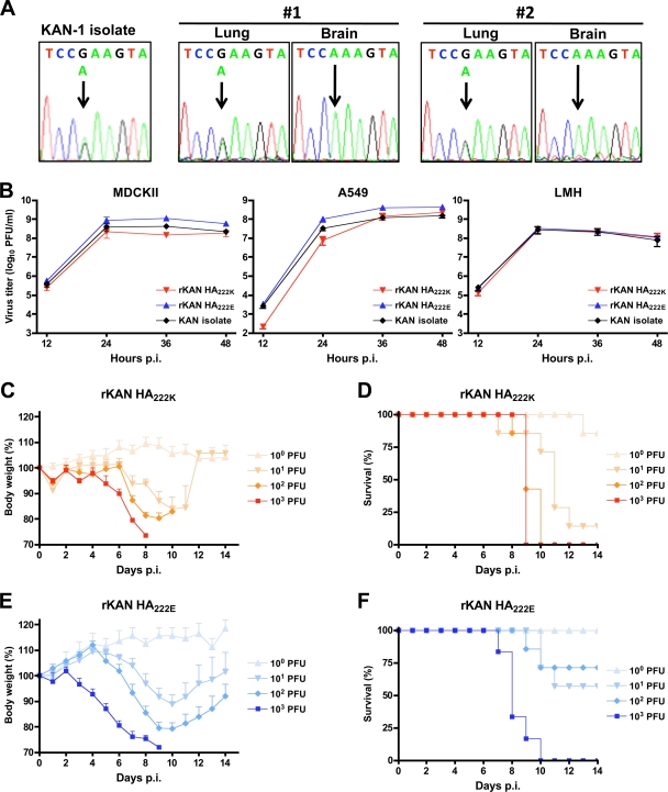
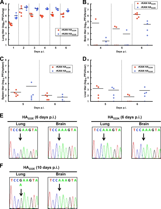
The virus variant with K222E represented 45% and 6% of the quasispecies present in the lung tissues of two out of three patients (9). The virus A/Thailand/KAN-1/2004 (KAN-1), isolated from the first patient in cell culture and passaged several times in MDCK cells, was kindly provided to us by Pilaipan Puthavathana (Mahidol University, Bangkok, Thailand). We sequenced the HA gene of the virus stock and indeed revealed the polymorphism at amino acid position 222 (Fig. 1A). Sequence analysis of all other open reading frames, including the neuraminidase-encoding gene segment, did not reveal any heterogeneity (data not shown). Because H5N1 viruses are known to be neurotropic (8, 18), we infected BALB/c mice with this virus stock, collected lung and brain tissue 5 days postinfection (p.i.), and determined the sequence of the viral HA. Whereas both HA variants were present in the lung, we only identified the HA222K variant in the brain (Fig. 1A), suggesting that the polymorphism in HA affects organ tropism. To confirm these findings, we used reverse genetics (7) to generate recombinant KAN-1 virus encoding either HA222K (rKAN-1 HA222K) or HA222E (rKAN-1 HA222E). For other gene segments, consensus sequences were used that were determined from the virus stock described above by sequencing overlapping reverse transcription (RT)-PCR products. Comparisons of the growth characteristics in various cell lines revealed that rKAN-1 HA222E replicated slightly more efficiently in MDCK cells and A549 human lung carcinoma cells than rKAN-1 HA222K, whereas both virus variants showed similar growth kinetics in avian hepatoma cells (LMH) (Fig. 1B). In BALB/c mice, infection with 100 PFU of either virus variant resulted in a pronounced weight loss; however, in contrast to mice infected with rKAN-1 HA222K, 70% of all mice survived infection with rKAN-1 HA222E (Fig. 1C to F). The 50% lethal doses (LD50) from the experiments whose results are shown in Fig. 1C to F were determined as described previously (16) and revealed a 67-fold difference between rKAN-1 HA222E (LD50 = 200 PFU) and rKAN-1 HA222K (LD50 = 3 PFU), suggesting that rKAN-1 HA222K is more pathogenic for mice. Surprisingly, determination of the viral titers in the lung showed that rKAN-1 HA222E replicated to higher titers than rKAN-1 HA222K at all time points investigated (Fig. 2A). On the contrary, at 6 days postinfection, lower viral titers could be detected in the brain of rKAN-1 HA222E-infected animals (Fig. 2B). Consistent with earlier reports on H5N1-infected mice (15), only low viral titers were observed for both virus variants in liver and spleen of mice at 5 and 6 days postinfection (Fig. 2C and D). Interestingly, sequencing analysis revealed that in all animals infected with rKAN-1 HA222E, the virus isolated from the brain was represented by a revertant with HA222K (Fig. 2E and data not shown). However, no reversion was found in the lung of rKAN-1 HA222E-infected animals. These results further highlighted the restriction of rKAN-1 HA222E to the lung. As expected, no changes in HA were observed in the lung and brain of mice infected with rKAN-1 HA222K (Fig. 2E and data not shown). In the lung of one mouse infected with 10 PFU, we also observed the appearance of the mutation E222K in HA and, as expected, only the HA222K variant in the brain (Fig. 2F). We therefore speculate that reversion to HA222K occurs in the lung, thereby allowing virus to spread to the brain.
FIG. 1.
Growth properties of KAN-1 variants with a polymorphism in HA. (A) Two 8-week-old female BALB/c mice (#1 and #2) were infected intranasally with 103 PFU of the virus isolate A/Thailand/KAN-1/2004. Five days postinfection (p.i.), the sequences of the HA genes from lung and brain tissue were determined and compared to the sequence obtained for the virus isolate used for infection. Sequences shown represent nucleotide positions 725 to 733 (aa 221 to 223) of the HA gene. Arrows indicate the site of the nucleotide polymorphism. (B) Cells were infected with a multiplicity of infection of 0.001 with rKAN-1 HA222K, rKAN-1 HA222E, or the virus isolate A/Thailand/KAN-1/2004 used in the experiment whose results are shown in panel A. (C to F) Eight-week-old female BALB/c mice were infected intranasally with either rKAN-1 HA222K (C and D) or rKAN-1 HA222E (E and F) using the indicated virus doses and were monitored for body weight loss and survival. Error bars show standard deviations.
FIG. 2.
Organ tropism of KAN-1 variants in mice. (A to D) Eight-week-old female BALB/c mice were infected intranasally with 1,000 PFU of either rKAN-1 HA222K or rKAN-1 HA222E. At the indicated time points p.i., virus titers were determined in lung (A), brain (B), spleen (C), and liver (D). Horizontal lines show the means of the results. (E) HA sequences obtained 6 days p.i. from lung and brain tissues of the BALB/c mice that were infected with either rKAN-1 HA222E or rKAN-1 HA222K as described for panels A to D. (F) HA sequences obtained from lung and brain tissues of a BALB/c mouse 10 days after infection with 10 PFU rKAN-1 HA222E as described for Fig. 1F. Arrows indicate the site of the nucleotide polymorphism.
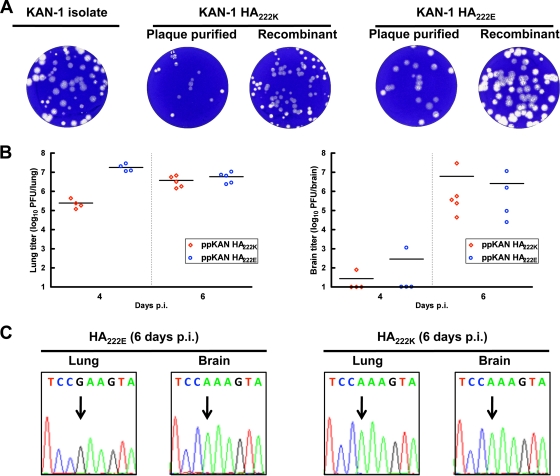
To verify these results with isolates of the original virus stock, we plaque purified two viruses (ppKAN-1 HA222E and ppKAN 1 HA222K). Both plaque-purified variants showed characteristic differences in their plaque sizes, which resembled those of the rescued viruses (Fig. 3A). With the exception of the polymorphism in HA, both plaque-purified isolates revealed complete sequence identity in all genes compared to the rescued viruses (data not shown). Similar to the results observed with the recombinant KAN-1 variants, the lung titers in mice infected with ppKAN-1 HA222E were significantly higher (P < 0.05, Student's t test) than the lung titers of ppKAN-1 HA222K at 4 days postinfection (Fig. 3B). At 6 days postinfection, high viral titers were observed in both ppKAN-1 HA222E- and ppKAN-1 HA222K-infected animals. Consistent with the results obtained with rKAN-1 HA222E, we only observed the HA222K variant in the brain of all the ppKAN-1 HA222E-infected animals (Fig. 3C and data not shown).
FIG. 3.
Growth properties of plaque-purified KAN-1 variants. (A) Comparison of the plaque sizes of the virus isolate A/Thailand/KAN-1/2004 and its plaque-purified and recombinant variants. (B) Eight-week-old female BALB/c mice were infected as described in the Fig. 1 legend with 1,000 PFU of the indicated plaque-purified viruses. At 4 and 6 days p.i., virus titers were determined in lung and brain. Horizontal lines show the means of the results. (C) HA sequences obtained from lung and brain tissues of two animals that were infected with either ppKAN-1 HA222E or ppKAN-1 HA222K 6 days p.i. as described for panel B. Arrows indicate the site of the nucleotide polymorphism.
To assess the effect of K-to-E substitution in the viral HA on the receptor-binding properties, we determined the affinity of the viruses for synthetic sialylglycopolymers (SGPs) bearing distinctive sialyloligosaccharide moieties as described previously (1, 5, 6, 12). The structures and designations of the oligosaccharide parts of the SGPs are shown in Table 1. rKAN-1 HA222K bound preferentially to Neu5Ac2-3Gal-terminated receptors and displayed the highest binding affinity for the sulfated trisaccharide Su-3′SLN, which is a typical binding pattern for Asian H5N1 viruses isolated from birds and humans in 1999 to 2004 (4, 17). The binding to other Neu5Ac2-3Gal-containing receptors varied depending on the structure of the oligosaccharide core. In particular, fucosylation of the GlcNAc residue of Su-3′SLN decreased the binding affinity (compare Su-3′SLN and Su-SLex), suggesting that the fucose moiety does not fit into the receptor-binding site (4). There was no binding to any of the Neu5Ac2-6Gal-terminated SGPs.
TABLE 1.
Association constants of virus complexes with sialylglycopolymers
| Structure of sialyloligosaccharide moiety | Abbreviation | Association constanta (mM−1 Neu5Ac) of HA variant: |
|
|---|---|---|---|
| 222K | 222E | ||
| Neu5Acα2-3Galβ1-3GalNAcα | 3′STF | 0.41 (0.069) | 0.16 (0.014) |
| Neu5Acα2-3Galβ1-3GlcNAcβ | SLec | 0.38 (0.093) | 0.21 (0.045) |
| Neu5Acα2-3Galβ1-3(6-HSO3)GlcNAcβ | Su-SLec | 0.47 (0.089) | 0.12 (0.02) |
| Neu5Acα2-3Galβ1-3(Fucα1-4)GlcNAcβ | SLea | 0.11 (0.089) | 0.07 (0.018) |
| Neu5Acα2-3Galβ1-4GlcNAcβ | 3′SLN | 0.29 (0.073) | 0.20 (0.032) |
| Neu5Acα2-3Galβ1-4(Fucα1-3)GlcNAcβ | SLex | 0.13 (0.031) | 0.1 (0.017) |
| Neu5Acα2-3Galβ1-4(6-HSO3)GlcNAcβ | Su-3′SLN | 2.1 (0.22) | 0.82 (0.28) |
| Neu5Acα2-3Galβ1-4(Fucα1-3)(6-HSO3)GlcNAcβ | Su-SLex | 0.46 (0.15) | 1.3 (0.29) |
| Neu5Acα2-6Galβ1-4GlcNAcβ | 6′SLN | <0.05b | <0.05 |
| Neu5Acα2-6Galβ1-4(6-HSO3)GlcNAcβ | Su-6′SLN | <0.05 | <0.05 |
| Neu5Acα2-6GalNAcα | S-Tn | <0.05 | <0.05 |
The constants were determined using solid-phase fetuin binding inhibition assays as described previously (1, 5, 6, 12) and are expressed in μM−1 with respect to sialic acid. The data are the mean values and standard deviations (in parentheses) from 2 to 4 replicate samples in one experiment that is representative of three independent experiments.
A value of <0.05 indicates that there was no significant inhibition in the fetuin binding inhibition assay at the highest concentration of SGP used.
rKAN-1 HA222E bound more weakly than rKAN-1 HA222K to most of the analogues tested. This result explains the reduced hemagglutinin activity of H5N1 virus with the K222E mutation as observed by others (2). We have looked into the prevalence of 222E in the HAs of H5 viruses in the influenza sequence database (Influenza Virus Resource on http://www.ncbi.nlm.nih.gov/genomes/FLU/FLU.html, accessed on 6 March 2010). Among about 2,500 available nonredundant H5 HA1 sequences, only three sequences of avian viruses and one sequence of the human isolate studied here had E at position 222. Nineteen HAs of the H5N2 viruses from South America and Japan contained 222Q. All other sequences contained either K or (much less frequently) R. This analysis suggests that a positively charged amino acid at position 222 is essential for the survival of H5N1 virus in avian and occasional mammalian host species.
It is interesting that rKAN-1 HA222E is less pathogenic in mice than the HA222K variant despite the higher lung titers of the former virus. We speculate that the higher receptor-binding affinity of HA222K allows efficient infection of cells of the central nervous system, resulting in enhanced pathogenicity. This is consistent with the findings that only the HA222K variant is found in the brain of KAN-1 infected mice and that the receptor distribution is different in brain and lung tissue (14). We speculate that the increased LD50 of the HA222E variant most likely reflects decreased probability of mutation to 222K at lower infection doses.
The reasons for the apparent selection of HA222E in two cases of human infection (9) are not clear. As noticed previously (11), a common feature of the human and swine viruses as compared to their avian precursors is the substantially decreased affinity of mammalian viruses for Neu5Ac2-3Gal-containing receptors. This feature may indicate that binding to such receptors is disadvantageous for the virus's replication in humans, for example, owing to neutralization by decoy receptors present on airway mucins (3). If this hypothesis is correct, it could provide an explanation for the accumulation in humans of the variant with the mutation 222E and decreased binding affinity to Neu5Ac2-3Gal-terminated oligosaccharides. This could also explain the higher viral lung titers in mice infected with rKAN-1 HA222E compared to the lung titers seen with infections with the HA222K variant.
Interestingly, despite its overall reduced binding affinity, rKAN-1 HA222E showed enhanced binding to one analogue tested, Su-SLex, a receptor determinant commonly recognized by poultry viruses with different HA subtypes (6, 10). It remains to be determined whether enhanced binding of HA222E to Su-SLex plays any specific role in H5N1 virus adaptation to humans or if this effect is merely coincidental with the mutation that serves to decrease viral binding to a majority of Neu5Ac2-3Gal-containing receptors.
Acknowledgments
We thank Pilaipan Puthavathana (Mahidol University, Bangkok, Thailand) for providing the virus isolate A/Thailand/KAN-1/2004 (KAN-1) and Geoffrey Chase for critical reading of the manuscript.
This work was funded by the Bundesministerium für Bildung und Forschung (BMBF, FluResearchNet) and the European Union FP6 projects FLUPATH and FLUINNATE.
Footnotes
Published ahead of print on 2 June 2010.
REFERENCES
- 1.Bovin, N. V. 1998. Polyacrylamide-based glycoconjugates as tools in glycobiology. Glycoconj. J. 15:431-446. [DOI] [PubMed] [Google Scholar]
- 2.Chutinimitkul, S., D. van Riel, V. J. Munster, J. M. van den Brand, G. F. Rimmelzwaan, T. Kuiken, A. D. Osterhaus, R. A. Fouchier, and E. de Wit. 14 April 2010. In vitro assessment of attachment pattern and replication efficiency of H5N1 influenza A viruses with altered receptor specificity. J. Virol. doi: 10.1128/JVI.02737-09. [DOI] [PMC free article] [PubMed]
- 3.Couceiro, J. N., J. C. Paulson, and L. G. Baum. 1993. Influenza virus strains selectively recognize sialyloligosaccharides on human respiratory epithelium; the role of the host cell in selection of hemagglutinin receptor specificity. Virus Res. 29:155-165. [DOI] [PubMed] [Google Scholar]
- 4.Gambaryan, A., A. Tuzikov, G. Pazynina, N. Bovin, A. Balish, and A. Klimov. 2006. Evolution of the receptor binding phenotype of influenza A (H5) viruses. Virology 344:432-438. [DOI] [PubMed] [Google Scholar]
- 5.Gambaryan, A. S., and M. N. Matrosovich. 1992. A solid-phase enzyme-linked assay for influenza virus receptor-binding activity. J. Virol. Methods 39:111-123. [DOI] [PubMed] [Google Scholar]
- 6.Gambaryan, A. S., A. B. Tuzikov, G. V. Pazynina, J. A. Desheva, N. V. Bovin, M. N. Matrosovich, and A. I. Klimov. 2008. 6-sulfo sialyl Lewis X is the common receptor determinant recognized by H5, H6, H7 and H9 influenza viruses of terrestrial poultry. Virol. J. 5:85. [DOI] [PMC free article] [PubMed] [Google Scholar]
- 7.Hoffmann, E., G. Neumann, Y. Kawaoka, G. Hobom, and R. G. Webster. 2000. A DNA transfection system for generation of influenza A virus from eight plasmids. Proc. Natl. Acad. Sci. U. S. A. 97:6108-6113. [DOI] [PMC free article] [PubMed] [Google Scholar]
- 8.Iwasaki, T., S. Itamura, H. Nishimura, Y. Sato, M. Tashiro, T. Hashikawa, and T. Kurata. 2004. Productive infection in the murine central nervous system with avian influenza virus A (H5N1) after intranasal inoculation. Acta Neuropathol. 108:485-492. [DOI] [PubMed] [Google Scholar]
- 9.Kongchanagul, A., O. Suptawiwat, P. Kanrai, M. Uiprasertkul, P. Puthavathana, and P. Auewarakul. 2008. Positive selection at the receptor-binding site of haemagglutinin H5 in viral sequences derived from human tissues. J. Gen. Virol. 89:1805-1810. [DOI] [PubMed] [Google Scholar]
- 10.Matrosovich, M., A. S. Gambarian, and H. D. Klenk. 2008. Receptor specificity of influenza viruses and its alteration during interspecies transmission. In H. D. Klenk, M. Matrosovich, and J. Stech (ed.), Avian influenza, vol. 27. Karger, Basel, Switzerland.
- 11.Matrosovich, M., A. Tuzikov, N. Bovin, A. Gambaryan, A. Klimov, M. R. Castrucci, I. Donatelli, and Y. Kawaoka. 2000. Early alterations of the receptor-binding properties of H1, H2, and H3 avian influenza virus hemagglutinins after their introduction into mammals. J. Virol. 74:8502-8512. [DOI] [PMC free article] [PubMed] [Google Scholar]
- 12.Matrosovich, M. N., A. S. Gambaryan, A. B. Tuzikov, N. E. Byramova, L. V. Mochalova, A. A. Golbraikh, M. D. Shenderovich, J. Finne, and N. V. Bovin. 1993. Probing of the receptor-binding sites of the H1 and H3 influenza A and influenza B virus hemagglutinins by synthetic and natural sialosides. Virology 196:111-121. [DOI] [PubMed] [Google Scholar]
- 13.Neumann, G., H. Chen, G. F. Gao, Y. Shu, and Y. Kawaoka. 2010. H5N1 influenza viruses: outbreaks and biological properties. Cell Res. 20:51-61. [DOI] [PMC free article] [PubMed] [Google Scholar]
- 14.Ning, Z. Y., M. Y. Luo, W. B. Qi, B. Yu, P. R. Jiao, and M. Liao. 2009. Detection of expression of influenza virus receptors in tissues of BALB/c mice by histochemistry. Vet. Res. Commun. 33:895-903. [DOI] [PubMed] [Google Scholar]
- 15.Nishimura, H., S. Itamura, T. Iwasaki, T. Kurata, and M. Tashiro. 2000. Characterization of human influenza A (H5N1) virus infection in mice: neuro-, pneumo- and adipotropic infection. J. Gen. Virol. 81:2503-2510. [DOI] [PubMed] [Google Scholar]
- 16.Reed, L. J., and H. Muench. 1938. A simple method for estimating fifty per cent endpoints. Am. J. Hyg. 27:493-497. [Google Scholar]
- 17.Stevens, J., O. Blixt, T. M. Tumpey, J. K. Taubenberger, J. C. Paulson, and I. A. Wilson. 2006. Structure and receptor specificity of the hemagglutinin from an H5N1 influenza virus. Science 312:404-410. [DOI] [PubMed] [Google Scholar]
- 18.Tanaka, H., C. H. Park, A. Ninomiya, H. Ozaki, A. Takada, T. Umemura, and H. Kida. 2003. Neurotropism of the 1997 Hong Kong H5N1 influenza virus in mice. Vet. Microbiol. 95:1-13. [DOI] [PubMed] [Google Scholar]


