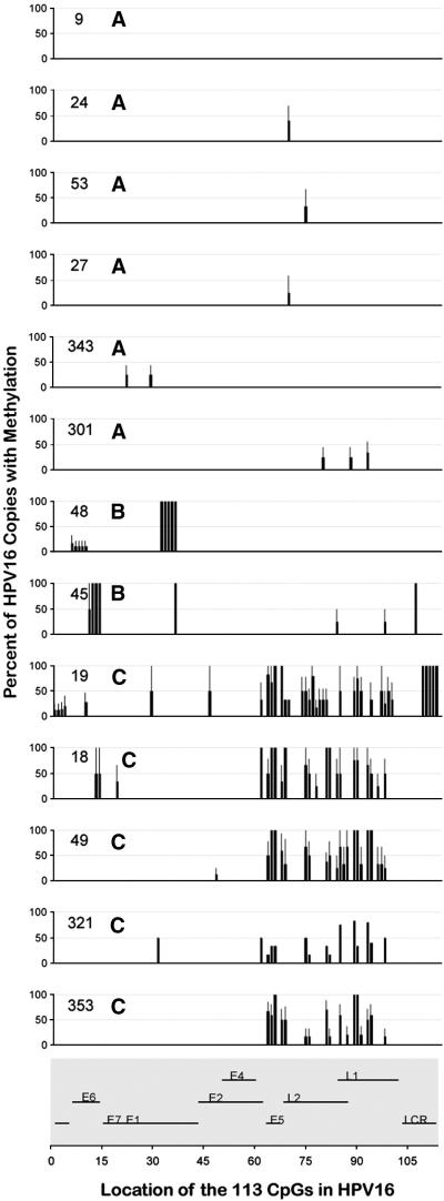
Figure 2.
HPV16 DNA methylation profiles of the individual cervical samples The HPV16 DNA methylation profile of each sample at each potential methylation site is shown. The numbers inside each plot are the sample numbers and the adjacent letters are the HPV16 DNA methylation pattern (A, B or C). Nucleotides other than the 113 CpGs are not shown. The organization of the HPV16 genome with sequential numbering of the CpGs is shown at the bottom of the figure.

