Abstract
Primary mesothelioma of the pericardium is a rare tumor and carries a dismal prognosis. This case report presents a 38-year-old man who suffered from recurrent pericardial fluid. Initial symptoms were unspecific, with dry cough and progressing fatigue. Pericardiocentesis was performed, but analyses for malignant cells and tuberculosis were negative. After recurrence a pericardiectomy was planned. At operation, partial resection of tumor tissue surrounding the heart was performed. Histopathologic examination including immunohistochemical staining for calretinin showed a biphasic mesothelioma. During the postoperative period the patient's condition ameliorated, but symptoms recurred and the patient died 3 months after diagnosis and 15 months after the first symptoms. At autopsy, the pericardium was transformed by the tumor that also expanded into the mediastinum and had set metastases to the liver. A review of 29 cases presented in the recent literature indicates a higher incidence of malignant pericardial mesothelioma among men than women. Median age was 46 (range, 19–76) years. In pleural mesotheliomas, exposure to asbestos is a known risk factor. However, in primary pericardial mesotheliomas the evidence for asbestos as an etiologic factor seems to be less convincing (3 exposed among 14 cases). Symptoms are often unspecific and cytologic examination of pericardial fluid is seldom conclusive (malignant cells demonstrated in 4/17 cases). Partial resection of the tumor can give a period of symptom reduction. Only a few patients have been treated with chemotherapy. Median survival of patients with pericardial mesotheliomas is approximately 6 months.
Key Words: Malignant mesothelioma, Pericardium, Review, Asbestos exposure
Introduction
Malignant diseases of the pericardium can be divided into primary and secondary tumors. Secondary tumors are more frequent, and in this group, metastases from lung or breast cancer are most common [1].
Malignant mesotheliomas are rare tumors [2] that can occur in any of the body cavities covered by mesothelium. They arise most frequently in pleura but also in peritoneum, pericardium or tunica vaginalis testis. In a large review of 4,710 cases by Hillerdal 1982 [3], pericardial mesotheliomas account for 0.7% of all malignant mesotheliomas. Our report presents one patient with pericardial mesothelioma and a review of 29 cases that appeared in English literature from 1993 through 2008.
Case Report
A 38-year-old metal worker, originally from Cyprus, was admitted to our clinic in October 2002 after over a year's history of recurring pericardial fluid, followed by pericardiectomy and a tumour diagnosis.
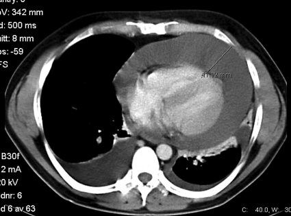
In November 2001, he was admitted to a local hospital due to cardiac tamponade. At that time he had a history of several weeks of dry cough and progressing fatigue. Pericardiocentesis with pericardial drain was performed. The fluid was clear and all analyses showed normal results, cultivations and PCR for tuberculosis were negative and cytological analysis of the fluid showed no malignant cells and only a small increase of eosinophilic cells. Chest X-ray was normal except for an increased heart volume; CT scan of the chest/abdomen showed pericardial fluid with a largest width of 4 cm, some amount of pleural fluid and some ascites (fig. 1). The radiological examination was interpreted as exudative pericarditis. A rheumatologist was consulted but could not find any explanation for the patient's condition. Even a small genealogy was made, taking Mediterranean fever into consideration, but with negative results. It finally became clear that the patient had had a bicycle accident 3 weeks prior to the first symptoms from which he had suffered a haematoma in the thoracic wall. Because of this the plausible diagnosis was post-cardiac trauma syndrome. Standard treatment with cortisone was initiated and the symptoms as well as the pericardial fluid disappeared. During the next 6 months the patient had 3 relapses, and in September 2002 he was referred to the University Hospital of Umeå for a pericardiectomy. At surgery, a thickened pericardium was opened and 1 l of filthy and hemorrhagic fluid was exploited. The heart was covered with a 0.5-cm thick layer that gave a constrictive hemodynamic. The layer was to some extent removed, but the area around the phrenic nerve was saved.
Fig. 1.
CT-scan of the thorax of the 38-year-old patient at presentation of symptoms.
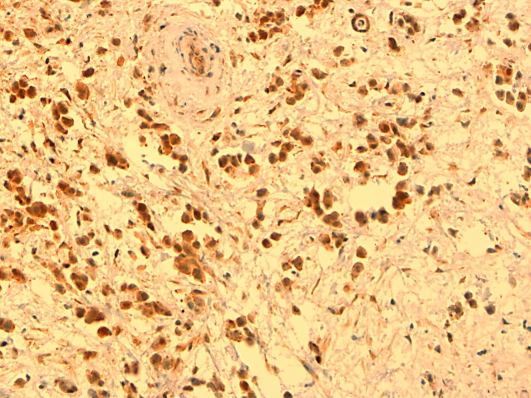
The histopathologic examination showed biphasic mesothelioma of the pericardium with positive immunohistochemical staining for calretinin (fig. 2). After surgery, the patient's symptoms diminished and in October 2002 he had recovered. At this time there was no indication for further postoperative treatment. One month later a follow-up CT scan showed pericardial fluid. The patient still had no symptoms. In the beginning of January 2003, his condition worsened with severe dyspnoea and he was admitted to the local hospital. Pleural fluid was stated and he was treated with corticosteroids and diuretics. Ten days later he was transported to our clinic for assessment of further treatment. However, his condition deteriorated and he died 3 months after diagnosis and 15 months after the first symptoms.
Fig. 2.
Immunohistochemical staining for calretinin (Zymed(r), polyclonal rabbit anti-calretinin antibody, Invitrogen, Carlsbad, Calif., USA) showing a biphasic mesothelioma (20X).
Autopsy revealed an advanced tumour that embraced the heart. The pericardium was highly transformed by the neoplasm, thus whitish and solid with a thickness of 3-4 cm in large areas. The myocardium was partly invaded by the tumour and the delimitation between the pericardium and the myocardium was vague. The tumour also expanded from the pericardium to the central parts of the mediastinum. Muddy fluid was noted around the heart. Metastatic tumour growth was found in the liver. A small focus of papillary thyroid cancer with spread to local lymph nodes was also found at autopsy.
Discussion and Literature Review
In 1994, Thomason et al. [4] presented a case report and reviewed 27 cases of primary pericardial mesothelioma that appeared in English literature from 1972 to 1992. After this no extent review has been made. Our review includes 29 cases in English literature from 1993 through 2008 where the diagnosis of primary pericardial mesothelioma was established [5,6,7,8,9,10,11,12,13,14,15,16,17,18,19,20,21,22,23,24,25,26,27,28,29]. It also includes the findings from our patient and the review, presented in table 1 and table 2. To provide an overview and comparison, the results from Thomason et al. [4] are presented adjacent to our data.
Table 1.
Clinical material, gender, age and asbestos exposure
| n | % | Thomason et al. [4], % | |
|---|---|---|---|
| Gender | |||
| Men | 22 | 73 | 71 |
| Women | 8 | 27 | 29 |
| Exposure to asbestos | |||
| Exposure | 3/14 | 21 | 33 |
| No known exposure | 11/14 | 79 | 67 |
| Not mentioned | 16/30 | 53 | 57 |
| n | Median (min.–max.) | Thomason et al. [4] | |
|---|---|---|---|
| Age, years | |||
| Men | 22 | 45.5 (19–70) | |
| Women | 8 | 52.5 (31–76) | |
| All | 30 | 46 (19–76) | 48 (12–77) |
Table 2.
Histopathologic and metastatic findings
| n | % | Thomason et al. [4], % | |
|---|---|---|---|
| Mesothelioma type | |||
| Biphasic | 7/24 | 29 | 35 |
| Epithelial | 13/24 | 54 | 35 |
| Sarcomatous | 4/24 | 17 | 30 |
| Not available | 6/30 | 20 | 28 |
| Effusion cytologic findings | |||
| Benign | 10/17 | 59 | 70 |
| Malignant | 4/17 | 23 | 20 |
| No fluid available | 3/17 | 18 | − |
| Inconclusive | − | − | 10 |
| Not available | 13/30 | 43 | 64 |
| Metastases | |||
| Lymphatic | 8/16 | 50 | |
| Hepatic | 2/16 | 13 | |
| None observed | 6/16 | 38 | |
| Not mentioned | 14/30 | 47 |
Table 1 illustrates that for 30 cases there is a male-female ratio of 3:1. The male domination correlates well with the review by Thomason et al. that reported a male dominance [4]. Andersen et al. [30] reported an even distribution between men and women. On the other hand, they used strict criteria for establishing the diagnosis primary pericardial mesothelioma which excluded cases with distant metastases and is thus not ideal for comparison. The youngest patient in this review was 19 years old and the eldest 76 years old; there is an even distribution of the age range with a median age of 46 years. This corresponds to the review by Thomason et al. in which over half of the patients were diagnosed between the 5th and the 7th decade [4]. An illustration of the age range on the basis of gender shows an even distribution of men and women. No similar illustration is presented in the study by Thomason et al. [4].
No obvious relationship between asbestos exposure and the development of pericardial mesothelioma has been established. Table 1 illustrates asbestos exposure. Three cases with known exposure to asbestos are reported [5,6,7]. For 11 of the patients no known exposure to asbestos is reported [8,9,10,11,12,13,14,15,16]. Most of the reviewed articles do not contain any information about asbestos exposure. Thomason et al. reported that a third of the patients were exposed to asbestos [4].
The histological pattern of pericardial mesothelioma is classified into 3 categories according to the World Health Organization: predominantly epithelial, predominantly fibrous (spindle cell) and biphasic (mixed) [31]. Table 2 summarizes the pathologic findings of the patients in this review. The histological pattern was reported in 24 patients and revealed about the same distribution of epithelial and biphasic pattern, 13 and 7 cases, respectively, while the fibrous or sarcomatoid pattern was rarer with only 4 cases. This result is similar to that of the study by Thomason et al., where the distribution was somewhat more even [4]. In the attempt to establish a diagnosis in these patients, cytological study of pericardial effusion is often made. Seventeen cases reported cytological findings from the effusion of pericardial fluid and only 24% of the cases presented malignant cells. The same result was demonstrated by Thomason et al. (20%) and this suggests cytological evaluation to be a poor method for the detection of mesothelioma [4].
The metastatic spread of mesothelioma is presented in table 2. According to Karadzic et al. [17], metastases are present in about 25-45% of the patients and involve regional lymph nodes, lungs and kidneys. Eren et al. [32] report a similar incidence of metastatic spread. In our study, 16 of the case reports contained information about metastatic spread, and 6 of them had no metastatic spread, 8 had regional lymph node involvement, 2 had liver metastases and 1 patient had lung metastases.
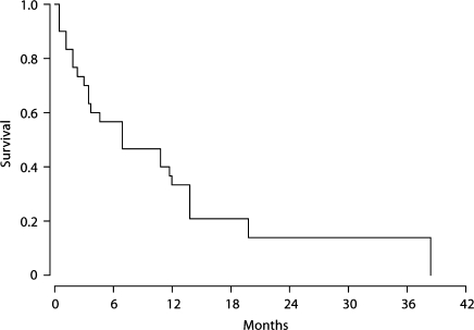
Survival distribution of the patients from the time when the first symptoms appeared is illustrated in fig. 3. Three patients were alive when the article was published [18], and for 2 patients no information on survival was presented [19]. The median survival time from first symptoms in this review is 6 months. This dismal prognosis correlates with earlier studies; Cohen [31] showed average disease duration of approximately 5 months. No similar diagram was presented in the study by Thomason et al. [4].
Fig. 3.
Kaplan-Meier plot of disease-specific survival of 30 patients with pericardial mesothelioma [5,6,7,8,9,10,11,12,13,14,15,16,17,18,19,20,21,22,23,24,25,26,27,28,29].
In summary, this review of the clinical characteristics of patients with primary pericardial mesothelioma shows that initial symptoms are unspecific. Whether exposure to asbestos is related to the disease or not is uncertain. Treatment options are limited and the prognosis is dismal.
Acknowledgements
This study was supported by grants from the Medical Faculty of the Umeå University. Dr. Thomas Höckenström, Department of Biomedicine, Pathology, Umeå University Hospital, is acknowledged for histopathologic evaluation. We also acknowledge the support by Björn Tavelin, Oncologic Center, Umeå University Hospital.
References
- 1.Kralstein J, Frishman WH. Malignant pericardial diseases: diagnosis and treatment. Cardiol Clin. 1987;5:583–589. [PubMed] [Google Scholar]
- 2.Murai Y. Malignant mesothelioma in Japan: analysis of registered autopsy cases. Arch Environ Health. 2001;56:84–88. doi: 10.1080/00039890109604058. [DOI] [PubMed] [Google Scholar]
- 3.Hillerdal G. Malignant mesothelioma 1982: review of 4,710 published cases. Br J Dis Chest. 1983;77:321–143. [PubMed] [Google Scholar]
- 4.Thomason R, Schlegel W, Lucca M, Cummings S, Lee S. Primary malignant mesothelioma of the pericardium. Case report and literature review. Tex Heart Inst J. 1994;21:170–174. [PMC free article] [PubMed] [Google Scholar]
- 5.Fujiwara H, Kamimori T, Morinaga K, Takeda Y, Kohyama N, Miki Y, Inai K, Yamamoto S. An autopsy case of primary pericardial mesothelioma in arc cutter exposed to asbestos through talc pencils. Ind Health. 2005;43:346–350. doi: 10.2486/indhealth.43.346. [DOI] [PubMed] [Google Scholar]
- 6.Oreopoulos G, Mickleborough L, Daniel L, De Sa M, Merchant N, Butany J. Primary pericardial mesothelioma presenting as constrictive pericarditis. Can J Cardiol. 1999;15:1367–1372. [PubMed] [Google Scholar]
- 7.Kaul TK, Fields BL, Kahn DR. Primary malignant pericardial mesothelioma: a case report and review. J Cardiovasc Surg (Torino) 1994;35:261–267. [PubMed] [Google Scholar]
- 8.Val-Bernal JF, Figols J, Gomez-Roman JJ. Incidental localized (solitary) epithelial mesothelioma of the pericardium: case report and literature review. Cardiovasc Pathol. 2002;11:181–185. doi: 10.1016/s1054-8807(02)00097-2. [DOI] [PubMed] [Google Scholar]
- 9.Hirano H, Maeda T, Tsuji M, Ito Y, Kizaki T, Yoshii Y, Sashikata T. Malignant mesothelioma of the pericardium: case reports and immunohistochemical studies including Ki-67 expression. Pathol Int. 2002;52:669–676. doi: 10.1046/j.1440-1827.2002.01404.x. [DOI] [PubMed] [Google Scholar]
- 10.Suman S, Schofield P, Large S. Primary pericardial mesothelioma presenting as pericardial constriction: a case report. Heart. 2004;90:e4. doi: 10.1136/heart.90.1.e4. [DOI] [PMC free article] [PubMed] [Google Scholar]
- 11.Lagrotteria DD, Tsang B, Elavathil LJ, Tomlinson CW. A case of primary malignant pericardial mesothelioma. Can J Cardiol. 2005;21:185–187. [PubMed] [Google Scholar]
- 12.Quinn DW, Qureshi F, Mitchell IM. Pericardial mesothelioma: the diagnostic dilemma of misleading imaging. Ann Thorac Surg. 2000;69:1926–1927. doi: 10.1016/s0003-4975(00)01204-2. [DOI] [PubMed] [Google Scholar]
- 13.Molina Garrido MJ, Mora Rufete A, Rodríguez-Lescure A, Cascón Pérez JD, Ardoy F, Guillén Ponce C, Carrato Mena A. Recurrent pericardial effusion as initial manifestation of primary diffuse pericardial malignant mesothelioma. Clin Transl Oncol. 2006;8:694–696. doi: 10.1007/s12094-006-0042-8. [DOI] [PubMed] [Google Scholar]
- 14.Small GR, Nicolson M, Buchan K, Broadhurst P. Pericardial malignant mesothelioma: a latent complication of radiotherapy? Eur J Cardiothorac Surg. 2008;33:745–747. doi: 10.1016/j.ejcts.2007.12.024. [DOI] [PubMed] [Google Scholar]
- 15.Santos C, Montesinos J, Castañer E, Sole JM, Baga R. Primary pericardial mesothelioma. Lung Cancer. 2008;60:291–293. doi: 10.1016/j.lungcan.2007.08.029. [DOI] [PubMed] [Google Scholar]
- 16.Vornicu M, Arora S, Achilleos A. Primary pericardial mesothelioma: a rare cardiac malignancy. Intern Med J. 2007;37:576–577. doi: 10.1111/j.1445-5994.2007.01409.x. [DOI] [PubMed] [Google Scholar]
- 17.Karadzic R, Kostic-Banovic L, Antovic A, Celar M, Katic V, Ilic G, Stojanovic J. Primary pericardial mesothelioma presenting as constrictive pericarditis. Arch Oncol. 2005;13:150–152. [Google Scholar]
- 18.Stein M, Neuman A, Dale J, Drumea K, Ben-Itzhak O, Bar-Shalom R, Goldscher D, Haim N. Cardiac tamponade as the initial manifestation of primary pericardial mesothelioma. Med Pediatr Oncol. 1995;24:208–212. doi: 10.1002/mpo.2950240313. [DOI] [PubMed] [Google Scholar]
- 19.Kaminaga T, Yamada N, Imakita S, Takamiya M, Nishimura T. Magnetic resonance imaging of pericardial malignant mesothelioma. Magn Reson Imaging. 1993;11:1057–1061. doi: 10.1016/0730-725x(93)90226-4. [DOI] [PubMed] [Google Scholar]
- 20.Ohnishi J, Shiotani H, Ueno H, Fujita N, Matsunaga K. Primary pericardial mesothelioma demonstrated by magnetic resonance imaging. Jpn Circ J. 1996;60:898–900. doi: 10.1253/jcj.60.898. [DOI] [PubMed] [Google Scholar]
- 21.Miyamoto Y, Nakano S, Shimazaki Y, Matsuda H, Fukuda H. Pericardial mesothelioma presenting as left atrial thrombus in a patient with mitral stenosis. Cardiovasc Surg. 1996;4:51–52. doi: 10.1016/0967-2109(96)83783-5. [DOI] [PubMed] [Google Scholar]
- 22.Peregud-Pogorzelska M, Kazmierczak J, Wojtarowicz A. Intracavitary mass as the initial manifestation of primary pericardial mesothelioma: a case report. Angiology. 2007;58:255–258. doi: 10.1177/0003319707300379. [DOI] [PubMed] [Google Scholar]
- 23.Kobayashi Y, Murakami R, Ogura J, Yamamoto K, Ichikawa T, Nagasawa K, Hosone M, Kumazaki T. Primary pericardial mesothelioma: a case report. Eur Radiol. 2001;11:2258–2261. doi: 10.1007/s003300100884. [DOI] [PubMed] [Google Scholar]
- 24.Eryilmaz S, Sirlak M, Inan MB, Erden E, Eren NT, Corapcioglu T, Akalin H. Primary pericardial mesothelioma. Cardiovasc Pathol. 2001;10:147–149. doi: 10.1016/s1054-8807(01)00073-4. [DOI] [PubMed] [Google Scholar]
- 25.Papi M, Genestreti G, Tassinari D, Lorenzini P, Serra S, Ricci M, Pasquini E, Nicolini M, Pasini G, Tamburini E, Fattori PP, Ravaioli A. Malignant pericardial mesothelioma. Report of two cases, review of the literature and differential diagnosis. Tumori. 2005;91:276–279. doi: 10.1177/030089160509100315. [DOI] [PubMed] [Google Scholar]
- 26.Yakirevich E, Sova Y, Drumea K, Bergman I, Quitt M, Resnick MB. Peripheral lymphadenopathy as the initial manifestation of pericardial mesothelioma: a case report. Int J Surg Pathol. 2004;12:403–405. doi: 10.1177/106689690401200415. [DOI] [PubMed] [Google Scholar]
- 27.Watanabe A, Sakata J, Kawamura H, Yamada O, Matsuyama T. Primary pericardial mesothelioma presenting as constrictive pericarditis: a case report. Jpn Circ J. 2000;64:385–388. doi: 10.1253/jcj.64.385. [DOI] [PubMed] [Google Scholar]
- 28.Doval DC, Pande SB, Sharma JB, Rao SA, Prakash N, Vaid AK. Report of a case of pericardial mesothelioma with liver metastases responding well to pemetrexed and platinum-based chemotherapy. J Thorac Oncol. 2007;2:780–781. doi: 10.1097/JTO.0b013e31811f3acd. [DOI] [PubMed] [Google Scholar]
- 29.Shimazaki H, Aida S, Iizuka Y, Yoshizu H, Tamai S. Vacuolated cell mesothelioma of the pericardium resembling liposarcoma: a case report. Human Pathol. 2000;31:767–770. doi: 10.1053/hupa.2000.7630. [DOI] [PubMed] [Google Scholar]
- 30.Andersen JA, Hansen BF. Primary pericardial mesothelioma. Dan Med Bull. 1974;21:195–200. [PubMed] [Google Scholar]
- 31.Cohen JL. Neoplastic pericarditis. Cardiovasc Clin. 1976;7:257–269. [PubMed] [Google Scholar]
- 32.Eren NT, Akar AR. Primary pericardial mesothelioma. Curr Treat Options Oncol. 2002;3:369–373. doi: 10.1007/s11864-002-0002-7. [DOI] [PubMed] [Google Scholar]


