Fig. 10.
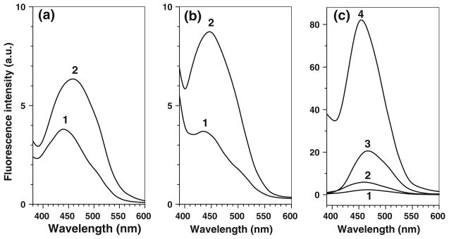
ABS-independent background (matrix) fluorescence originating from oxidative protein modification. a and b Spectra of samples containing 100 μg of (a) digested or (b) non-digested C2C12 proteins (1) before and (2) after 1 h incubation in 0.1 M PBS (pH 9) with 10 μM K3Fe(CN)6. c Fluorescence spectra of 10 (curves 1,3,4) and 2 μM (curve 2) kynurenin in 0.1 M PBS, pH 9 (curve 1), 50% PBS/50% ethanol (curves 2 and 3) and 100% ethanol (curve 4). Excitation wavelength was set at 360 nm

