Abstract
We describe a patient in which an osteochondroma, which resulted from hereditary multiple exostoses, limited flexion of the proximal interphalangeal (PIP) joint at birth. The tumor grew over the original distal head of the proximal phalanx, and the early appearance of a second ossification center on the base of the middle phalanx was observed. The mass was removed surgically when the patient was 17 months old. There was an improvement in the range of motion at a follow-up evaluation 3 years later. The tumor shape and the growth of the affected PIP joint are examined in detail.
Keywords: Hereditary multiple exostosis, Osteochondroma, Growth of tumor
Osteochondroma limiting the movement of the proximal interphalangeal (PIP) joint at birth is a rare tumorous condition of the bone. Here, we report a patient with hereditary multiple exostosis with a tumor mass on the palmar side of the PIP joint. The shape and the serial change of this tumor including surrounding structures were examined in detail. We describe a case with osteochondroma-limited flexion of the PIP joint at birth and discuss the serial changes of this tumor including the structures around the tumor.
Case Report
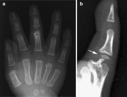
A 24-day-old boy was not able to flex the PIP joint of his right middle finger. He could not flex his PIP joint more than 5° as shown by stress plain radiographic examination. A mass was palpable on the palmer aspect of the PIP joint, and radiographs showed a bony prominence attached to the distal end of the proximal phalanx, making the head look like it was divided into two parts (Fig. 1). Although the tumor had to be removed by surgery, the operation was postponed because it is optimal to perform surgery when patients are 1 year old. When the patient was 11 months old, the tumor had branched to the intra-articular region and had grown over the original distal head of the proximal phalanx. Furthermore, the early appearance of a second ossification center on the base of the middle phalanx was observed in plain radiographs (Fig. 2). The 3D shape of the tumor was clearly demonstrated by 3D-computed tomography (3D-CT; Fig. 3). A magnetic resonance imaging (MRI) examination revealed that the tumor had progressed into the intra-articular and extra-articular spaces and had a cartilage cap (Fig. 4). A skeletal survey showed multiple tumors in other extremities. His mother, older brother, and younger sister also had multiple tumors in the extremities, including the fingers, and plain radiographs confirmed this. Based on the radiological findings and the family history, the patient was diagnosed with hereditary multiple exostosis. At the age of 17 months, the osteochondroma was removed surgically by a palmar approach. The extra-articular portion, a large palmar protuberance, was resected first (Fig. 5a). The intra-articular portion that covered the distal head of the proximal phalanx was resected next (Fig. 5b). Normal cartilage was seen under the tumor. Histopathologic findings revealed a cartilage cap and cancellous bone, consistent with an osteochondroma.
Figure 1.
Plain radiograph of the right hand at 1 month after birth. A bony prominence (white arrow) attached to the distal end of the proximal phalanx makes the head appear as if it is divided into two parts. a Anterior–posterior view. b Lateral view.
Figure 2.
Plain radiograph when the patient was 11 months old. The tumor branches to the intra-articular region, and the early appearance of the second ossification center of the base of middle phalanx is recognized (white arrow). a Anterior–posterior view. b Lateral view.
Figure 3.
The 3D shape of the tumor of the right middle finger was clearly demonstrated by 3D-CT. a From volar view. b From lateral view. c From distal view.
Figure 4.
The high intensity area by proton-enhanced MRI shows a cartilaginous region including a cartilage cap (arrow).
Figure 5.
A schematic drawing of the surgery: removal of tumor by a volar approach. The extra-articular portion was removed first (arrow A). The intra-articular portion that covered the distal head of the proximal phalanx was removed secondly (arrow B). Normal cartilage was seen under the tumor.
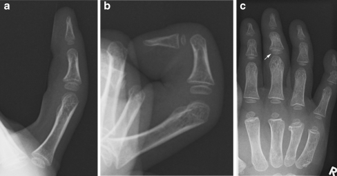
Passive flexion of 80° was possible after the operation. Three years after the surgical treatment, although there was a slight deformity of the distal head of the proximal phalanx, as seen in plain radiographs (Fig. 6), the patient could flex his PIP joint up to 80° as an active range of motion. Recurrence of the osteochondroma was not observed.
Figure 6.
Plain radiograph 3 years after the operation. There is a slight deformity of the distal head of the proximal phalanx. Note the overgrowth of the second ossification center of the base of middle phalanx (white arrow). a Active extension. b Active flexion. c Anterior–posterior view.
Discussion
Hereditary multiple exostosis is a bone dysplasia characterized by multiple cartilaginous exostosis principally involving long and flat bones. However, several authors have stated that involvement of the hand is rare and that proximal phalanges and metacarpals are frequent sites of hand exostoses. Karr et al. [3] stated that all hand exostoses located at the distal end of the proximal or middle phalanx will result in rotational or angular deformities if left untreated. On the other hand, Cates and Burgess [1] described that 47 exostoses occurred at the distal end of the proximal or middle phalanx in 22 patients and radiographic rotational or angular deformities developed in only three. None of these three was symptomatic, and none required operation. We treated our case with surgical intervention at 17 months of age, but minor deformity of the distal head of the proximal phalanx remained. It may have been optimal to operate on this case at an earlier age.
Exostosis that blocks movement of an interphalangeal joint in the hand has been reported in several cases in the past [2–7]. The youngest case reported was a 7-month-old boy who was not able to flex the proximal interphalangeal joint by solitary exostosis, as reported by Kojima et al. [4]. In our patient, the tumor was confirmed with plain radiographs just after birth. Afterward, the tumor branched into the intra-articular region of the middle phalanx and had an early appearance of a second ossification center in plain radiographs. No previous reports in the literature describe a situation similar to our case.
In summary, we report an infant with an unusual exostosis of the proximal phalanx. We recommend surgical intervention for hereditary multiple exostosis cases at an early age, if the joint movement is blocked.
References
- 1.Cates HE, Burgess RC. Incidence of brachydactyly and hand exostosis in hereditary multiple exostosis. J Hand Surg Am. 1991;16:127–132. doi: 10.1016/S0363-5023(10)80027-9. [DOI] [PubMed] [Google Scholar]
- 2.Hayashi J, Ikuta Y, Murakami T, et al. An experience with 16 cases of exostosis in hand. J Jpn Soc Surg Hand. 1987;4:697–701. [Google Scholar]
- 3.Karr MA, Aulicino PL, DuPuy TE, et al. Osteochondromas of the hand in hereditary multiple exostosis: report of a case presenting as a blocked proximal interphalangeal joint. J Hand Surg Am. 1984;9:264–268. doi: 10.1016/s0363-5023(84)80157-4. [DOI] [PubMed] [Google Scholar]
- 4.Kojima T, Yanagawa H, Tomonari H. Solitary osteochondroma limiting flexion of the proximal interphalangeal joint in an infant: a case report. J Hand Surg Am. 1992;17:1057–1059. doi: 10.1016/S0363-5023(09)91061-9. [DOI] [PubMed] [Google Scholar]
- 5.Moore JR, Curtis RM, Wilgis EFS. Osteocartilaginous lesions of the digits in children: an experience with 10 cases. J Hand Surg Am. 1983;8:309–315. doi: 10.1016/s0363-5023(83)80167-1. [DOI] [PubMed] [Google Scholar]
- 6.Murase T, Morimoto H, Tada K, et al. Pseudomallet finger associated with exostosis of the phalanx: a report of 2 cases. J Hand Surg Am. 2002;27:817–820. doi: 10.1053/jhsu.2002.35305. [DOI] [PubMed] [Google Scholar]
- 7.Stern PJ, Phillips D. Phalangeal osteochondroma: an unusual cause of swan-neck deformity. J Hand Surg Am. 1986;11:70–72. doi: 10.1016/s0363-5023(86)80106-x. [DOI] [PubMed] [Google Scholar]


