PAPA syndrome (pyogenic sterile arthritis, pyoderma gangrenosum, and acne) is an autosomal dominant, autoinflammatory disorder characterized by destructive inflammation of the skin and joints. Single amino acid substitutions in the gene encoding the Pombe Cdc15 homology (PCH) family member PSTPIP1 result in PAPA syndrome (1). PSTPIP1 is an adaptor protein expressed primarily in hematopoietic cells and is involved in cytoskeletal organization in part through its interactions with the phosphatase PTP-PEST, and Wiskott-Aldrich syndrome protein (WASp) (2, 3). Interestingly, PAPA syndrome causing mutations in PSTPIP1 disrupt binding to PTP-PEST (1) with unknown consequences on cytoskeletal organization and leukocyte motility.
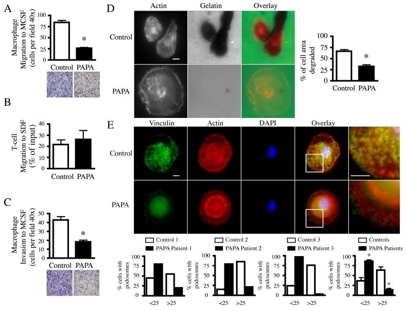
Highly motile leukocytes such as macrophages and dendritic cells form dynamic actin-containing adhesive and invasive structures known as podosomes, which facilitate migration and invasion (4). To determine if macrophages from PAPA patients display altered invasive motility and cytoskeletal organization, peripheral blood mononuclear cells were isolated from healthy volunteers and four patients with PAPA syndrome. Two of the PAPA patients were siblings and had PSTPIP1 A230T mutations. The other two PAPA patients were unrelated and had the PSTPIP1 A230T or the E250Q mutation, respectively. All mutations were confirmed by DNA sequence analysis (data not shown). Monocytes were differentiated into macrophages with 20 ng/ml human MCSF and used for experiments on day 7 using previously described methods (5). Macrophages were cultured as recommended by the American Type Culture Collection (ATCC). First we examined the directed migration of control and patient macrophages to the chemokine, MCSF. Macrophages isolated from two different patients with PAPA syndrome displayed significantly impaired chemotaxis to MCSF as compared to control macrophages (Figure 1A). In contrast to macrophage migration, T cell migration to SDF-1 was not altered in T cells isolated from PAPA patients (Figure 1B). These findings suggest that, although PSTPIP1 is expressed in both T cells and macrophages, patients with PAPA syndrome exhibit a specific defect in macrophage directed migration. To further characterize the function of macrophages from patients with PAPA syndrome, we examined the ability of PAPA patient macrophages to undergo invasive migration into gels and to degrade the extracellular matrix. We found that PAPA patient macrophages displayed significantly impaired invasion across matrigel invasion chambers (Figure 1C) as well as a decreased capacity of PAPA patient macrophages to degrade fluorescent conjugated gelatin-coated coverslips (Figure 1D). To determine if cytoskeletal regulation was altered in PAPA patient macrophages, we examined the organization of the actin cytoskeleton and podosome formation in macrophages isolated from PAPA patients. Control and PAPA patient macrophages were cultured on gelatin-coated coverslips and we examined podosome formation by staining for vinculin and actin (Figure 1E). Control macrophages showed robust polarized podosome formation with vinculin ring-like structures surrounding an actin core (Figure 1E). In contrast, PAPA patient macrophages showed a significant defect in podosome formation with many cells forming no podosomes and the cells that formed podosomes generally contained fewer than control (Figure 1E). This trend was observed in the three different PAPA patients tested (Figure 1E). In addition, PAPA patient macrophages that did form podosomes often showed abnormal architecture with increased numbers and size of vinculin-containing focal complexes (Figure 1E and data not shown). These findings indicate that PAPA patients display a specific defect in macrophage migration and invasion that may result from a switch from the formation of dynamic podosome adhesions, to more firm focal complexes.
Figure 1. PAPA patient macrophages exhibit decreased invasive migration and podosome formation.
(A) Primary control and PAPA macrophages (1 × 10⁵) or (B) T-cells (2 × 10⁵) were plated on coated (10 μg/ml fibronectin or 10 μg/ml fibrinogen, respectively) transwell membranes in culture media and assayed for their ability to migrate for (A) 24 hr or (B) 3 hr through the membrane to culture media containing 20 ng/ml MCSF or SDF, respectively. Macrophage migration was quantified by counting the number of adherent macrophages on the bottom of the transwell membrane from six separate fields at 40x magnification. A representative field at 10x magnification is shown below. T-cell migration was quantified by flow cytometry and is shown as the percentage of cell input that migrated. Data are mean ± SEM of two independent experiments. *, p < 0.001 compared with control cells by two-tailed, paired, Student’s t test. (C) Primary control and PAPA macrophages (1 × 10⁵) were plated in culture media on Matrigel-coated membranes and assayed for their ability to invade for 48 hr through the membrane to 20 ng/ml MCSF. Macrophage invasion was quantified by counting the number of adherent macrophages on the bottom of the membrane from six separate fields at 40x magnification. A representative field at 10x magnification is shown below. Data are mean ± SEM of three independent experiments. *, p < 0.001 compared with control cells by two-tailed, paired, Student’s t test. (D) Primary control and PAPA macrophages were cultured for 22 hr on Oregon-Green 488 gelatin coated coverslips as previously described (9). Cells were fixed and permeabilized in 4% paraformaldehyde, 0.25 mg/ml saponin for 10 min, quenched with 0.15 M glycine for 10 min and blocked with 10% heat-inactivated FBS containing 0.25 mg/ml saponin. Cells were stained with rhodamine phalloidin as previously described (9). Areas of degradation appear black on the gelatin. Quantification of degradation is shown as a percentage of total cell area from 71 control and 65 PAPA macrophages. Data are mean ± SEM of two independent experiments. *, p < 0.0001 compared with control cells by two-tailed, paired, Student’s t test. Bar 10 μm. (E) Primary control or PAPA macrophages were cultured on 10 μg/ml fibronectin coated coverslips, fixed and permeabilized as in (D) and stained with anti-vinculin (green), rhodamine phalloidin (red) and DAPI (blue) as previously described (9). The white-boxed region of each overlay was magnified 3x and is shown to the right. Bars 10 μm. Images were acquired at 100x and processed as previously described (9). The percentage of control or PAPA macrophages that contained less than or greater than 25 podosomes was quantified for three different PAPA patients (graphs show individual and averaged data). Podosomes were quantified from at least 50 control or PAPA cells in each experiment. Data are mean ± SEM of five independent experiments. *, p < 0.007 compared with control cells by two-tailed, paired, Student’s t test.
Defects in mononuclear cell podosome formation and migration have been reported in patients with WASp mutations and the primary immunodeficiency disorder Wiskott Aldrich Syndrome (WAS) (6-8). The similarities in macrophage morphology from patients with both PAPA syndrome and WAS, suggest that PSTPIP1 may regulate macrophage podosome formation through its interaction with WASp. Future studies will be needed to determine how PSTPIP1 regulates podosome formation and to examine if PAPA-associated mutations alter signaling through WASp. This is the first report to suggest that patients with a chronic inflammatory disease can also exhibit impaired macrophage migration and podosome formation. It is interesting, that unlike patients with WAS, PAPA patients show no evidence of immunodeficiency but instead exhibit chronic tissue inflammation and destruction. It is intriguing to speculate that abnormal macrophage trafficking and retention of macrophages within tissues because of impaired migration may contribute to the development of chronic inflammation through neutrophil recruitment, a hallmark of PAPA syndrome. Taken together, these findings suggest that defects in macrophage migration and podosome formation can contribute to the pathogenesis of both primary immunodeficiency disorders and chronic inflammatory disease.
References
- 1.Wise CA, Gillum JD, Seidman CE, Lindor NM, Veile R, Bashiardes S, et al. Mutations in CD2BP1 disrupt binding to PTP PEST and are responsible for PAPA syndrome, an autoinflammatory disorder. Hum Mol Genet. 2002;11(8):961–9. doi: 10.1093/hmg/11.8.961. [DOI] [PubMed] [Google Scholar]
- 2.Cote JF, Chung PL, Theberge JF, Halle M, Spencer S, Lasky LA, et al. PSTPIP is a substrate of PTP-PEST and serves as a scaffold guiding PTP-PEST toward a specific dephosphorylation of WASP. J Biol Chem. 2002;277(4):2973–86. doi: 10.1074/jbc.M106428200. [DOI] [PubMed] [Google Scholar]
- 3.Badour K, Zhang J, Shi F, Leng Y, Collins M, Siminovitch KA. Fyn and PTP-PEST-mediated regulation of Wiskott-Aldrich syndrome protein (WASp) tyrosine phosphorylation is required for coupling T cell antigen receptor engagement to WASp effector function and T cell activation. J Exp Med. 2004;199(1):99–112. doi: 10.1084/jem.20030976. [DOI] [PMC free article] [PubMed] [Google Scholar]
- 4.Calle Y, Burns S, Thrasher AJ, Jones GE. The leukocyte podosome. Eur J Cell Biol. 2006;85(3-4):151–7. doi: 10.1016/j.ejcb.2005.09.003. [DOI] [PubMed] [Google Scholar]
- 5.Tsuboi S. Requirement for a complex of Wiskott-Aldrich syndrome protein (WASP) with WASP interacting protein in podosome formation in macrophages. J Immunol. 2007;178(5):2987–95. doi: 10.4049/jimmunol.178.5.2987. [DOI] [PMC free article] [PubMed] [Google Scholar]
- 6.Linder S, Nelson D, Weiss M, Aepfelbacher M. Wiskott-Aldrich syndrome protein regulates podosomes in primary human macrophages. Proc Natl Acad Sci U S A. 1999;96(17):9648–53. doi: 10.1073/pnas.96.17.9648. [DOI] [PMC free article] [PubMed] [Google Scholar]
- 7.Calle Y, Chou HC, Thrasher AJ, Jones GE. Wiskott-Aldrich syndrome protein and the cytoskeletal dynamics of dendritic cells. J Pathol. 2004;204(4):460–9. doi: 10.1002/path.1651. [DOI] [PubMed] [Google Scholar]
- 8.Notarangelo LD, Ochs HD. Wiskott-Aldrich Syndrome: a model for defective actin reorganization, cell trafficking and synapse formation. Curr Opin Immunol. 2003;15(5):585–91. doi: 10.1016/s0952-7915(03)00112-2. [DOI] [PubMed] [Google Scholar]
- 9.Cortesio CL, Chan KT, Perrin BJ, Burton NO, Zhang S, Zhang ZY, et al. Calpain 2 and PTP1B function in a novel pathway with Src to regulate invadopodia dynamics and breast cancer cell invasion. J Cell Biol. 2008;180(5):957–71. doi: 10.1083/jcb.200708048. [DOI] [PMC free article] [PubMed] [Google Scholar]


