Abstract
Protective immunity requires a diverse, polyclonal B cell repertoire. We demonstrate that affinity maturation of the humoral response to a hapten is impaired when pre-existing clonally restricted cells recognizing the hapten are dominant in the B cell repertoire. B1- 8i+/− mice, which feature a high frequency of B cells with nitrophenyl (NP) binding specificity, respond to NP-haptenated proteins with the production of NP-specific antibodies, but affinity maturation is impaired due to insufficient generation of high affinity antibody producing cells. We manipulated the frequency of NP-specific B cells by adoptive transfer of B1-8 B cells into naïve, wild-type recipients. Remarkably, when 104 B1-8 B cells were transferred, these cells supported efficient affinity maturation and plasma cell differentiation. In contrast, when 106 B1-8 cells were transferred, affinity maturation did not occur. These data indicate that restricting the frequency of clonally related B cells is required to support affinity maturation.
Keywords: B cells, antibodies, rodent, transgenic/knockout mice
Affinity maturation of the antibody response is a hallmark of adaptive immunity. Affinity maturation is a process by which antibodies (Ab)4 produced in response to immunization show increased binding strength to their target antigen (Ag) over time. This process involves both clonal selection (1), and somatic hypermutation (SHM) (2). A broad range of studies using cellular and molecular approaches are providing an increasingly complete appreciation of the molecular mechanisms underlying these processes.
CD4+ T cells are activated by encounter with proteolytically processed antigen presented on APC (3–5). In contrast, naïve B cells can be primed by either soluble (6) or membrane bound unprocessed Ag (7). Priming of Ag specific B and T cells is thought to occur in the B cell and T cell zones of secondary lymphoid tissues (8). Within 1–3 days after immunization, IgM+ Antibody Secreting Cell (ASC) clusters appear in bridging channels between the red pulp and the Periarteriolar Lymphoid Sheathes (PALS) of the spleen or in the medullary cords of lymph nodes (9). The bridging channel ASC are thought to arise largely from marginal zone B (MZB) cells (10). In contrast to activated MZB cells, Ag primed follicular B cells and T cells relocate to and interact at the junction between the B cell and T cell zones. There, B cells present processed Ag to CD4+ TH cells (11). Class switch recombination (CSR) also occurs at the B cell/T cell interface (12). Activated Ag specific B and T cells subsequently migrate to the follicles and enter into germinal center (GC) reactions (8, 13). Within the GC structure, GC B cells interact intimately with the local cellular microenvironment and here it is thought that the GC B cells undergo SHM, affinity-based selection and differentiation into memory B cells and long-lived ASC (8).
When C57BL/6 mice are immunized with (4-hydroxy-3-nitrophenyl)acetyl (NP)- haptenated proteins, B cells expressing the VH186.2 gene paired with a λ1-light chain (LC) predominate in the repertoire of responding cells. The B1-8i mouse strain was created by targeted insertion of a VH186.2(DFL16.2)JH2 gene into the heavy chain (HC) locus of a 129/Ola ES cell (14). B cells that co-express this B1-8 HC transgene with an endogenous λ1-LC express the a allotypic variant of the HC and confer specificity to the hapten NP. Because not all λ-LC bearing B cells that pair with the B1-8 HC have the same NP-binding affinity, the λ-LC bearing B cells in the B1-8i strain do not act as a true monoclonal population. Nevertheless, because the predominant λ LC in mice is λ1, we considered this NP-binding population to be functionally monoclonal.
In wild-type (WT) mice, as the immune response progresses, increasing numbers of high affinity ASC and memory B cells are detected in secondary lymphoid tissues and bone marrow. Clonal proliferation of high affinity B cells is thought to be the result of competition for growth signals between high affinity B cells, low affinity B cells, and B cells with no affinity to the Ag. This competition is defined as inter-clonal and is widely accepted as a mechanism for selection of high affinity B cells within the GC. We demonstrate that, in addition to inter-clonal competition between B cells that express different BCR, elevated numbers of B cells expressing similar or identical Ig genes undergo intra-clonal competition. When elevated, this form of competition results in the reduction of the high affinity antibody response and alters the pathways that lead to generation of high affinity ASC.
METHODS
Mice
C57BL/6, C57BL/6(Ig κ−/−), and C57BL/6(CD45.1) mice were purchased from the Jackson Laboratories (Bar Harbor, ME) and 129/Ola mice were purchased from Harlan (Indianapolis, IN). The B1-8i+/− knock-in mouse strain was produced by Sonoda, et al. (14), and was generously provided by Frederick Alt (Harvard Medical School). It was backcrossed to the C57BL/6 strain for at least 10 generations. Previous studies have demonstrated that the B1-8 knock-in allele affectively excludes the endogenous allele (14). Therefore we used heterozygous mice in all subsequent studies. Mice were immunized i.p. with 50 μg of NP-KLH (Biosearch Technologies) precipitated in alum (Sigma-Aldrich) and boosted with 25 μg NP-KLH at day 25. All immunization protocols followed this timeline and dose unless stated in the text. Mice were housed in a barrier facility and maintained under protocols approved by the Institutional Animal Care and Usage Committee at the University of Alabama at Birmingham.
Antibodies and Reagents
Monoclonal mouse anti-IgMa-PE, anti-IgMb-FITC, anti-B220-PerCP, anti-B220-allophycocyanin-Cy7, anti-IgG1a-biotin, anti-IgG1b-biotin, anti-CD138-PE, and anti-MHC-II-PE, were purchased from BD-Biosciences (San Jose, CA). Anti-CD22-PE-Cy5 was purchased from Abcam (Cambridge, MA) and anti-CD38-FITC was purchased from eBioscience (San Diego, CA). Monoclonal rat anti-mouse IgM-horseradish peroxidase (HRP), anti-IgG1-HRP and streptavidin-HRP were purchased from Southern Biotech (Birmingham, AL). Polyclonal goat anti-mouse IgM, goat anti mouse IgG, and mouse IgG1 antibodies were purchased from Sigma-Aldrich (St. Louis, MO). NP-allophycocyanin (NP-APC) was made by coupling NP-Osu (Biosearch Technologies; Nuvato, CA.) with allophycocyanin (ProZyme; San Leandro, CA) in N,-N-dimethylformamide.
Immunofluorescence Microscopy
Freshly isolated tissues were embedded in Tissue Tek O.C.T compound (Fisher Scientific; Hampton, NJ) and frozen by floating the tissues on liquid nitrogen chilled 2-methylbutane. 8–10 μm sections were cut on a cryostat (Leica; Bannockburn, IL), air dried on Superfrost Plus slides (Fisher Scientific) and fixed for 10 minutes in acetone at 4°C prior to storage at −20°C until further use. Non-specific binding was blocked using a combination of 10% rabbit and 10% goat serum together with a biotin blocking kit (Vector Laboratories; Burlingame, CA), followed by staining with primary and secondary antibodies or 4-hydroxy-3-iodo-5-nitrophenylacetyl-biotin (NIP-biotin; Biosearch Technologies; Nuvato, CA). Signals due to bound NIP were enhanced using streptavidin-HRP Alexa Fluor 350 Tyramide Signal Amplification System (Invitrogen Corporation; Carlsbad, CA).
Flow Cytometry
Single cell suspensions from spleen tissues were stained with the indicated Abs or NP-APC. For adoptive transfer experiments, purified B cells were prepared by incubation with MACS anti-CD43 beads and fractionation with the AutoMACS (Miltenyi Biotec; Bergisch Gladbach, Germany) according to the manufacturer’s protocols. For intracellular staining, cells were incubated with mAb 24.G2 and anti-mouse IgG1 to block Fc receptors and surface IgG1. The cells were fixed and permeabilized using CytoFix-Cytoperm (BD Biosciences), and then intracellular IgG1 was stained according to the manufacturers guidelines. Cytometric data were acquired using an LSR II (BD Biosciences) and analyzed using FLOWJo software (Treestar; Ashland, OR).
ELISA
Immunlon plates (Thermo Labsystems; Milford, MA) were coated with 10 μg/ml of NP24BSA or NP2BSA (Biosearch Technologies) overnight at 4°C. The plates were washed with 0.05% Tween20 in phosphate buffered saline (PBS-Tween20) and blocked for 1 hour at 37°C with blocking buffer consisting of 1% BSA in PBS-Tween20. All subsequent incubations were at 37°C for 1 hour. Sera were diluted in blocking buffer, added to the NP-BSA coated plates, and incubated for 1 hour. Unbound antibodies were removed by washing and bound antibodies were detected using anti-mouse IgM-HRP or anti-mouse IgG1-HRP. For allele-specific ELISA, NP-reactive IgG1 antibodies that were produced by WT B cells were detected by anti IgG1b-biotin, while NP-reactive IgG1 antibodies produced by B1-8 B cells were detected with anti-IgG1a-biotin followed by streptavidin-HRP. Unbound antibodies were removed by washing and 2,2′-azino-bis-(3-ethylbenzthiazoline-6-sulfonic acid) (ABTS; Roche Applied Sciences; Indianapolis, IN) substrate was added. The reaction product was detected at OD405. Serial dilutions of mouse sera were performed to determine the working dilutions, and dilutions that produced optical densities that were within the linear range for the assay were used. In the indicated experiments, the concentrations of antibodies in mouse sera were normalized as follows. ELISA plates were coated with polyclonal goat anti-mouse IgM or IgG followed by incubation with mouse IgM or IgG subclass isotype control antibodies. Secondary HRP-conjugated detection antibodies were added as described above. The concentrations of the standard antibodies were converted into arbitrary units (A.U.). The antibody concentrations in experimental samples were similarly converted to A.U. based on the dilutions that yielded O.D. measurements in the linear range.
ELISpot
MAHA (Millipore; Billerica, MA) plates were coated overnight at 4°C with 10 μg/ml of NP24BSA or NP2BSA, then washed and incubated for 1 hour at 37°C with blocking buffer. BM cells were harvested and suspended in RPMI 1640 supplemented with 10% FBS. Cells were added to each well at the concentrations described in the text and cultured for 12 hours at 37°C in a humidified chamber under 5% CO2. All subsequent blocking, washing and incubation with primary and secondary antibodies were performed as described for ELISA. Bound anti-mouse HRP-conjugated antibodies were detected with 3-amino-9-ethylcarbazole (Moss Inc.; Pasadena, MA). Spots were systematically counted using the CTL ImmunoSpot Reader and analyzed with ImmunoSpot software (Cellular Technologies Ltd.; Cleveland, OH).
Lymphocyte Adoptive Transfer
For adoptive transfer experiments, the B1-8i+/− mice were crossed with Igκ−/− and CD45.1 mice, also on the C57BL/6 background. Recipient C57BL/6 mice were prepared for adoptive transfer by exposure to 500R from a cesium source. Two days after irradiation, the recipients were infused i.v. with a mixture of 1 × 10⁸ spleen cells from C57BL/6 mice, and either 104 or 106 purified B cells from either C57BL/6.CD45.1, or B1-8+/− x κ−/− x CD45.1 mice. The purified B cells were prepared by depletion of CD43+ cells with CD43 micro beads followed by magnetic associated cell sorting with the AutoMACS Separator (Miltenyi Biotec). Two days after adoptive transfer, the mice were immunized i.p. with NP-KLH as described above.
Nucleotide Sequencing
Spleen RNA was harvested using the RNeasy kit (Qiagen; Valencia, CA). All subsequent reagents for purification of RNA and sequencing were purchased from Invitrogen Corporation unless stated otherwise. cDNA was prepared using SuperScript III reverse transcriptase. VH186.2 sequences were amplified from γ1 heavy chain transcripts using Pfu50 polymerase and two rounds of PCR with the following nested primer pairs: Vh186.2-F 5′gatggagctgtatcatgctcttcttggcag3′, IgG1-R 5′gctgctcagagtgtagaggtcagactgc-3′, Vh186.2-Fnest 5′atcgatttggcagcaacagctacagg3′, IgG1-Rnest 5′ccggcctcaccatggagttagtttgg3′. The amplified products were purified using the ImmunPure PCR cleanup kit, cloned into the PCR-Blunt II TOPO vector using the Zero-Blunt TOPO cloning kit, and transformed into DH5α competent cells. Kanamycin resistant colonies were picked and plasmids were purified with the Wizard SV 96 plasmid purification system (Promega; Madison, WI). Sequence analysis was performed at the Genomics Core Facility of the Howell and Elizabeth Heflin Center for Human Genetics of the University of Alabama at Birmingham using the BigDye Terminator v3.1 Cycle Sequencing Ready Reaction kit (Cat# 4336919) and an Applied Biosystems 3730 Capillary Sequencer (Applied Biosystems; Foster City, CA). All sequences were analyzed using the Vector NTI Advance Software (Invitrogen).
Statistical Analysis
All ELISA and ELISpot data were analyzed by performing one tailed t-test assuming unequal variance. Significance was defined as P < 0.05.
RESULTS
NP-reactive B cells in B1-8i+/− mice participate in the GC reaction
To identify how frequently mature NP reactive B cells are found in the peripheral lymphoid tissues of the B1-8i+/− mouse strain, we analyzed spleen lymphocytes for NP binding by flow cytometry. In B1-8i+/− mice, B cells that bind NP represent over 1.5% of splenic lymphocytes (Fig. 1a). The specificity of binding with NP-APC was demonstrated by pre-incubating spleen cells with 15 ng/ml of NP24BSA. This completely blocked subsequent binding of NP-APC, whereas pre-incubation with BSA alone did not. Approximately 5% of murine B cells express λ-LC, with approximately half of these expressing λ1. This correlates with the observed 2.5% of NP-binding splenic B cells in naïve mice (Fig. 1b, right panel). In contrast, NP-binding is undetectable in naïve C57BL/6 splenic B cells (Fig. 1b, left panel). The fact that NP-reactive B cells were present in substantial numbers in B1-8i+/− mice permitted the detection of Ag-induced changes in B cell localization in the spleen. Following immunization with NP-KLH + alum, NP-reactive B cells which were initially distributed evenly in the B cell zones of the spleen re-localized to form foci at the T cell-B cell interface (Fig, 2a) and subsequently entered into GC reactions (Fig. 2c) in a fashion similar to previous reports (12). These changes in distribution of the NP-reactive cells were antigen-specific as no changes in the distribution of NP-reactive cells were detected when B1-8i+/− mice were immunized with KLH + alum alone (Figure 2b, right panels).
Figure 1. Enrichment of NP-specific B cells in B1-8i+/− knock-in mice.
Spleen cells were harvested from naïve C57BL/6 (WT) and B1-8i+/− mice. NP-reactive cells were detected with allophycocyanin conjugated NP (NP-APC). a) (top) 1.6 % of splenic lymphocytes from B1-8i+/− mice demonstrated binding to NP-APC. The numbers of NP-binding cells in naive C57BL/6 mice were below the levels of detection. Preincubation of cells from B1-8i+/− mice with 15 ng NP24BSA completely blocked the binding of NP-APC (bottom right). b) Naïve B220+ lymphocytes from C57BL/6 and B1- 8i+/− mice. NP-specific B cells were below the level of detection by FACS in WT mice and enriched to 2.5% of the B220+ gate in B1-8i+/− mice. Greater than 90% of B cells expressed the B1-8 HC transgene (IgMa), and fewer than 10% expressed endogenous heavy chain genes (IgMb). All NP-binding B cells express the B1-8 HC transgene.
Figure 2. B1-8 NP-binding B cells redistribute to the B cell-T cell interface and enter germinal centers in response to immunization with NP-KLH.
a) C57BL/6 and B1-8i+/− mice were immunized with NP-KLH + alum and sacrificed at the indicated times. The location of NP-specific B cells was demonstrated by binding of biotinylated NIP followed by counterstaining with the streptavidin-HRP Alexa Fluor 350 Tyramide Signal Amplification System. b) Specificity of the anti-NP response. Serial sections prepared from spleens of naïve B1-8i+/− mice were preincubated with BSA or NP24BSA prior to incubation with biotinylated NIP (left panels). NIP binding was blocked by preincubation with NP24BSA. B1-8i+/− mice were immunized with non- haptenated KLH + alum and spleens were harvested on days 3, 5, and 10, and NIP-binding cells were identified by incubation of sections with biotinylated NIP (right panels). NIP-binding cells showed minimal relocalization following immnization with KLH. NIP-binding cells were detected as in panel a. c) 10 days post immunization, GCs in both WT and B1-8i+/− mice contained predominately λ-LC+ cells.
Affinity maturation is impaired in B1-8i+/− mice
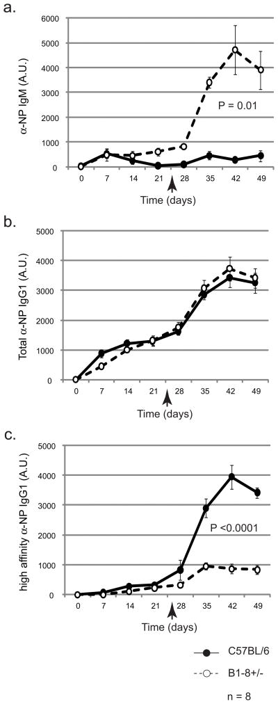
To address how the presence of increased numbers of NP-specific B cells affected the serum Ab response, we measured this response in immunized B1-8i+/− and C57BL/6 mice using an ELISA. During the primary response to NP-KLH, a 4-8-fold induction of NP-specific IgM was measured in B1-8i+/− serum. The response peaked 21 days post-immunization (Fig. 3a). C57BL/6 mice responded similarly; however, the response peaked at 7 days post-immunization. Thereafter, the serum IgM titers declined. In the secondary response, the serum anti-NP IgM response in WT-littermates was indistinguishable from the primary response. In contrast, in B1-8i+/− mice, the secondary response resulted in IgM levels that were increased 20-fold above baseline levels.
Figure 3. Affinity maturation is suppressed in B1-8i+/− mice.
Mice were immunized i.p. with NP-KLH + alum on day 0 and boosted at the time indicated by the arrow. a) Measurement of NP-specific IgM by ELISA indicates the anti-NP IgM response is elevated in B1-8i+/− mice. b) Induction of isotype class switching and the anti-NP IgG1 response occur to similar levels in B1-8i+/− mice compared to WT mice. c) The high affinity anti-NP IgG1 response was severely reduced in B1-8i+/− mice. n represents the number of mice in each experimental group. The experiment was repeated 3 times with similar results. Error bars represent standard errors of the mean (SEM).
Previous reports have established techniques to use steady state ELISAs with differentially haptenated target antigens to estimate the relative affinities of NP-reactive IgGs (15-18). Using this approach, we detected total NP-reactive IgG1 Abs using ELISA plates that were coated with NP24BSA. In contrast, by using plates that were coated with the same concentration of BSA carrying only 2 NP residues for every BSA (NP2BSA) we detected IgG1s with only relatively high affinities for the hapten. It should be indicated clearly that this technique actually detects the average avidity of the antibody for the hapten antigen; however, in the case of bivalent IgG antibodies, this measurement closely estimates affinity (16). Using this strategy, NP2BSA efficiently captures antibodies that bind with either high avidity or high affinity. Antibodies with low avidity or low affinity manifest higher off rates from NP2BSA than from NP24BSA and are not detected using the sparsely-haptenated target antigen (15, 17).
The serum anti-NP IgG1 titer of B1-8i+/− mice was similar to that of WT-littermates (Fig. 3b). Serum anti-NP IgG2b, IgG2c and IgG3 were also similar between the two strains (data not shown). These data were consistent with previous reports that demonstrated that the increase in NP-specific B cells in B1-8i mice does not correlate directly with the levels of serum anti-NP IgGs (19). Interestingly, after a booster immunization, B1-8i+/− mice produced 4-fold less high affinity anti-NP IgG1 Ab than C57BL/6 mice (Fig. 3c). This defect in affinity maturation persisted even after secondary and tertiary booster immunizations (data not shown).
High B cell clonal abundance interferes with affinity maturation
Immunization of B1-8i+/− mice with NP-KLH resulted in the re-distribution of NP reactive cells in a fashion that was similar to that described previously for adoptively transferred antigen specific B cells in the lymph nodes (6) and antigen specific B cells in the spleen (12). Furthermore, the fact that specific immunization induced the production of NP-reactive IgM and IgG antibodies demonstrated that B1-8i+/− mice were capable of responding to a primary and a secondary immunization and that their ability to undergo isotype switching was preserved; however, the dramatic impairment of affinity maturation indicated that there is disturbed regulation of the humoral response in this transgenic model. There is precedent for impaired lymphocyte regulation in mice carrying abundantly expressed antigen receptors. For example, primary immunization of T cell receptor transgenic mice such as those of the DO11.10 strain leads to abnormally high levels of cell death and thymic involution (20). In our experiments, we have considered that the aberrant affinity maturation may have been due to a B cell intrinsic defect. Alternatively, the abnormal response may not represent an intrinsic B cells impairment, but rather may represent the natural expression of normal immunoregulatory pathways.
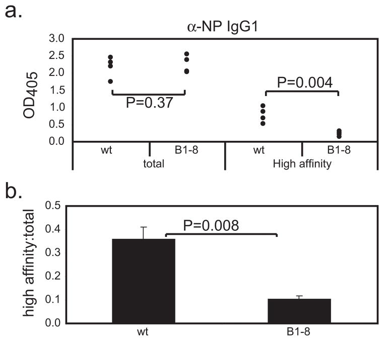
A major distinction between B1-8i+/− mice and WT mice is the proportion of recirculating B cells that has specificity for the hapten NP. We hypothesized that the large numbers of NP-specific precursor B cells in the B1-8i+/− mice (Fig. 1a and 1b) may result in increased competition for limiting signals that were required for affinity maturation of the antibody response to the hapten. It is well established that affinity maturation and isotype switching both require T cell help (13). We considered that the impaired affinity maturation we observed in the B1-8i+/− mice might have been due to a disproportionately smaller T helper cell population compared to the large number of NP-specific B cells in this strain. However, when we expanded the available pool of KLH specific TH cells by priming B1-8i+/− mice with KLH prior to immunization with NP-KLH, we did not restore affinity maturation (Fig. 4). This indicated that T cell help was not the critical limiting factor for affinity maturation in B1-8i+/− mice when immunized with NP-haptenated carrier proteins.
Figure 4. The induction of a high affinity Ab response is not restored in B1-8i+/− mice by priming with the T cell Ag.
C57BL/6 or B1-8i+/− mice were immunized with 50 μg of KLH precipitated in alum. Three weeks after priming, the mice were re-immunized i.p. with 50 μg of NP-KLH + alum. Sera were collected 14 days after immunization and serum anti-NP Ab was measure by ELISA. All sera were diluted 1:200. This working concentration was determined to yield measurements within the linear range of the assay for all samples tested. a) Total anti-NP IgG1 titers were similar in WT and B1-8i+/− mice. Despite priming, B1-8i+/− mice showed impaired ability to undergo affinity maturation. b) The ratio of serum high affinity anti-NP IgG1 to total serum anti-NP IgG1 was reduced in B1- 8i+/− mice. Each data point represents one mouse. This experiment was repeated 3 times with similar results.
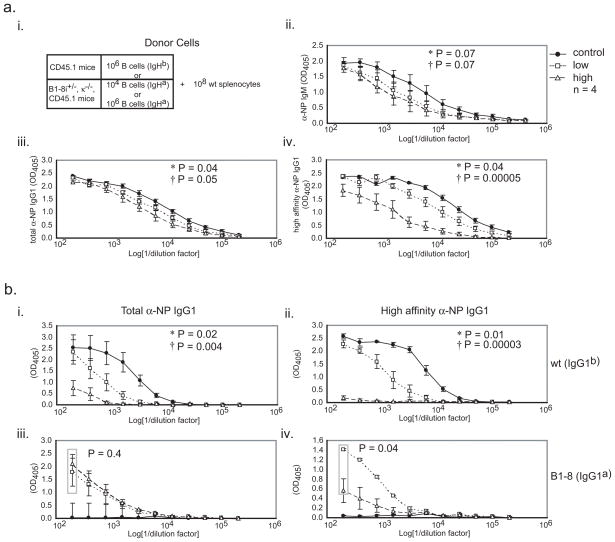
To test whether the frequency of Ag-specific B cells affects affinity maturation, we used adoptive transfer of different numbers of B1-8i+/− B cells into naïve C57BL/6 mice to regulate the starting numbers of NP-reactive cells (Fig. 5a i). 1×10⁸ spleen cells from C57BL/6 mice and varying numbers of either CD43-depleted CD45.1+ B cells from WT congenic donors or CD43-depleted B1-8i+/−κ−/− CD45.1+ B cells were adoptively transferred i.v. into sub lethally irradiated recipients. In naïve B1-8i+/− mice, approximately 2.5% of spleen B cells were NP-reactive (Fig. 1b). This represents approximately 1×10⁶ NP-specific B cells out of an average of approximately 5×10⁷ total B cells in the adult mouse spleen. Therefore, adoptive transfer of 106 B1-8i+/− k−/− CD45.1+ B cells into naïve irradiated recipient mice produced recipients with NP-specific B cells at a level similar to that detected in the B1-8i+− mice. In contrast, adoptive transfer of 104 donor B1-8i+/− B cells yielded recipients in which NP-reactive transgenic B cells represented approximately 0.025% of the total B cell population, a frequency closer to the estimated frequency in WT adult mice.
Figure 5. Attenuation of affinity maturation is a consequence of high clonal abundance in B1-8i+/− mice.
a) (i) Sera collected 14 days after a booster immunization of mice that had received adoptive transfer of 108 WT spleen lymphocytes with either 106 WT polyclonal B cells (control), 104 (low) or 106 (high) B1-8,κ−/−,CD45.1+ B cells. (ii) NP-specific IgM, (iii) and total anti-NP IgG1 were similar in all experimental groups. (iv) Mice that received adoptive transfer of 106 B1-8,κ−/−,CD45.1 B cells produced 10 fold lower titer of high affinity anti-NP IgG1 than control mice whereas mice that received lower numbers (104 cells/mouse) produced a high affinity anti-NP IgG1 response. b) (i) B1-8 B cells suppressed the WT total anti-NP IgG1 response. (ii) Affinity maturation of the WT response was also disrupted in a cell density dependent manner. (iii) B1-8 B cells produced total anti-NP IgG1 in a fashion independent of clonal abundance. (iv) Induction of a high affinity NP-IgG1 response by the B1-8 B cells was observed when B1-8 B cells were transferred at low cell numbers (104 per mouse). * P values comparing control mice and mice that received 104 B1-8 B cells; † P values comparing WT mice and mice that received 106 B1-8 B cells. 4 mice were used in each experimental group. The adoptive transfer experiment was performed 3 times with similar results.
B1-8i+/− mice responded to immunization with NP-KLH with an approximately 8-fold higher titer of hapten-specific IgM compared to the titer in WT mice (Fig. 3a). Unlike B1-8i+/− mice, the levels of NP-specific IgM in the sera of mice that received either adoptively transferred WT B cells, 104 B1-8i+/− B cells, or 106 B1-8+/− B cells all were similar following immunization with 50 μg of NP-KLH (Fig. 5a ii). The higher levels of serum IgM in immunized WT mice compared to the levels in the mice that received adoptively transferred WT or B1-8i+/− B cells may have been due to the steady input of naive B1-8i+/− B cells that occurs in an ongoing fashion in the B1-8i+/− mouse strain. The induced levels of total anti-NP serum IgG1 were similar in all groups of mice despite a 100-fold difference in the starting frequencies of B1-8i+/− B cells between the mice that received the low and high numbers of adoptively transferred NP-reactive cells (Fig. 5a iii). This was consistent with the observation that the numbers of NP-specific B cells do not correlate with the titers of NP-specific IgG1 (19) (Fig. 3b). Strikingly, mice that received the high frequency of adoptively transferred NP-specific B cells produced 11-fold less high affinity anti-NP IgG1 than control mice. In contrast, mice that received the low frequency of adoptively transferred B1-8i+/− B cells produced levels of high affinity anti-NP IgG1 only 2 fold lower than mice reconstituted with WT B cells (Fig. 5a iv). Together, these data demonstrated that total production of Ag-specific serum Ab was independent of the starting numbers of Ag-specific B cells, but that production of high affinity Ab was dependent on maintenance of an appropriately low frequency of Ag-specific B cells.
To determine the contributions of the transgenic and the WT B cells to the NP-specific Ab response, we performed allele specific ELISA. When mice that received adoptive transfer of NP-specific B1-8i+/− B cells were immunized with NP-KLH, the high affinity anti-NP Ab response from the endogenous WT cells was suppressed in a B1-8i+/−cell number-dependent manner (Fig. 5b i and 5b ii). The titer of total anti-NP IgG1a was similar regardless of the numbers of B1-8i+/− donor B cells (Fig. 5b iii). When B1-8i+/− B cells were present in high numbers, affinity maturation was inhibited (Fig. 5b iv). In contrast, affinity maturation was observed when low numbers of B1-8i+/− B cells were transferred. These data established that B1-8i+/− B cells were proficient to undergo affinity maturation when their population density was low. The inverse correlation between the numbers of B1-8i+/− B cells transferred and the amount of anti-NP Ab produced by the WT B cells represents inter-clonal competition between the functionally monoclonal B1-8i+/− B cells and the diverse WT B cell repertoire. Although the frequency of B1-8i+/− cells did not have a large affect on either the amount of IgM or the total (high affinity + low affinity) IgG1 produced, affinity maturation was attenuated when the B1-8i+/− B cell frequency was high. These data indicate that the density of the responding cell population does not affect the overall quantity of the Ab response but does significantly alter the quality of the serum Ab.
Clonal selection is modulated by the density of the responding B cell population
To investigate whether the defect in affinity maturation observed in the B1-8i+/− mice was due to aberrant SHM, we sequenced theVH186.2-γ1 transcripts from immunized C57BL/6 and B1-8i+/− mice. Affinity maturation was tracked by the accumulation of the affinity-determining tryptophan to leucine mutation at codon 33 (W33L) in the sequence of individual clones. This single point mutation of the germ line W to L results in a 10-fold increase in affinity of the Ab for NP (21). 70-80% of unique VH186.2-γ1 transcripts amplified from spleens of immunized C57BL/6 mice contained the high affinity W33L mutation in contrast to 50% of transcripts from B1-8i+/− spleens. Furthermore, the ratio of replacement to silent (R:S) mutations in the Complementary Determining Regions 1 (CDR1) and CDR2 from C57BL/6 mice was 3.2 versus 1.5 from B1-8i+/− mice. These data indicate that selection of high affinity Ab expressing B cells is impaired in immunized B1-8i+/− mice.
VH186.2-γ1 sequences were also analyzed in the adoptive transfer model. During the generation of the B1-8i+/− strain, a silent T to C mutation was engineered into the heavy chain cryptic Recombination Signal Sequence (cRSS) at nucleotide 292 to prevent VH gene replacement (14). This mutation and the DFL16.1-JH2 rearrangement distinguished the B1-8 sequences from the non-transgenic sequences. For mice carrying only WT HC genes, the mean percentage of unique sequences carrying the W33L mutation was 58%, (35–77%) (Table I). 62% of sequences from B1-8 B cells in the mice that received adoptive transfer of 104 B1-8 B cells carried the W33L mutation, ranging from 43–76%. In contrast, mice that received adoptive transfer of 106 B1-8 B cells manifested a lower distribution of clones carrying the high affinity mutation (22–58%, with a mean of 41%). Although the accumulation of point mutations was only modestly reduced (data not shown), these data suggest that when there are abnormally high numbers of B cells with the same antigenic specificity, the frequency of transcripts that accumulate mutations resulting in a high affinity Ab product is reduced. Interestingly, similar to C57BL/6 mice, the mean R:S ratios of mutations in the heavy chains were 2.2 and 2.5 respectively for the control mice and the mice that received adoptive transfer of the low frequency of B1-8 B cells, indicating a strong selection for replacement mutation within the CDRs. The mean R:S ratio in mice that received the high frequency of B1-8 B cells was 1.4, confirming in the adoptive transfer system that reduced selection is observed when the frequency of clonally related B cells is high.
Table I.
Summary of VH186.2-γ1 Sequences4
| Group | Mouse # | Total sequences determined | Unique sequences | % unique | R:S | Mean R:S | %W33L | Mean %W33L |
|---|---|---|---|---|---|---|---|---|
| Control | 1 | 20 | 15 | 86.06 | 2.27 | 2.38 | 66.67 | 58.14 |
| 2 | 23 | 22 | 2.63 | 77.27 | ||||
| 3 | 21 | 17 | 2.35 | 35.29 | ||||
| 4 | 15 | 14 | 1.66 | 42.86 | ||||
| Low 104 B1-8 | 1 | 28 | 22 | 80.49 | 2.4 | 2.77 | 68.18 | 62.43 |
| 2 | 24 | 22 | 3.61 | P=0.1 | 72.73 | |||
| 3 | 30 | 22 | 2.28 | 63.64 | ||||
| High 106 B1-8 | 1 | 21 | 17 | 63.86 | 1.67 | 1.34 | 50 | 40.92 |
| 2 | 20 | 12 | 1.5 | P<0.0005 | 58.33 | |||
| 3 | 21 | 9 | 1.33 | 22.22 | ||||
| 4 | 21 | 15 | 0.88 | 31.25 | ||||
VH186.2-γ1 cDNAs were amplified from total spleen RNA from WT mice that had received either 104 (low) or 106 (high) B1-8 B cells prior to two immunizations with NP-KLH adsorbed to alum as in Figure 5. The cDNA sequences were analyzed for the presence of the B1-8-specific sequence tags, somatic mutations, the ratio of replacement mutations to silent mutations (R:S), and the presence of the affinity defining codon 33 tryptophan to leucine (W33L) mutation. 3–4 mice were analyzed from in each group (column 2), and the total numbers of sequences determined and the numbers of sequences that were defined as unique because they carried a unique pattern of mutations are shown (column 3 and 4 respectively). For the groups of mice that received adoptively transferred B1-8 cells, only the B1-8-specific sequences are reported. The percents of the total determined sequences that were unique are shown in column 5. The mean ratio of replacement to silent mutations (R:S) in the unique sequences in each individual mouse (column 6) and the mean R:S of all the sequence from all mice in each group (column 7) are shown. The percentage of the sequences carrying the W33L mutation in each individual mouse (column 8) and the mean % of all the W33L sequences for all mice in each experimental group (column 9) is also displayed.
Intra-clonal competition suppresses formation of high affinity ASC
Analysis of the serum Abs produced after immunization indicated that production of high affinity anti-NP IgG1 was dramatically reduced in B1-8i+/− mice and mice that received 106 B1-8 B cells. In contrast, DNA sequencing indicated that approximately half of the VH186.2-γ1 transcripts amplified from spleens of these mice were from cells that potentially could produce high affinity Ab because the included the high affinity encoding W33L mutation. Because each of the sequences analyzed displayed a unique nucleotide sequence, the possibility that the discrepancy between the measurements of hapten-binding avidity and the presence of the high affinity determining codon 33 mutation might be due to PCR artifact representing contamination of the samples with plasmid DNA is very unlikely. A more likely alternative explanation for the discrepancy between the sequence data and the ELISA data is that the sequenced transcripts were from a pool of total spleen cells, representing the entire expressed VH186.2-γ1 repertoire including transcripts for secreted Ab from ASC and membrane bound Ab from memory or activated B cells. In contrast, the serum Abs measured by ELISA derives from cells driven to produce secreted Ab.
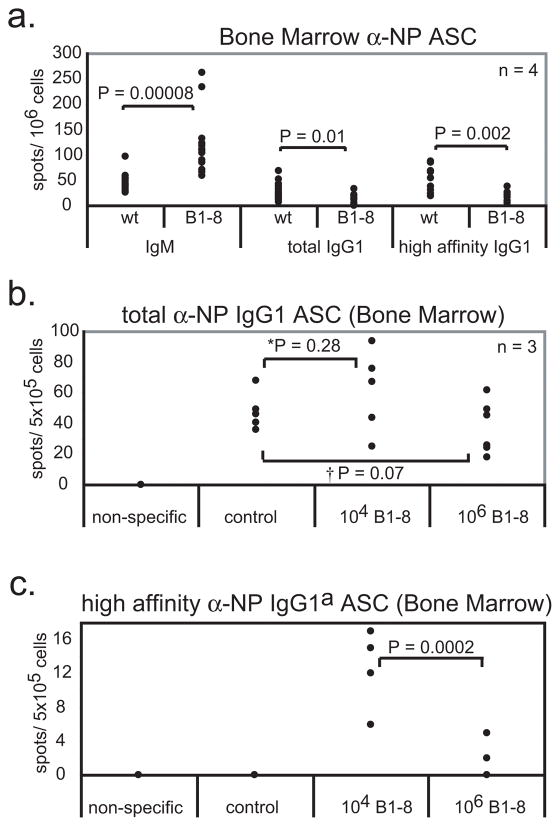
To determine whether cells in the B1-8i+/− mice were competent to produce NP-specific antibodies, particularly high affinity antibodies, we used ELISpot assays to identify high affinity and total anti-NP IgG1-producing cells in BM of immunized mice. There were 3-fold greater numbers of high affinity IgG1 ASC in BM of C57BL/6 mice compared to BM of B1-8i+/− mice (Fig. 6a). Interestingly, when WT mice were sub-lethally irradiated and reconstituted with WT splenocytes plus 106 B1-8 B cells, then immunized, the numbers of high affinity anti-NP IgG1 ASC in BM were also reduced (Figs. 6b and 6c). In contrast, reconstitution of mice with WT splenocytes plus 104 B1-8 B cells prior to immunization showed a near normal high affinity ASC response. This suggested that the transcripts carrying the high affinity determining W33L mutation that we observed in the immunized B1-8i+/− mice and in WT mice that had received 106 adoptively transferred B1-8 B cells were unlikely to contribute to the long-lived ASC pool and circulating Ab.
Figure 6. The ASC repertoire is largely limited to low affinity Ab when B1-8 B cells are abundant.
a) ELISpot assays were performed using C57BL/6 or B1-8i+/− BM to test for the presence of long-lived ASC. a) B1-8i+/− mice produced ~4 fold more NP-specific IgM secreting cells than WT mice, 1.8 fold less total NP-specific IgG1 ASC and ~3 fold less high affinity NP-specific IgG1 ASC. b-c) BM from mice adoptively transferred with WT B cells or B1-8+/− B cells. b) The frequency of long-lived total NP IgG1 ASC is not affected by B1-8 population density. c) B1-8 B cells from mice adoptively transferred with the high frequency of B cells do not produce high affinity IgG1 ASC. Each data point represents one mouse. The experiment was repeated 3 times with similar results.
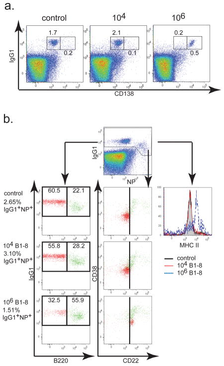
Under normal conditions, the pool of long-lived ASC elicited in response to a systemic infection resides primarily in the BM and is composed largely of high affinity ASC while the pool of memory B cells in the BM is thought to be composed of a mixture of high and low or primarily low affinity cells (22). Our observation that the numbers of high affinity ASC are reduced in the BM of B1-8i+/− mice indicated that the mechanisms governing ASC and/or memory B cell development are disrupted in these animals. To address whether the effector ASC and memory B cell populations are altered when B cells are subjected to intra-clonal competition, we examined BM from control mice, and from mice that had received adoptive transfer of either 104 or 106 B1-8 B cells prior to primary and booster immunization. Long-lived intracellular IgG+CD138+ ASC make up 1.7% of the CD43+ fraction in WT mice compared to 2.1% in mice that received the low numbers of adoptively transferred cells. Although there was an increase in the numbers of CD138hi ASCs in the mice that received 106 adoptively transferred cells, the % of long lived ASC in these animals was significantly reduced compared to that in WT mice and mice that had received 104 adoptively transferred cells (Fig. 7a).
Figure 7. The formation of long-lived ASC is suppressed when B1-8 B cells are over-represented.
BM cells were recovered from adoptive transfer mice 14 days after the booster immunization. a) CD43+ MACS sorted cells from control mice or from mice that received 104 cells or 106 cells were stained for CD138 and intracellular IgG1 to detect IgG1 ASC. b) CD43− BM cells were gated for NP-binding and intracellular IgG1 staining (top) and analyzed for B220 (left), CD22 and CD38 (center) and MHC II expression (right). Numbers to the left indicates the percent of IgG1+ NP-binding in CD43− cell fractions. Numbers inside the gates indicate the percent of gated cells that are B220lo/−(red) and B220hi (green). B220lo/− plasma and B cell blasts (red) down-regulate expression of CD22. B220hi (green) up-regulate CD38 and CD22 expression (middle). Control mice (grey filled) and mice transferred with 104 B1-8 B cells (red) expressed equal levels of MHC II while IgG1+ NP-binding cells from mice transferred with 106 B1-8 B cells (blue-dashes) up-regulate expression of MHC II.
We have demonstrated that the development of long-lived ASC can be influenced and the numbers of ASC can be suppressed when unusually high numbers of antigen-specific B cells are present. Previous studies from Marzo and colleagues have established that following immunization, large numbers of clonally restricted, antigen specific CD8 T cells revert from an effector memory CD8+ T cell phenotype into a central memory CD8+ T cell phenotype (23). We hypothesize that the large numbers of NP-specific B cells present in the B1-8i+/− strain in similar fashion may be impaired for differentiation into the high affinity long-lived ASC compartment. This impairment may not be restricted to the long-lived ASC compartment alone, but may also be observed in the memory compartment as well. By definition, memory B cells have previously encountered Ag and most have class switched. Functionally, memory B cells are thought to be more potent APC compared to naïve B cells. Following Ag re-stimulation, they up-regulate expression of MHC II molecules and other activation molecules more rapidly than naïve cells. In the CD43− cell population, IgG1+NP-bindingcells from both WT mice and mice that had received 104 B1-8 B cells were found to express surface MHC II at similar levels. In contrast, IgG1+NP-binding cells from mice that had received 106 adoptively transferred B1-8 B cells showed substantially up-regulated expression of class II MHC (Fig. 7b). IgG1+ low NP-binding cells were predominately B220lo/−. This population consisted of a mixture of low affinity ASC and B cell blasts. The IgG1+high NP-binding population expressed high levels of B220 and was composed of high affinity, activated and memory B cells that were CD38hiCD22int/hi. While mice that received adoptive transfer of 106 B1-8 B cells had reduced numbers of B cell blasts and ASC, these mice did show enrichment of the high affinity B220hiCD38hi CD22int/hi activated and memory phenotype B cells (Fig. 7b).
High Ag dose restores affinity maturation in B1-8i+/− mice
The process of affinity maturation of the Ab response requires delivery of a combination of BCR-dependent signals that activate Ag-specific B cells together with T cell-dependent signals that provide B cell help. Since our initial studies showed that pre- activation of carrier-specific TH cells by priming with the carrier protein failed to restore affinity maturation, we suspected that T cell help, per se, was not limiting for the affinity maturation response. We therefore tested whether Ag dependent signals were limiting by immunizing mice with an increased dose of the Ag (150 μg NP-KLH, rather than the 50μg dose used originally). While the titer of total NP-specific IgG1 remained unchanged in WT and B1-8i+/− mice, immunization with the higher Ag dose induced high titers of high affinity NP-specific IgG1 in the B1-8i+/− mice (Fig. 8).
Figure 8. Additional immunizing Ag restores affinity maturation in B1-8i+/− mice.
C57BL/6 mice and B1-8i+/− mice were immunized with the indicated dose of Ag precipitated with a constant amount of alum and boosted 25 days after the initial immunization. Sera were collected and analyzed for high affinity anti-NP IgG1 at the designated time points. a) Induction of total anti-NP IgG1 was similar in all mice irrespective of Ag dose. b) Induction of high affinity Ab was restored in B1-8i+/− mice immunized with 150 μg NP-KLH + alum. The experiment was repeated 3 times with similar results.
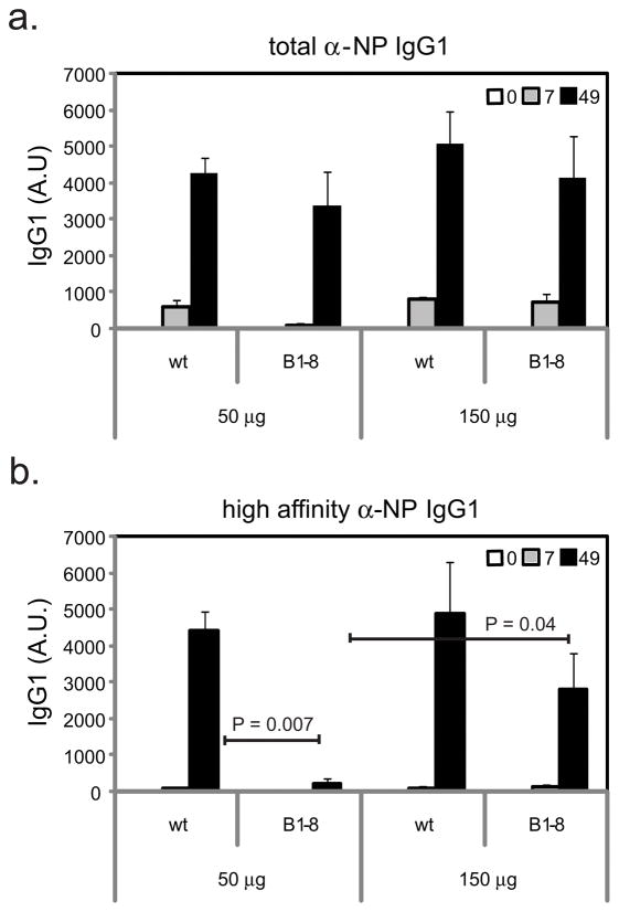
In order to test whether immunization with high dose antigen restored the production of high affinity antibodies in the adoptive transfer model, we analyzed mice that received 106 WT B cells or 106 B1-8 x κ−/− B cells by adoptive transfer prior to immunization with either 50 μg or 150 μg of NP-KLH + alum. Mice treated in this fashion accumulated similar levels of total serum anti-NP IgG1 when immunized and boosted with either the low or the high dose of antigen (Fig. 9a, black bars). In contrast, raising the dose of immunizing antigen from 50 μg to 150 μg increased the high affinity IgG1 response nearly three-fold (Fig. 9a, white bars). To determine whether the B1-8 B cells contributed to the high affinity antibody production, we performed allele-specific ELISA. As previously shown, when B1-8 B cells are present at high cell density (following adoptive transfer of 106 cells), immunization with 50 μg of NP-KLH + alum resulted in suppression of the production of high affinity antibody (Fig. 5b iv) and also suppression of the generation of high affinity ASC (Fig. 6c). While the B1-8 B cells produced detectable high affinity NP-reactive IgG1a (Fig. 9b, white bars) when mice were immunized with 50 μg of NP-KLH, this represented only 30% of the total NP reactive Ab present in the blood. In contrast, when mice that had received adoptive transfer of 106 B1-8 B cells were immunized with 150 μg of NP-KLH, a robust affinity matured Ab response was observed (Fig. 9b, white bars). Thus, in the adoptive transfer model, as well as in the B1-8 mouse strain itself, immunization with a high dose of Ag restored the high affinity antibody response.
Figure 9. Immunization with an increased dose of Ag induces the production of high affinity Ab in mice that received adoptive transfer of high numbers of B1-8 B cells.
C57BL/6 mice were conditioned by adoptive transfer of either 106 WT or 106 B1-8 B cells, then were immunized with either 50 μg or 150 μg of NP-KLH + alum and boosted 25 days after the primary immunization. a) Sera were collected on day 35 and analyzed for either total (black bar) or high affinity (white bar) NP-reactive IgG1 by ELISA. b) Total (black bar) and high affinity (white bar) anti-NP IgG1a antibodies derived from the adoptively transferred B1-8 cells were measured by ELISA. Data represent the means of 2 independent experiments with 3 mice per group in each experiment. Error bars represent standard errors of the means.
These data have identified Ag-dependent signaling as a key resource that B cells compete for in the induction of a high affinity Ab response. When the numbers of naïve, clonally related, Ag-specific cells are high and when the relative amount of the Ag is limited, then affinity maturation is attenuated. This is in sharp contrast to the situation in mice with a diverse polyclonal B cell repertoire in which cells compete for limiting Ag and clones with high affinity BCRs expand.
DISCUSSION
The normal T cell-dependent humoral immune response is characterized by activation of Ag-specific B cells and T cells, expansion of the pool of responding B cells to permit production of substantial amounts of circulating antibodies, switching to isotypes appropriate for elimination of the immunizing Ag, and affinity maturation to permit the immune response to eliminate the Ag even as its concentration becomes very limited. In our current study, we have observed in the B1-8i+/− Ig HC transgenic mouse strain that affinity maturation is impaired when the number of Ag-specific naïve B cells is inappropriately large. We saw no evidence of failure to activate the SHM process. Rather, we observed that there was failure to select high affinity B cell variants as a likely cause of the impaired generation of high affinity ASC. This impairment of affinity maturation in the B1-8i+/− mice was not a B cell intrinsic defect. Affinity maturation could be re-established either by reducing the number of Ag-specific naïve B cells by adoptive transfer into a non-transgenic host, or by immunizing with an increased amount of Ag.
In addition to having large numbers of B cells that are poised to respond to immunization with NP-haptenated Ag, the B1-8i+/− strain differs from WT mice in that it has elevated levels of circulating NP-binding serum Abs that are present prior to deliberate immunization with NP-haptenated Ag (data not shown). We have considered that the elevated levels of anti-NP IgM in naïve B1-8i+/− mice might affect the quality of the Ab response following immunization. Previous studies have demonstrated that infusion of Ag-specific Ab into naïve mice can alter the quality of the humoral response following systemic immunization. Harte and colleagues found that injection of Ag-specific IgM prior to immunization enhanced the endogenous Ab response to subsequent immunization (24–26). This enhancement of the humoral response was dependent on the formation of immune complexes by the passively transferred IgM (27). Arguing against a role for elevated levels of anti-NP IgM in the attenuated affinity maturation in the B1-8i+/−mice is our observation that affinity maturation was attenuated both in B1-8i+/− mice and in WT mice that received adoptive transfer of 106 B1-8 B cells (Fig. 5). Prior to immunization the mice that received adoptive transfer of B1-8 B cells had levels of anti-NP IgM and IgG equivalent to the levels observed in naïve WT mice that are competent to exhibit normal levels of affinity maturation.
Studies by Freitas et al. showed that when WT mice were reconstituted with a mixture of WT and BCR-transgenic BM, the initial growth curves of WT and transgenic B cells were similar. Once steady-state was reached, the WT B cells became the predominant population (28). The B cell population could be shifted towards greater representation of the transgenic B cells if the mice were immunized with the Ag specific to the transgenic BCR (29). In other experiments, mice with conditional ablation of surface Ig had diminishing numbers of peripheral B cells, indicating that BCR signaling is necessary for B cell survival in the periphery (30).
In our adoptive B cell transfer experiments, we anticipated that irradiation of the recipient mice created niches that support the engraftment of all donor cells equivalently, irrespective of their clonal specificity (28) (and unpublished data). Furthermore, we anticipated that immunization with NP-haptenated carrier proteins should favor selection of the NP-specific transgenic cells over the WT donor cell population. Our data indicated that when the numbers of cells from an individual B cell clone are high, this leads to failure to select cells carrying somatically mutated high affinity Ig HC. Our data support the concept that inter-clonal competition selected for the NP-specific B1-8 B cells over the predominately Ag non-specific WT polyclonal B cells. Competition for limited resources was high because of the large representation of NP-specific B1-8 B cells in the total B cell pool.
Cells compete for limiting resources that are needed for growth or survival, including cytokines, mitogens, or local cellular niches such as the GC. In our studies, increasing the dose of immunizing Ag was able to restore affinity maturation in B1-8i+/− mice in a fashion similar to reducing the B1-8 B cell clonal abundance by adoptive transfer into WT mice. Batista and colleagues have demonstrated that B cells are capable of acquiring membrane bound Ag, internalizing the Ag and subsequently presenting the processed Ag to TH cells (7). This ability to acquire membrane bound Ag displayed on target cells provides a possible mechanism for affinity based selection of B cells. When B1-8 B cells or NP-specific B cells are rare and infrequent, the cells are able to test their affinity and undergo affinity-based selection efficiently. B cells with low affinity have the potential to interact with the membrane bound Ag, but are at a disadvantage and unable to acquire and internalize the Ag as efficiently as high affinity cells. These cells likely receive very low-level stimulation, are negatively selected in the GC, and undergo apoptosis if the Ab is non-specific or auto-reactive. However, when B1-8B cells are abundant, a fraction of the cells may capture and internalize the Ag, preventing the remaining B1-8 B cells from contacting the now more limited amounts of Ag. Thus, in the setting of high numbers of Ag-specific cells, the acquisition of Ag by a minority population of cells may create an environment in which the majority of cells (which have not encountered the Ag) are at a selective disadvantage, resulting in an overall reduced high affinity response.
B cell activation requires BCR cross-linking. This process is enhanced when the antigenic particle is displayed at high density. Immunization with virus like particles (VLP) that displayed low, medium or high densities of an antigenic peptide induced similar IgM responses to the peptide and was not dependent of epitope density. Alternatively, the IgG response to the peptide was severely reduced when VLP with low-density peptide were used to immunize mice. Anti-peptide IgG was induced only when high density peptide VLP were used to immunize mice (31). This indicated that the induction of an IgG response requires multiple antigen-capturing events and cross-linking of the BCR. Increasing the numbers of antigen specific B cells skews the ratio of B cell to antigen, which can result in insufficient activation or selection via the reduction of cross-linking events. The lack of Ag based selection may result in a loss of apoptotic signals, further altering the balance of high affinity and low affinity B cells and the pathway to memory or ASC differentiation. Mice that are re-immunized with Ag shortly after the primary immunization have increased numbers of apoptotic bodies within the GC (32). This observation is in keeping with our finding that increasing the amount of Ag can rescue affinity maturation, by enhancing Ag-dependent selection in the GC. Our data show that isotype switching can occur in the context of failed selection for production of high affinity antibodies. Specifically, we find that high intra-clonal competition results in failure of somatically mutated cells to progress to ASC. Our observation that immunization with increased amounts of Ag can restore affinity maturation suggests that this intra-clonal competition is based at least in part on Ag being a limiting resource for the subsequent selection of high affinity B cell clones to generate high affinity ASC when B1-8 B cells are present in large numbers.
When the VH186.2 sequences of IgG1 transcripts were compared between NP- KLH immunized and boosted control mice and mice that received 106 B1-8 B cells, we observed that there were small numbers of unique sequences and a high frequency of sequences that were identical in the mice that received transgenic B cells (Table I). These data suggested that although B cell activation and class switching occurred, the repertoire of VH gene diversity generated by SHM was very limited. It is formally possible that recovery of a large number of identical sequences was the result of selective PCR amplification of a limited subset of the expressed VH sequences; however, because recovery of large numbers of identical sequences was restricted to mice that received adoptive transfer of large numbers of B1-8 B cells, and because the amplified transcripts were from B cells that were activated and class switched, we conclude, rather, that recovery of multiple identical HC sequences was the result of there being a limited number of mutations in the VH genes of the pool of activated B cells. This indicated that in the setting of high intra-clonal competition there was reduced efficiency of somatic hypermutation. This was in the context of the total level of NP-specific serum IgG being similar in the B1-8i+/− mice and the WT mice, as were the numbers of total NP specific IgG producing ASC. Although CSR and SHM are mechanistically linked by Activation Induced Cytidine Deaminase (AID) (33), studies from the laboratories of Honjo (34) and Nussenzweig (35) have independently demonstrated that these processes can occur separately. Our data further underscore the fact that CSR and affinity SHM are not obligately linked.
The mechanisms governing the formation of the memory and long-lived ASC compartments remain controversial. The stochastic model suggests that BCR independent signals such as IL-4 and IL-5 drive ASC differentiation (36). In contrast, studies comparing GC B cells with high affinity BCR to cells with low affinity BCR demonstrated that high affinity cells expressed Blimp-1 whereas low affinity cells did not (37). Also, analysis of VH gene sequences supported the hypothesis that the memory B cell pool expresses relatively low affinity BCR while the ASC compartment in the BM produces Ab with relatively high affinity (22). These data support a differential signaling model for determination of memory cell and ASC formation.
In the setting of high intra-clonal competition, we found that the numbers of high affinity ASC were reduced, and a large fraction of the high NP-binding B220+ cells displayed a memory cell-like phenotype with enhanced expression of CD22 and MHC II. The relative frequency of these NP-specific memory-like cells compared to ASC in immunized B1-8i+/− mice exceeded that in immunized WT mice, and also in immunized mice that were recipients of 104 adoptively transferred NP-binding B1-8 B cells. Additional studies will determine whether these phenotypically memory-like cells express functional memory. Nonetheless, the data indicate that a low Ag-specific precursor B cell population is optimal for establishment of a high affinity Ab response. The reduction of high affinity ASC correlates with a shift towards generation of memory-like B cells albeit, with a reduction in the numbers of total NP ASC and memory cells. Together, these data indicate that there were insufficient signals or growth factors to support the development of high affinity ASC. These signals most likely originate from cells that reside within the GC structure.
Our data demonstrate clearly that the normal differentiation pathway that guides isotype switched B cells through the process of SHM, selection of high affinity variants, and progression either to ASC formation or B memory cell formation is dependent on maintaining limited intra-clonal competition. Our studies have been facilitated by the well-characterized B1-8i+/− B cell receptor transgenic mouse strain (14) in which the precursor frequency of naïve B cells specific for the small hapten NP can be manipulated over a broad range. The extent to which the mechanisms identified here will dramatically impact the humoral responses to complex, multi-epitope protein antigens remains to be tested. With this caveat in mind, our data highlight the need to be aware of the stoichiometry under which B cells are activated in vivo. Furthermore, our data establish the presence of previously unrecognized control points that govern important B cell differentiation processes that follow class switching, leading to the expression of an affinity selected antibody response.
Acknowledgments
We thank E. Lefkowitz for helpful discussions regarding analysis of somatic mutation and clonal selection, K. Rajewsky for permitting us access to the B1-8i+/− mouse strain, and C. Zindl, M. Cooper, P. Burrows, R. Carter, J. Kearney, and C.T. Weaver for helpful discussions and for critique the manuscript.
Footnotes
This work was supported by National Institute of Allergy and Infectious Diseases—National Institutes of Health Training Grant T32 AI007493 (to T.L.L.).
Abbreviations used in this paper: Ab, antibody; ASC, antibody secreting cells; Ag, antigen; APC, antigen-presenting cell; BM, bone marrow; ELISA, enzyme-linked immunosorbent assay; ELISpot, enzyme-linked immunospot assay; HC, heavy chain; Ig, immunoglobulin; i.p., intraperitoneally; i.v., intravenously; KLH, keyhole limpet hemocyanin; LC, light chain; MZB, marginal zone B cells; NP, (4-hydroxy-3-nitrophenyl)acetyl; PALS, periarteriolar lymphoid sheath; SHM, somatic hyper mutation.
References
- 1.Nossal GJ, Lederberg J. Antibody production by single cells. Nature. 1958;181:1419–1420. [PubMed] [Google Scholar]
- 2.Li Z, Woo CJ, Iglesias-Ussel MD, Ronai D, Scharff MD. The generation of antibody diversity through somatic hypermutation and class switch recombination. Genes Dev. 2004;18:1–11. doi: 10.1101/gad.1161904. [DOI] [PubMed] [Google Scholar]
- 3.Banchereau J, Steinman RM. Dendritic cells and the control of immunity. Nature. 1998;392:245–252. doi: 10.1038/32588. [DOI] [PubMed] [Google Scholar]
- 4.Itano AA, Jenkins MK. Antigen presentation to naive CD4 T cells in the lymph node. Nat Immunol. 2003;4:733–739. doi: 10.1038/ni957. [DOI] [PubMed] [Google Scholar]
- 5.von Andrian UH, Mackay CR. T-cell function and migration. Two sides of the same coin. N Engl J Med. 2000;343:1020–1034. doi: 10.1056/NEJM200010053431407. [DOI] [PubMed] [Google Scholar]
- 6.Pape KA, Catron DM, Itano AA, Jenkins MK. The humoral immune response is initiated in lymph nodes by B cells that acquire soluble antigen directly in the follicles. Immunity. 2007;26:491–502. doi: 10.1016/j.immuni.2007.02.011. [DOI] [PubMed] [Google Scholar]
- 7.Batista FD, Iber D, Neuberger MS. B cells acquire antigen from target cells after synapse formation. Nature. 2001;411:489–494. doi: 10.1038/35078099. [DOI] [PubMed] [Google Scholar]
- 8.McHeyzer-Williams LJ, McHeyzer-Williams MG. Antigen-specific memory B cell development. Annu Rev Immunol. 2005;23:487–513. doi: 10.1146/annurev.immunol.23.021704.115732. [DOI] [PubMed] [Google Scholar]
- 9.Jacob J, Kelsoe G. In situ studies of the primary immune response to (4-hydroxy-3-nitrophenyl)acetyl. II. A common clonal origin for periarteriolar lymphoid sheath-associated foci and germinal centers. J Exp Med. 1992;176:679–687. doi: 10.1084/jem.176.3.679. [DOI] [PMC free article] [PubMed] [Google Scholar]
- 10.Oliver AM, Martin F, Kearney JF. IgMhighCD21high lymphocytes enriched in the splenic marginal zone generate effector cells more rapidly than the bulk of follicular B cells. J Immunol. 1999;162:7198–7207. [PubMed] [Google Scholar]
- 11.Lanzavecchia A. Antigen-specific interaction between T and B cells. Nature. 1985;314:537–539. doi: 10.1038/314537a0. [DOI] [PubMed] [Google Scholar]
- 12.Pape KA, Kouskoff V, Nemazee D, Tang HL, Cyster JG, Tze LE, Hippen KL, Behrens TW, Jenkins MK. Visualization of the genesis and fate of isotype-switched B cells during a primary immune response. J Exp Med. 2003;197:1677–1687. doi: 10.1084/jem.20012065. [DOI] [PMC free article] [PubMed] [Google Scholar]
- 13.Tarlinton D. B-cell memory: are subsets necessary? Nat Rev Immunol. 2006;6:785–790. doi: 10.1038/nri1938. [DOI] [PubMed] [Google Scholar]
- 14.Sonoda E, Pewzner-Jung Y, Schwers S, Taki S, Jung S, Eilat D, Rajewsky K. B cell development under the condition of allelic inclusion. Immunity. 1997;6:225–233. doi: 10.1016/s1074-7613(00)80325-8. [DOI] [PubMed] [Google Scholar]
- 15.Chen Z, Koralov SB, Gendelman M, Carroll MC, Kelsoe G. Humoral immune responses in Cr2−/− mice: enhanced affinity maturation but impaired antibody persistence. J Immunol. 2000;164:4522–4532. doi: 10.4049/jimmunol.164.9.4522. [DOI] [PubMed] [Google Scholar]
- 16.Herzenberg LA, Black SJ, Tokuhisa T, Herzenberg LA. Memory B cells at successive stages of differentiation. Affinity maturation and the role of IgD receptors. J Exp Med. 1980;151:1071–1087. doi: 10.1084/jem.151.5.1071. [DOI] [PMC free article] [PubMed] [Google Scholar]
- 17.Shimizu T, Oda M, Azuma T. Estimation of the relative affinity of B cell receptor by flow cytometry. J Immunol Methods. 2003;276:33–44. doi: 10.1016/s0022-1759(03)00068-1. [DOI] [PubMed] [Google Scholar]
- 18.Smith KG, Weiss U, Rajewsky K, Nossal GJ, Tarlinton DM. Bcl-2 increases memory B cell recruitment but does not perturb selection in germinal centers. Immunity. 1994;1:803–813. doi: 10.1016/s1074-7613(94)80022-7. [DOI] [PubMed] [Google Scholar]
- 19.Shih TA, Meffre E, Roederer M, Nussenzweig MC. Role of BCR affinity in T cell dependent antibody responses in vivo. Nat Immunol. 2002;3:570–575. doi: 10.1038/ni803. [DOI] [PubMed] [Google Scholar]
- 20.Murphy KM, Heimberger AB, Loh DY. Induction by antigen of intrathymic apoptosis of CD4+CD8+TCRlo thymocytes in vivo. Science. 1990;250:1720–1723. doi: 10.1126/science.2125367. [DOI] [PubMed] [Google Scholar]
- 21.Allen D, Simon T, Sablitzky F, Rajewsky K, Cumano A. Antibody engineering for the analysis of affinity maturation of an anti-hapten response. EMBO J. 1988;7:1995–2001. doi: 10.1002/j.1460-2075.1988.tb03038.x. [DOI] [PMC free article] [PubMed] [Google Scholar]
- 22.Smith KG, Light A, Nossal GJ, Tarlinton DM. The extent of affinity maturation differs between the memory and antibody-forming cell compartments in the primary immune response. EMBO J. 1997;16:2996–3006. doi: 10.1093/emboj/16.11.2996. [DOI] [PMC free article] [PubMed] [Google Scholar]
- 23.Marzo AL, Klonowski KD, Le Bon A, Borrow P, Tough DF, Lefrancois L. Initial T cell frequency dictates memory CD8+ T cell lineage commitment. Nat Immunol. 2005;6:793–799. doi: 10.1038/ni1227. [DOI] [PMC free article] [PubMed] [Google Scholar]
- 24.Harte PG, Cooke A, Playfair JH. Specific monoclonal IgM is a potent adjuvant in murine malaria vaccination. Nature. 1983;302:256–258. doi: 10.1038/302256a0. [DOI] [PubMed] [Google Scholar]
- 25.Lehner P, Hutchings P, Lydyard PM, Cooke A. II. IgM-mediated enhancement: dependency on antigen dose, T-cell requirement and lack of evidence for an idiotype-related mechanism. Immunology. 1983;50:503–509. [PMC free article] [PubMed] [Google Scholar]
- 26.Powell R, Hutchings P, Cooke A, Lydyard PM. Antibody mediated regulation of immune responses. I. Enhancement of specific antibody responses through IgM antibodies. Immunol Lett. 1982;4:253–258. doi: 10.1016/0165-2478(82)90047-5. [DOI] [PubMed] [Google Scholar]
- 27.Morgan EL, Weigle WO. Polyclonal activation of murine B lymphocytes by immune complexes. J Immunol. 1983;130:1066–1070. [PubMed] [Google Scholar]
- 28.Freitas AA, Rosado MM, Viale AC, Grandien A. The role of cellular competition in B cell survival and selection of B cell repertoires. Eur J Immunol. 1995;25:1729–1738. doi: 10.1002/eji.1830250636. [DOI] [PubMed] [Google Scholar]
- 29.McLean AR, Rosado MM, Agenes F, Vasconcellos R, Freitas AA. Resource competition as a mechanism for B cell homeostasis. Proc Natl Acad Sci U S A. 1997;94:5792–5797. doi: 10.1073/pnas.94.11.5792. [DOI] [PMC free article] [PubMed] [Google Scholar]
- 30.Lam KP, Kuhn R, Rajewsky K. In vivo ablation of surface immunoglobulin on mature B cells by inducible gene targeting results in rapid cell death. Cell. 1997;90:1073–1083. doi: 10.1016/s0092-8674(00)80373-6. [DOI] [PubMed] [Google Scholar]
- 31.Jegerlehner A, Storni T, Lipowsky G, Schmid M, Pumpens P, Bachmann MF. Regulation of IgG antibody responses by epitope density and CD21-mediated costimulation. Eur J Immunol. 2002;32:3305–3314. doi: 10.1002/1521-4141(200211)32:11<3305::AID-IMMU3305>3.0.CO;2-J. [DOI] [PubMed] [Google Scholar]
- 32.Han S, Zheng B, Dal Porto J, Kelsoe G. In situ studies of the primary immune response to (4-hydroxy-3-nitrophenyl)acetyl. IV. Affinity-dependent, antigen-driven B cell apoptosis in germinal centers as a mechanism for maintaining self-tolerance. J Exp Med. 1995;182:1635–1644. doi: 10.1084/jem.182.6.1635. [DOI] [PMC free article] [PubMed] [Google Scholar]
- 33.Muramatsu M, Kinoshita K, Fagarasan S, Yamada S, Shinkai Y, Honjo T. Class switch recombination and hypermutation require activation-induced cytidine deaminase (AID), a potential RNA editing enzyme. Cell. 2000;102:553–563. doi: 10.1016/s0092-8674(00)00078-7. [DOI] [PubMed] [Google Scholar]
- 34.Ta VT, Nagaoka H, Catalan N, Durandy A, Fischer A, Imai K, Nonoyama S, Tashiro J, Ikegawa M, Ito S, Kinoshita K, Muramatsu M, Honjo T. AID mutant analyses indicate requirement for class-switch-specific cofactors. Nat Immunol. 2003;4:843–848. doi: 10.1038/ni964. [DOI] [PubMed] [Google Scholar]
- 35.Barreto V, Reina-San-Martin B, Ramiro AR, McBride KM, Nussenzweig MC. C-terminal deletion of AID uncouples class switch recombination from somatic hypermutation and gene conversion. Mol Cell. 2003;12:501–508. doi: 10.1016/s1097-2765(03)00309-5. [DOI] [PubMed] [Google Scholar]
- 36.Hasbold J, Corcoran LM, Tarlinton DM, Tangye SG, Hodgkin PD. Evidence from the generation of immunoglobulin G-secreting cells that stochastic mechanisms regulate lymphocyte differentiation. Nat Immunol. 2004;5:55–63. doi: 10.1038/ni1016. [DOI] [PubMed] [Google Scholar]
- 37.Phan TG, Paus D, Chan TD, Turner ML, Nutt SL, Basten A, Brink R. High affinity germinal center B cells are actively selected into the plasma cell compartment. J Exp Med. 2006;203:2419–2424. doi: 10.1084/jem.20061254. [DOI] [PMC free article] [PubMed] [Google Scholar]


