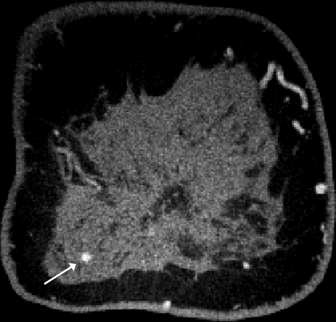
Figure 2c:

Images obtained in a 57-year-old woman. (a) Mediolateral oblique magnification mammogram shows a group of indeterminate microcalcifications (arrow). (b) Precontrast coronal, (c) postcontrast coronal, (d) postcontrast sagittal, and (e) postcontrast transverse breast CT images show the same focus of DCIS (arrow) that enhanced 50.2 HU.
