Abstract
Many patients with stage I non-small cell lung carcinoma will develop recurrence following surgical excision. Sox2 is a marker of embryonic stem cell pluripotency that is associated with aggressive tumor behavior and is expressed in a subset of lung adenocarcinomas. We hypothesized that Sox2 expression may provide prognostic information in early stage lung adenocarcinomas. We evaluated formalin-fixed paraffin embedded tissue from 104 stage I lung adenocarcinomas resected between 1997 and 2000. Sox2 expression was analyzed by immunohistochemistry and compared to clinicopathologic features, time-to-progression (TTP) and overall survival (OS). Sox2 expression was detected in 50% of cases and was more frequent in tumors from older and male patients but not significantly associated with smoking status, tumor stage, grade, or histologic subtype. Compared to Sox2-negative tumors, Sox2 expression predicted a shorter TTP (49% versus 82% at 5 years; P = 0.0006) and shorter OS (54% versus 79% at 5 years; P = 0.004). By multivariate analysis, Sox2 expression predicted a greater risk of progression among men (hazard ratio [HR] 5.6; 95% CI 2.3 to 13.8) and women (HR 2.1; 95% CI 0.8 to 5.7). Sox2 expression was associated with significantly shorter OS among men (HR 2.5; 95% CI 1.2 to 5.1), but not in women. Sox2 appears to be an independent predictor of poor outcome in stage I lung adenocarcinomas and may help stratify patients at increased risk for recurrence.
Keywords: Sox2, prognosis, lung, adenocarcinoma, immunohistochemistry, biomarkers
INTRODUCTION
Lung cancer is among the most common and morbid cancers in the United States.(15, 16) The majority of lung cancers are non-small cell carcinomas (NSCLC), the most common types of which are adenocarcinoma (ACA) and squamous cell carcinoma (SCC). While SCC has been decreasing in incidence with changes in smoking patterns, ACA is increasing in incidence, particularly among women.(25) NSCLC mortality is predicted in large part by tumor stage, with a median survival of more than 8 years for stage I and less than 1 year for stage IV patients.(9) Although surgery can be curative for early stage disease,(6) a significant subset of these patients develop metastases.
Several groups have attempted to identify markers of more aggressive tumor behavior using gene expression profiling;(7, 11, 20–22) however, this approach is hampered by the lack of overlap between gene signatures identified by individual groups, a problem thought to be related to tumor heterogeneity and varying statistical approaches.(5) Immunohistochemistry is a potentially more cost-effective and faster way to identify tumor signatures via their protein expression profiles.(29, 30) However, to date there is no single IHC marker that is used routinely for prognostic purposes in early stage NSCLC.
Sox2 is a transcription factor that is involved in the maintenance of embryonic stem cell pluripotency(14, 27) and in multiple developmental processes, including lung branching morphogenesis.(13) In tumor gene expression studies, analyses of genes known to be active in development and differentiation have shown that Sox2 is overexpressed in certain poorly differentiated subtypes of cancer.(3, 8) A cancer therapy outcome predictor algorithm that incorporates genomic analysis of "stemness" pathways, including the Nanog/Oct4/Sox2 (NOS) pathway, demonstrates high prognostic accuracy in retrospective studies of patients with multiple tumor types, including lung cancer.(10)
Expression of Sox2 protein has not been extensively studied in lung cancer; however, we have recently demonstrated that Sox2 is strongly and diffusely expressed in ~90% of pulmonary SCC and ~20% of ACA.(23) Given the association between NOS pathway gene expression and aggressive tumor types, and given the fact that Sox2 is expressed in a subset of ACA, we hypothesized that Sox2 may be associated with poor prognosis in lung ACA. The purpose of this retrospective study was to evaluate the prognostic value of Sox2 expression in surgically resected stage I lung ACA.
MATERIALS AND METHODS
Study Population
We collected 151 consecutive cases of surgically resected Stage I lung ACA (T1 N0 M0 or T2 N0 M0) in the period ranging from 1997–2000 from the pathology files of Brigham and Women's Hospital (BWH), following approval by the Partners Institutional Review Board (Partners IRB # 2009P001426 and 2006P001921). Diagnoses were confirmed, tumors were graded and subtyped according to WHO criteria,(26) and staged according to the American Joint Committee on Cancer criteria by three pathologists (LMS, JAB, LRC).(9) On review, five tumors were re-classified as adenosquamous or large cell neuroendocrine carcinomas and were excluded from further analysis.
Patient demographic and outcome data were retrieved from the electronic medical records; 24 patients were lost to follow-up and were excluded. Four patients with concurrent cancer diagnoses (not including skin squamous cell and basal cell carcinomas) were excluded. Of the remaining patients, 14 had insufficient tissue for immunohistochemical studies, leaving 104 cases for the final analysis.
Immunohistochemistry
Sox2 expression was evaluated on 37 ACA included in a tissue microarray (TMA) that was previously described(1) and 67 ACA using whole tissue sections. Briefly, a TMA of formalin-fixed, paraffin-embedded tumor containing three 0.6 mm cores from each sample was constructed. The areas included on the TMA were carefully selected to ensure that heterogeneous tumors were well-represented. Immunohistochemistry for Sox2 (rabbit polyclonal antibody; 1:500 dilution; Novus Biologicals, Littleton, CO, USA), TTF-1 (Clone 8G7G3/1; 1:1000 dilution; Dako, Carpinteria, CA), Nanog (goat polyclonal; 1:500 dilution; R&D Systems, Minneapolis, MN) and Oct3/4 (rabbit polyclonal; 1:150 dilution; Santa Cruz Biotechnology, Santa Cruz, CA) were performed following heat-induced epitope retrieval in citrate buffer (pH 6) using a pressure cooker.
Nuclear staining was considered positive. We graded the immunoreactivity by two methodologies: (1) semiquantitatively with 0 as no staining; 1+, <5% tumor cells staining; 2+, 5–25% tumor cells staining; 3+, 26–50% tumor cells staining; and 4+, >50% tumor cells staining; and (2) as negative or positive utilizing a 5% cut-off.
Statistical Analysis
Comparison of clinicopathologic characteristics and Sox2 expression was performed using the Wilcoxon rank-sum and Fisher’s exact tests. The median potential follow-up time using censored data was 96 months. Time-to-progression was computed from time of surgery to time of clinical diagnosis of recurrent carcinoma; patients who died without documentation of recurrence were censored. Overall survival was calculated from time of surgery to time of death from any cause or to time of last follow-up, at which point the data were censored. The logrank (Mantel-Cox) test and Kaplan-Meier estimation were used to examine the time to progression and overall survival. The Cox proportional hazards model was used to perform regression analysis of the failure time data. In addition to controlling for stage, which is prognostic for NSCLC, the multivariate analysis was adjusted for the clinicopathologic characteristics that were associated with Sox2 expression. All reported P-values are based on a two-sided hypothesis. The data analysis was conducted primarily using GraphPad Prism 5 (GraphPad Software, Inc., La Jolla, CA) and SAS 9.1 (SAS Institute Inc., Cary, NC).
RESULTS
Patient Demographics and Pathologic Characteristics
Patient demographic and surgical and tumor pathologic data are detailed in Table 1. There were relatively more women in the study. The majority of patients were smokers. 50% of patients were stage IA and 50% of patients were stage IB. 82% of cases had mixed-subtype histology. Acinar pattern was most commonly the predominant subtype (51%), followed by papillary (18%), solid (17%), and bronchioloalveolar (13%). Of the 19 cases that contained a single histologic subtype, 7 (37%) were acinar, 5 (26%) were papillary, and 7 (37%) were solid. None of the cases were pure bronchioloalveolar carcinomas.
Table 1.
Clinicopathologic features of 104 patients with stage I lung adenocarcinoma.
| Clinicopathologic Parameter |
n (%) |
|---|---|
| Total | 104 (100) |
| Age | |
| Median Age (Range) | 68 (36–91) |
| Sex | |
| Male | 38 (37) |
| Female | 66 (63) |
| Tobacco history* | |
| Smoker | 85 (87) |
| Never smoker | 13 (13) |
| Surgical procedure | |
| Lobectomy | 64 (62) |
| Wedge resection | 40 (38) |
| Stage† | |
| IA | 52 (50) |
| IB | 51 (50) |
| Tumor grade | |
| Well | 19 (18) |
| Moderate | 55 (53) |
| Poor | 30 (29) |
| Tumor heterogeneity | |
| Heterogeneous | 85 (82) |
| Homogeneous | 19 (18) |
| Predominant histologic subtype | |
| Acinar | 53 (51) |
| Bronchioloalveolar | 14 (13) |
| Papillary | 19 (18) |
| Solid | 18 (17) |
Due to rounding, not all percentages total 100.
Smoking history was unavailable for 6 patients.
Distinction between stage IA and IB could not be made in one case.
Sox2 Expression in Lung Adenocarcinoma
Using the semiquantitative scoring system, 36 (35%) cases had no Sox2 staining (0), 16 (15%) cases had <5% tumor cells staining (1+), 14 (13%) cases had 5–25% tumor cells staining (2+), 20 (19%) cases had 26–50% tumor cells staining (3+), and 18 (17%) had >50% tumor cells staining (4+). This is consistent with previously reported data.(23) Using the 5% cut-off, Sox2 expression was observed in 17 of 37 (46%) cases evaluated by TMA and in 35 of 67 (52%) cases evaluated as whole tissue sections (P=0.68). Overall, 52 (50%) cases were considered positive using this cut-off (see below). Representative images of Sox2 expression in low and high grade tumors are shown in Figure 1.
Figure 1.
Lung adenocarcinomas (ACA) show variable Sox2 expression. (A) Well-differentiated ACA with papillary features and (B) Sox2 expression. (C) Well-differentiated ACA with acinar features and (D) absent Sox2 expression. (E) Poorly differentiated ACA with predominantly solid pattern and (F) Sox2 expression. (G) Poorly differentiated ACA with predominantly solid pattern and (H) absent Sox2 expression. Normal bronchial airways are present as positive internal controls in panels D and H.
TTF-1 was positive in 92 (88%) cases. Oct3/4 and Nanog protein expression was very weak and was detected in only rare cells in 8% and 7% of cases, respectively. Based on a 5% cut-off, both markers were considered negative in all cases.
Patient clinical characteristics were correlated with Sox2 expression (Table 2). Median age was higher in patients with Sox2-positive tumors (69 vs. 64 years, P = 0.05). Patients older than 60 years were more likely than those less than 60 years to have Sox2-positive tumors (58% vs. 35%, P = 0.04). Sox2-positive tumors were more frequent in men (61%) than in women (44%); however, this difference was not statistically significant (P=0.15). There was no significant difference in Sox2 expression based on smoking status or resection type.
Table 2.
Sox2 expression according to clinical characteristics in 104 stage I lung adenocarcinomas.
| Patient Characteristic | n | Sox2 negative (%) |
Sox2 positive (%) |
P value | |
|---|---|---|---|---|---|
| Age | |||||
| Median | 64 | 69 | 0.05 | ||
| Age ≤60 | 37 | 24 (69) | 13 (35) | 0.04 | |
| Age >60 | 67 | 28 (42) | 39 (58) | ||
| Sex | 0.15 | ||||
| Men | 38 | 15 (39) | 23 (61) | ||
| Women | 66 | 37 (56) | 29 (44) | ||
| Smoking history* | 0.38 | ||||
| Positive | 85 | 40 (47) | 45 (53) | ||
| Negative | 13 | 8 (62) | 5(38) | ||
| Surgical procedure | 0.84 | ||||
| Lobectomy | 64 | 31 (48) | 33 (52) | ||
| Wedge resection | 40 | 21 (53) | 19 (48) | ||
Due to rounding, not all percentage totals equal 100.
Smoking history was unavailable for 6 patients.
Tumor pathologic characteristics were correlated with Sox2 expression (Table 3). There was no difference in the average size of Sox2-negative and Sox2-positive tumors (2.4 cm vs. 2.5 cm). Tumors with pleural invasion were more likely to show Sox2 expression than those without pleural invasion (61% vs. 44%); however, this difference was not statistically significant (P = 0.14). There was no significant difference in Sox2 expression based on tumor stage, grade, or dominant histologic subtype. Of note, both adenocarcinomas in our study that had prominent mucinous features were strongly Sox2-positive.
Table 3.
Sox2 expression according to pathologic characteristics in 104 stage I lung adenocarcinomas.
| Tumor Characteristic | n | Sox2 negative (%) |
Sox2 positive (%) |
P value | |
|---|---|---|---|---|---|
| AJCC classification* | 0.70 | ||||
| T1 | 52 | 27 (52) | 25 (48) | ||
| T2 | 51 | 24 (47) | 27 (53) | ||
| Pleural invasion† | 0.14 | ||||
| Present | 36 | 14 (39) | 22 (61) | ||
| Absent | 64 | 36 (56) | 28 (44) | ||
| Grade | 0.24 | ||||
| Well | 19 | 11 (58) | 8 (42) | ||
| Moderate | 55 | 23 (42) | 32 (58) | ||
| Poor | 30 | 18 (60) | 12 (40) | ||
| Predominant subtype | 0.72 | ||||
| Acinar | 53 | 25 (47) | 28 (53) | ||
| BAC | 14 | 6 (43) | 8 (57) | ||
| Papillary | 19 | 10 (53) | 9 (47) | ||
| Solid | 18 | 11 (61) | 7 (39) | ||
| TTF-1 expression | 0.76 | ||||
| Positive | 92 | 45 (49) | 47 (51) | ||
| Negative | 12 | 7 (58) | 5 (42) | ||
T classification could not be determined in one case.
The presence of pleural invasion could not be assessed in four cases.
One Sox2-negative and two Sox2-positive tumors had positive surgical margins, none of whom received adjuvant therapy. Margins could not be assessed in two cases. Three patients with Sox2-negative tumors and three patients with Sox2-positive tumors received adjuvant therapy (four patients received local radiotherapy only; one patient with a Sox2-negative tumor received both radiotherapy and chemotherapy and one patient with a Sox2-positive tumor received chemotherapy only).
Survival Analysis
Univariate survival analysis was performed for each cut-off in the semiquantitative Sox2 scoring system; >5% tumor cell staining proved to be the strongest predictor of survival (data not shown), and this cut-off was therefore used for the analyses.
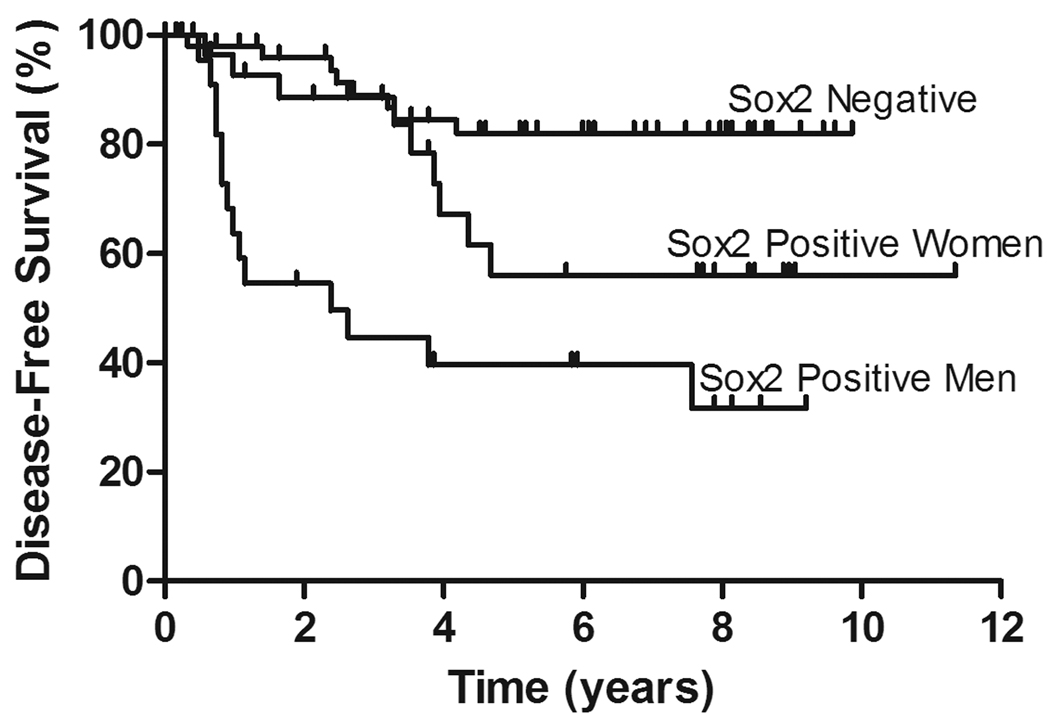
By univariate analysis, Sox2 expression predicted a shorter time to progression (P = 0.0006). At five years, 82% of the patients with Sox2-negative ACA and 49% of patients with Sox2-positive ACA were disease-free. Exploring the interaction between Sox2 expression and patient sex, men and women with Sox2-negative tumors had virtually identical TTP curves (P = 0.59). Although Sox2-positive ACA are associated with shorter time-to-progression among both genders (Figure 2), men with Sox2-positive tumors had significantly shorter TTP than did women (P = 0.03). Accounting for the sex differences as well as controlling for age and stage, the risk of progression was more than five times higher (HR 5.6; 95% CI 2.3–13.8) among male patients with Sox2-positive tumors (P = 0.0002). In contrast, women with Sox2-positive tumors were twice as likely to progress (HR 2.1; 95% CI 0.8–5.7) compared to all those with Sox2-negative tumors (P = 0.12), but this difference was not statistically significant.
Figure 2.
Time to progression in patients with lung adenocarcinoma based on Sox2 expression and gender.
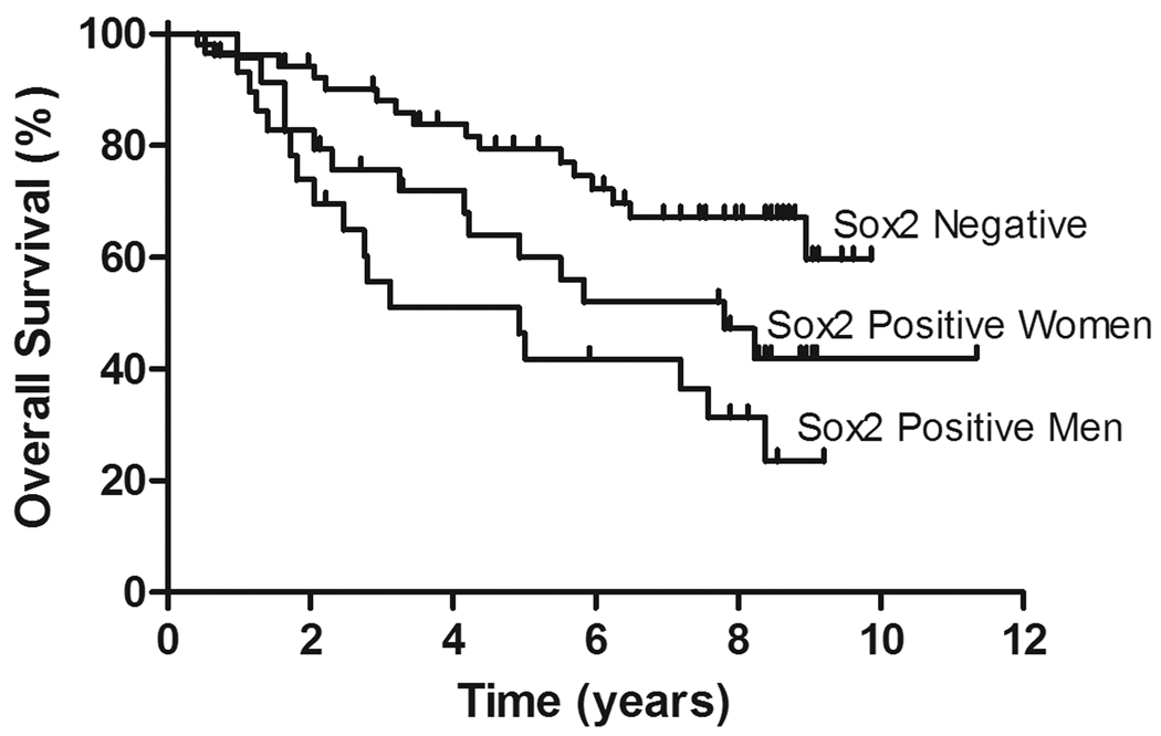
By univariate analysis, Sox2 expression was also a significant predictor of a shorter overall survival (P = 0.004). The overall survival at five years was 79% in the Sox2-negative group and 54% in the Sox2-positive tumors (Figure 3). After allowing for differential sex effects as well as adjusting for age and stage, Sox2 expression is associated with more than double the risk of dying (HR 2.5; 95% CI 1.2–5.1) among male patients compared to all those with Sox2-negative tumors (P = 0.01). In contrast, the risk of death is increased moderately (HR 1.6; 95% CI 0.8–3.2) for female patients, but this difference was not statistically significant (P = 0.2).
Figure 3.
Overall survival in patients with lung adenocarcinoma based on Sox2 expression and gender.
Of the 52 patients with Sox2-positive tumors, 23 (44%) had clinically documented progression: 11 patients developed metastatic disease and 12 patients had a local recurrence. During the same period, of the 52 patients with Sox2-negative tumors, eight (15%) had progression: five patients developed metastatic disease and three had a local recurrence.
Discussion
There is a pressing need for additional markers to provide prognostic information in early stage lung cancer, as the current surgical-pathologic staging system cannot predict which stage I lung cancers will be cured by surgery alone and which will recur. Patients with more aggressive tumors may benefit from adjuvant therapy, if they can be identified at the time of surgical resection. A significant effort has been made to identify markers that can provide this prognostic information in the form of gene expression and immunohistochemical analysis. Most of the currently available models for prognostication in early stage lung cancer are not in widespread use, and, in the case of gene expression analysis, are prohibitively expensive. In Sox2, we have identified a single immunohistochemical marker that is easily interpreted, inexpensive, and highly associated with shorter time to progression and overall survival in stage I lung adenocarcinomas.
We examined expression of the transcription factors Nanog, Oct3/4 and Sox2 in surgically resected lung adenocarcinomas. Prior studies have demonstrated increased gene expression of Nanog, Oct3/4 and Sox2 and their downstream targets in aggressive tumor types.(3, 10) Of note, in our study only Sox2 showed significant protein expression in tumor cells. This discrepancy may be a function of the different methodologies employed, but also raises the possibility that Sox2 is acting independently of its usual cofactors in lung cancer, or alternatively that the sensitivity of the available antibodies when used for immunohistochemistry may be too low to detect the levels of Nanog and Oct3/4 expressed in this tumor type.
We correlated Sox2 expression with patient characteristics, tumor pathology and survival. Other than a higher frequency observed with older patient age and male sex, Sox2 expression was not significantly associated with any other clinical or pathologic variables, including histologic subtype, grade, T classification, or TTF-1 expression. Although not statistically significant, it is interesting to note that Sox2 expression was more commonly associated with pleural invasion. Preliminary studies of Sox2 in melanoma have suggested that Sox2 expression may be associated with local tumor progression.(19)
Sox2 was strongly prognostic, even after adjusting for other clinical variables including age and gender. Sox2 expression was significantly associated with shorter TTP and OS by univariate and multivariate analyses. Of note, men with Sox2-expressing tumors were more likely to progress than were women; their risk of death was about double compared to all those with Sox2-negative tumors.
Possible pitfalls include use of this marker in large cell neuroendocrine carcinomas or squamous cell carcinomas that may have been misdiagnosed as adenocarcinomas; because ~80% of LCNEC and >90% of pulmonary squamous cell carcinomas show Sox2 expression,(23) it is unlikely that this marker would have prognostic value in these cases. The utility of this marker in tumors of mixed adenosquamous histology has not been examined.
The biologic significance of Sox2 overexpression in lung ACA is not yet known. The lack of correlation between Sox2 and TTF-1 expression in these tumors suggests that its expression is not related to tumor differentiation pathways. Of note, SOX2 has been identified as an oncogene that undergoes amplification in lung and esophageal squamous cell carcinomas.(2) Large scale copy number gains have been reproducibly detected in several regions of the NSCLC genome, including at 3q, 5p and 8q.(12, 28) Sox2 is located at 3q26. Comparative genomic hybridization studies have demonstrated that >90% of squamous cell carcinomas and ~20% of adenocarcinomas have copy number gain involving 3q26,(4) essentially the same proportions that have been demonstrated to have high level Sox2 expression at the protein level.(23) Of note, these studies have found high level amplification only in SCC.(4) Epigenetic regulation of Sox2 has been shown to drive its expression in neural systems, with effects on cell proliferation and differentiation.(24) These observations warrant additional studies to determine the molecular mechanisms of Sox2 expression in ACA. Correlation between Sox2 expression and the mutation status of established predictive markers such as EGFR and KRAS will be of particular interest.
To date there is no single IHC marker that is used routinely for prognostic purposes in early stage NSCLC. A recent report has identified a combination of clinical parameters and immunohistochemical (IHC) marker panels that provide independent prognostic information in stage IB NSCLC;(30) however, the proposed algorithms are relatively complex and time-consuming. Sox2 is currently used in clinical practice to aid in identification and subtyping of germ cell tumors,(28–30) but it has limited known application outside of that context. It now appears that it can effectively identify those early stage lung adenocarcinomas that are more likely to recur in the first five years following surgery. The findings in this retrospective study require prospective validation. In this study, the strength of the association between Sox2 expression and poor outcomes is on par with that of other established poor prognostic markers, including lymphovascular invasion, solid histology, and tumor size.(17, 18) However, Sox2 expression appears to be independent of these and other pathologic features, suggesting that it may serve as a novel prognostic marker, enabling the identification of those high-risk patients who may benefit from adjuvant therapy.
Acknowledgments
Financial support: This work was supported in part by Dana-Farber/Harvard Cancer Center Specialized Programs of Research Excellence (SPORE) in Lung Cancer 2P50 CA090578-06 (LRC). The authors have no financial disclosures.
Footnotes
Publisher's Disclaimer: This is a PDF file of an unedited manuscript that has been accepted for publication. As a service to our customers we are providing this early version of the manuscript. The manuscript will undergo copyediting, typesetting, and review of the resulting proof before it is published in its final citable form. Please note that during the production process errors may be discovered which could affect the content, and all legal disclaimers that apply to the journal pertain.
Presented in part at the 99th annual meeting of the United States and Canadian Academy of Pathology (USCAP) in Washington, D.C., March 20–26, 2010.
REFERENCES
- 1.Barletta JA, Perner S, Iafrate AJ, et al. Clinical significance of TTF-1 protein expression and TTF-1 gene amplification in lung adenocarcinoma. J Cell Mol Med. 2008;13:1977–1986. doi: 10.1111/j.1582-4934.2008.00594.x. [DOI] [PMC free article] [PubMed] [Google Scholar]
- 2.Bass AJ, Watanabe H, Mermel CH, et al. SOX2 is an amplified lineage-survival oncogene in lung and esophageal squamous cell carcinomas. Nat Genet. 2009;41:1238–1242. doi: 10.1038/ng.465. [DOI] [PMC free article] [PubMed] [Google Scholar]
- 3.Ben-Porath I, Thomson MW, Carey VJ, et al. An embryonic stem cell-like gene expression signature in poorly differentiated aggressive human tumors. Nat Genet. 2008;40:499–507. doi: 10.1038/ng.127. [DOI] [PMC free article] [PubMed] [Google Scholar]
- 4.Bjorkqvist AM, Husgafvel-Pursiainen K, Anttila S, et al. DNA gains in 3q occur frequently in squamous cell carcinoma of the lung, but not in adenocarcinoma. Genes Chromosomes Cancer. 1998;22:79–82. [PubMed] [Google Scholar]
- 5.Boutros PC, Lau SK, Pintilie M, et al. Prognostic gene signatures for non-small-cell lung cancer. Proc Natl Acad Sci U S A. 2009;106:2824–2828. doi: 10.1073/pnas.0809444106. [DOI] [PMC free article] [PubMed] [Google Scholar]
- 6.Chang MY, Sugarbaker DJ. Surgery for early stage non-small cell lung cancer. Semin Surg Oncol. 2003;21:74–84. doi: 10.1002/ssu.10024. [DOI] [PubMed] [Google Scholar]
- 7.Chen HY, Yu SL, Chen CH, et al. A five-gene signature and clinical outcome in non-small-cell lung cancer. N Engl J Med. 2007;356:11–20. doi: 10.1056/NEJMoa060096. [DOI] [PubMed] [Google Scholar]
- 8.Chen Y, Shi L, Zhang L, et al. The molecular mechanism governing the oncogenic potential of SOX2 in breast cancer. J Biol Chem. 2008;283:17969–17978. doi: 10.1074/jbc.M802917200. [DOI] [PubMed] [Google Scholar]
- 9.Edge SB, Byrd DR, Compton CC, et al., editors. AJCC Cancer Staging Manual. New York: Springer; 2009. [Google Scholar]
- 10.Glinsky GV. "Stemness" genomics law governs clinical behavior of human cancer: implications for decision making in disease management. J Clin Oncol. 2008;26:2846–2853. doi: 10.1200/JCO.2008.17.0266. [DOI] [PubMed] [Google Scholar]
- 11.Hassan KA, Chen G, Kalemkerian GP, et al. An embryonic stem cell-like signature identifies poorly differentiated lung adenocarcinoma but not squamous cell carcinoma. Clin Cancer Res. 2009;15:6386–6390. doi: 10.1158/1078-0432.CCR-09-1105. [DOI] [PMC free article] [PubMed] [Google Scholar]
- 12.Huang YT, Heist RS, Chirieac LR, et al. Genome-wide analysis of survival in early-stage non-small-cell lung cancer. J Clin Oncol. 2009;27:2660–2667. doi: 10.1200/JCO.2008.18.7906. [DOI] [PMC free article] [PubMed] [Google Scholar]
- 13.Ishii Y, Rex M, Scotting PJ, et al. Region-specific expression of chicken Sox2 in the developing gut and lung epithelium: regulation by epithelial-mesenchymal interactions. Dev Dyn. 1998;213:464–475. doi: 10.1002/(SICI)1097-0177(199812)213:4<464::AID-AJA11>3.0.CO;2-Z. [DOI] [PubMed] [Google Scholar]
- 14.Ivanova N, Dobrin R, Lu R, et al. Dissecting self-renewal in stem cells with RNA interference. Nature. 2006;442:533–538. doi: 10.1038/nature04915. [DOI] [PubMed] [Google Scholar]
- 15.Jemal A, Siegel R, Ward E, et al. Cancer statistics, 2009. CA Cancer J Clin. 2009;59:225–249. doi: 10.3322/caac.20006. [DOI] [PubMed] [Google Scholar]
- 16.Jemal A, Thun MJ, Ries LA, et al. Annual report to the nation on the status of cancer, 1975–2005, featuring trends in lung cancer, tobacco use, and tobacco control. J Natl Cancer Inst. 2008;100:1672–1694. doi: 10.1093/jnci/djn389. [DOI] [PMC free article] [PubMed] [Google Scholar]
- 17.Jones DR, Daniel TM, Denlinger CE, et al. Stage IB nonsmall cell lung cancers: are they all the same? Ann Thorac Surg. 2006;81:1958–1962. doi: 10.1016/j.athoracsur.2005.12.054. discussion 1962. [DOI] [PubMed] [Google Scholar]
- 18.Kwiatkowski DJ, Harpole DH, Jr., Godleski J, et al. Molecular pathologic substaging in 244 stage I non-small-cell lung cancer patients: clinical implications. J Clin Oncol. 1998;16:2468–2477. doi: 10.1200/JCO.1998.16.7.2468. [DOI] [PubMed] [Google Scholar]
- 19.Laga AC, Lai CY, Zhan Q, et al. Expression of the embryonic stem cell transcription factor SOX2 in human skin: relevance to melanocyte and merkel cell biology. Am J Pathol. 176:903–913. doi: 10.2353/ajpath.2010.090495. [DOI] [PMC free article] [PubMed] [Google Scholar]
- 20.Lau SK, Boutros PC, Pintilie M, et al. Three-gene prognostic classifier for early-stage non small-cell lung cancer. J Clin Oncol. 2007;25:5562–5569. doi: 10.1200/JCO.2007.12.0352. [DOI] [PubMed] [Google Scholar]
- 21.Lu Y, Lemon W, Liu PY, et al. A gene expression signature predicts survival of patients with stage I non-small cell lung cancer. PLoS Med. 2006;3:e467. doi: 10.1371/journal.pmed.0030467. [DOI] [PMC free article] [PubMed] [Google Scholar]
- 22.Shedden K, Taylor JM, Enkemann SA, et al. Gene expression-based survival prediction in lung adenocarcinoma: a multi-site, blinded validation study. Nat Med. 2008;14:822–827. doi: 10.1038/nm.1790. [DOI] [PMC free article] [PubMed] [Google Scholar]
- 23.Sholl LM, Long KB, Hornick JL. Sox2 expression in pulmonary non-small cell and neuroendocrine carcinomas. Appl Immunohistochem Mol Morphol. 2010;18:55–61. doi: 10.1097/PAI.0b013e3181b16b88. [DOI] [PubMed] [Google Scholar]
- 24.Sikorska M, Sandhu JK, Deb-Rinker P, et al. Epigenetic modifications of SOX2 enhancers, SRR1 and SRR2, correlate with in vitro neural differentiation. J Neurosci Res. 2008;86:1680–1693. doi: 10.1002/jnr.21635. [DOI] [PubMed] [Google Scholar]
- 25.Toh CK. The changing epidemiology of lung cancer. Methods Mol Biol. 2009;472:397–411. doi: 10.1007/978-1-60327-492-0_19. [DOI] [PubMed] [Google Scholar]
- 26.Travis W, Brambilla E, Muller-Hermelink H, et al., editors. Pathology and Genetics of Tumours of the Lung, Pleura, Thymus, and Heart. Lyon: IARCPress; 2004. [Google Scholar]
- 27.Wang J, Rao S, Chu J, et al. A protein interaction network for pluripotency of embryonic stem cells. Nature. 2006;444:364–368. doi: 10.1038/nature05284. [DOI] [PubMed] [Google Scholar]
- 28.Weir BA, Woo MS, Getz G, et al. Characterizing the cancer genome in lung adenocarcinoma. Nature. 2007;450:893–898. doi: 10.1038/nature06358. [DOI] [PMC free article] [PubMed] [Google Scholar]
- 29.Zhu CQ, Shih W, Ling CH, et al. Immunohistochemical markers of prognosis in non-small cell lung cancer: a review and proposal for a multiphase approach to marker evaluation. J Clin Pathol. 2006;59:790–800. doi: 10.1136/jcp.2005.031351. [DOI] [PMC free article] [PubMed] [Google Scholar]
- 30.Zhu ZH, Sun BY, Ma Y, et al. Three immunomarker support vector machines-based prognostic classifiers for stage IB non-small-cell lung cancer. J Clin Oncol. 2009;27:1091–1099. doi: 10.1200/JCO.2008.16.6991. [DOI] [PubMed] [Google Scholar]


