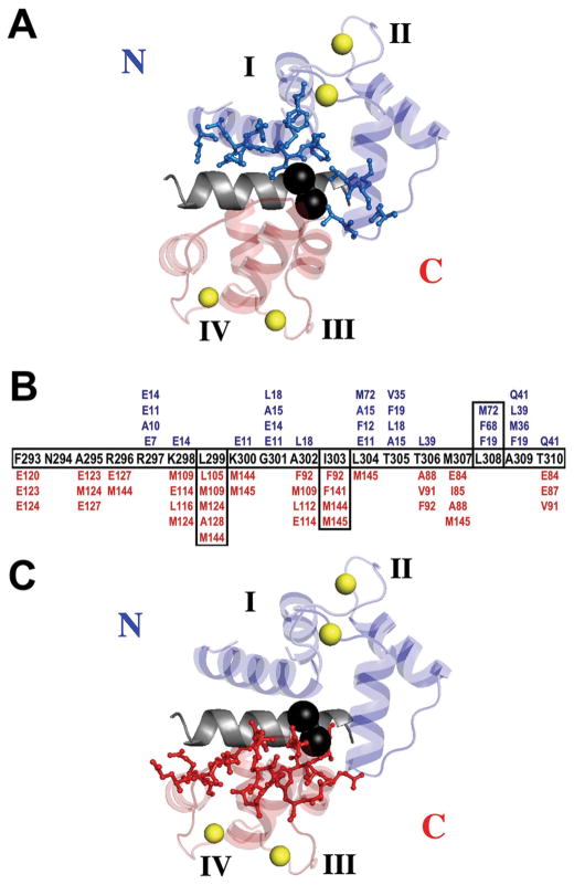
Figure 10.
Ribbon diagram of CaM bound to CaMBD of Ca2+/CaM-dependent kinase II (1CDM.pdb) 20. CaM residues in the N-domain (A) and C-domain (C) within 4.5 Å of CaMKIIp are shown in blue (N-domain) and red (C-domain) ball-and-stick; CaMKIIp T305 and T306 are shown as black spheres (made with PyMol, DeLano Scientific LLC). (B) Sequence map showing CaM residues that are within 4.5 Å of each residue of the CaMKII CaMBD peptide calculated with CSU66.

