Abstract
Purpose
ABCR protein in the rod outer segment is thought to facilitate movement of the all-trans retinal photoproduct of rhodopsin bleaching out of the disk lumen. We investigated the extent to which ABCR deficiency affects post-bleach recovery of the rod photoresponse in ABCR-deficient (abcr−/−) mice.
Methods
Electroretinographic (ERG) a-wave responses were recorded from abcr−/− mice and two control strains. Using a bright probe flash, we examined the course of rod recovery following fractional rhodopsin bleaches of ~10−6, ~3×10⁻⁵, ~0.03 and ~0.30–0.40.
Results
Dark-adapted abcr−/− mice and controls exhibited similar normalized near-peak amplitudes of the paired-flash-ERG-derived, weak-flash response. Response recovery following ~10−6 bleaching exhibited an average exponential time constant of 319, 171 and 213 ms, respectively, in the abcr−/− and the two control strains. Recovery time constants determined for ~3×10⁻⁵ bleaching did not differ significantly among strains. However, those determined for the ~0.03 bleach indicated significantly faster recovery in abcr−/− (2.34 ± 0.74 min) than in the controls (5.36 ± 2.20 min, and 5.92 ± 2.44 min). Following ~0.30–0.40 bleaching, the initial recovery in the abcr−/− was on average faster than in controls.
Conclusions
By comparison with controls, abcr−/− mice exhibit faster rod recovery following a bleach of ~0.03. The data suggest that ABCR in normal rods may directly or indirectly prolong all-trans retinal clearance from the disk lumen over a significant bleaching range, and that the essential function of ABCR may be to promote the clearance of residual amounts of all-trans retinal that remain in the disks long after bleaching.
Keywords: ABCR, ABCA4, electroretinography, dark adaptation, rods
INTRODUCTION
The ATP-binding cassette transporter protein known as ABCR, or ABCA4, facilitates processing of the all-trans retinal photoproduct of rhodopsin bleaching in rod photoreceptors (1–7). In an ATP-dependent reaction, ABCR moves N-retinylidene-phosphatidylethanolamine (N-ret-PE), a complex formed between all-trans retinal and phosphatidylethanolamine, from the luminal to cytosolic side of the rod disk membrane. The all-trans retinal in the rod cytosol is then enzymatically reduced to all-trans retinol for further processing in the retinoid visual cycle that enables rhodopsin regeneration in bleached rods (8–11). As all-trans retinal can interact with opsin to form a metarhodopsin II- (MII)-like signaling state (12–15), the clearance of all-trans retinal out of the disk (i.e., from the vicinity of opsin) promotes shut-off of the phototransduction cascade.
The impairment of ABCR activity leads to the build-up of N-ret-PE in the rods and to the resulting accumulation, in the retinal pigment epithelium (RPE), of retinoid-based components of lipofuscin that produce atrophy of the RPE (16–18). Furthermore, mutations in ABCR are associated with Stargardt disease and other retinal degenerations (19–26). However, relatively little information is available as to how ABCR deficiency affects recovery of the rod electrophysiological response following rhodopsin bleaching illumination, i.e., following the generation of all-trans retinal. In electroretinographic (ERG) experiments on 16–20 week-old abcr−/− mice, Weng et al. (1) investigated recovery of the ERG a-wave after illumination that bleached about 45% of the rhodopsin. These investigators found that a-wave recovery in abcr−/− mice was substantially slower than that exhibited by wildtype mice of similar age, consistent with a delayed clearance of all-trans retinal from the disk lumen in mice lacking ABCR. For example, a-wave recovery at 30 min after the bleach amounted to about 75% in wildtype mice but only about 50% in abcr−/− mice.
The signaling activity of the MII-like complex formed by all-trans retinal and opsin far exceeds that due to free opsin (i.e., opsin devoid of chromophore), and may be as great as ~10% of that of MII generated in the phototransduction process (13,14,27,28). Furthermore, studies of both single rod photocurrents and the in vivo, massed rod response show that in dark-adapted mouse rods, photoactivation of as little as ~100 rhodopsins per rod, i.e., a fractional bleach of ~10−6 (29), produces a rod photocurrent response of near-saturating peak amplitude (see, e.g., 30). On the basis of these findings, and of previous ERG data obtained with ~45% bleaching (1), we reasoned that abcr−/− mice might exhibit a detectable delay in rod recovery at fractional bleaches well below ~45%. The present study was undertaken to test this possibility. Preliminary results have been reported (31,32).
METHODS
All procedures conformed to the principles embodied in the ARVO Statement for the Use of Animals in Ophthalmic and Vision Research. Experiments were conducted on mice of ages 3 weeks to 3–4 months that were maintained on a light/dark cycle (12 hr light/12 hr dark or 14 hr light/10 hr dark) at an ambient illumination of ~2–19 lux. The abcr/&mice; mice were derived from breeding pairs that were generously provided by Dr. G. H. Travis (University of California at Los Angeles). Two wildtype (i.e., abcr+/+) strains were used as controls. The first of these was a C57-derived strain that, like abcr−/−, possesses the leucine variant at amino acid position 450 of the retinal pigment epithelium (RPE) protein RPE65 (Dr. G. H Travis and Dr. R. Radu, personal communication; and ref. 33). Breeding pairs of these mice (N10, C57BL/6J) were generously provided by Dr. M. Danciger (Loyola Marymount University). The second was strain C57BL/6J (Jackson Laboratories, Bar Harbor, ME, USA), which is known to possess the methionine variant at amino acid position 450 of RPE65 (34). As noted below (Discussion), the investigation of controls possessing both the leucine 450 and methionine 450 variants was of importance to the present study in light of RPE65’s role in the retinoid visual cycle (35–37). These control strains possessing the leucine and methionine RPE65 variants will henceforth be termed C57-leu450 and C57-met450, respectively (cf. 33).
All mice were dark-adapted overnight before the experiment. Equipment and procedures used for single- and paired-flash ERG recording were similar to those described (38–40). Briefly, under dim red light, the mouse was anesthetized with an intraperitoneal injection of ketamine and xylazine [0.15 and 0.01 mg (g body wt) −1, respectively]. Pupil dilation was carried out using 2.5% phenylephrine HCl, and 1% tropicamide, and the cornea was anesthetized using proparacaine HCl (0.5%). Drops of a moistening/lubricating agent (Tears Naturale Forte; Alcon, Fort Worth TX) were periodically applied to the corneal surface. The mouse’s body temperature was maintained in the range of 37.5 – 38.5 °C with use of a heating pad positioned beneath the animal. Boosts of anesthetic (approximately 1/6 of the initial dose) were delivered subcutaneously at approximately 20-min intervals beginning ~40 min after the initial dose. Responses to full-field test flashes (green light of duration about 20 μs) and to probe flashes (white light of duration about 1.7 ms) were obtained with use of a stainless steel recording electrode positioned on the cornea, a stainless steel reference electrode placed in the mouth, and a platinum subdermal needle ground electrode positioned in the nape of the neck. Responses were amplified (bandpass: 0.3 – 3000 Hz), sampled at 100 kHz, stored in a computer, and subsequently analyzed using Matlab software (manufacturer information: Mathworks, Natick, MA). Paired-flash ERG determinations of the rod response to a given test stimulus (i.e., in the present experiments, to a bleaching illumination of defined strength) used the methodology described previously (38–41). In the paired-flash method, a bright (i.e., rod-saturating) probe flash is presented at a defined time following the test stimulus. The bright probe flash, which rapidly drives the rods to saturation, produces an a-wave response that essentially titrates the prevailing level of rod circulating current. As referenced to the “probe-alone” response obtained from the fully dark-adapted eye (i.e., in the absence of recent presentation of the test stimulus), the probe response obtained in the paired-flash trial yields the prevailing “derived” amplitude of the rod response to the test stimulus (see, e.g., pp. 519–520 of ref. 41).
ERG experiments investigating rod recovery were conducted on a total of 18 abcr−/−, 17 C57-leu450 and 20 C57-met450 mice, age ranges for which were 23–116, 23–95, and 23–86 days, respectively. The initial phase of each experiment consisted of a series of single- and paired-flash measurements on the dark-adapted mouse. In the single-flash trials we recorded the a-wave response to a bright probe flash of fixed strength (773 scotopic candela seconds per square meter; sc cd s m−2) to determine the peak amplitude (Apeak) and time-to-peak (tpeak) of this response. The derived rod response to a weak test flash (0.3 sc cd s m−2) at a fixed post-test-flash time (t = 86 ms) was determined in paired-flash trials, using a test-probe interval (tprobe) of 80 ms, a probe flash of strength 773 sc cd s m−2, and a determination time (tdet) of 6 ms (38). Consecutive experimental runs were separated by a dark-adaptation period of ≥2 min. The normalized amplitude, at t = 86 ms, of the dark-adapted derived response to the weak test flash was obtained from the relation (38)
| (1a) |
| (1b) |
where A(t) is the derived response amplitude, AmoD is the amplitude (at 6 ms) of the response to the probe flash in a probe-alone trial under dark-adapted conditions, and Am(t) is the amplitude of the probe response in the paired-flash trial.
Following completion of the dark-adapted characterization, a bleaching stimulus was delivered, and the time course of recovery of the derived response A(t) to this bleaching light was investigated through analysis of the response to the 773 sc cd s m−2 probe flash. The present study employed four different bleaching stimuli: (1) the weak test flash (0.3 sc cd s m−2) used in the dark-adapted characterization described above; (2) a single bright flash of strength sufficient to produce rod saturation (7.9 sc cd s m−2) (38); (3) a series of 20 flashes (each of strength 477 sc cd s m−2) delivered over a period of about 100 s [cumulative bleaching energy: (20)(477 sc cd s m−2) = 9.5×10³ sc cd s m−2]; and (4) a 2-min exposure to intense full-field green light from a microscope illuminator positioned above the eye under investigation (42). The recording electrode was withdrawn during the 2-min illumination period. In experiments of types 1 and 2, the bleaching light was presented in each experimental run (38), and the experiment typically involved two determinations at a given value of tprobe. Some experiments involved the investigation of recovery from both the sub-saturating (type 1) and saturating (type 2) bleaching flashes. Unless otherwise indicated, values of A(t) determined after the bleaching illumination were normalized to the dark-adapted probe-alone amplitude AmoD determined early in the experiment. For recoveries from the 20-flash and 2-min bleaching stimuli, the 6-ms determination time tdet was negligible and was ignored. Conclusion of the bleaching illumination defined time zero in each experiment or (in type 1 and type 2 experiments) each experimental run. For the 20-flash and 2-min bleaching illuminations, the bleaching light was presented only once in the experiment, and the recovery time course was determined by presentations of the probe flash alone at varying times. In all four types of experiment, probe responses obtained after the bleaching illumination were analyzed to yield Am(t), and the derived response A(t) was obtained through eq. 1. In experiments of types 3 and 4, both the post-bleach times of measurement and the overall post-bleach period of investigation differed somewhat among experiments. To permit ANOVA of individual sets of recovery data in the type 3 experiments, consecutive values of the determined, normalized derived response A(t)/AmoD were linearly interpolated to yield minute-by-minute values of A(t)/AmoD. For sets of recovery data in the type 4 experiments, determined values of A(t)/AmoD were grouped within 3-min bins to permit ANOVA (see Results).
For low extents of rhodopsin bleaching (i.e., those relevant to the present experiments of types 1–3), the fractional bleach B produced by a bleaching stimulus of strength L (in sc cd s m−2) is approximately given by B = aL/Ro, where Ro is the population of rhodopsin molecules in the dark-adapted (i.e., unbleached) rod, and a is the number of photoisomerizations (R*) produced by a flash of unit strength. Previous estimates of a (based on different experimental approaches) have ranged from 100 R* (sc cd s m−2) −1 (38) to 490–580 R* (sc cd s m−2) −1 (29). Taking Ro = 7×10⁷ (29) and a ~250 R*, the fractional bleaches corresponding with the bleaching illuminations in the present type 1–3 experiments (0.3 sc cd s m−2, 7.9 sc cd s m−2, and 9.5×10³ sc cd s m−2, respectively) are ~10−6, ~3×10⁻⁵ and ~0.03, respectively. Procedures used to determine the extent of rhodopsin bleaching by the 2-min illumination (type 4 experiment) followed those described (39,42). The anesthetized mouse was killed by cervical dislocation immediately after the bleach, and the retinas and RPEs were isolated. The retina and RPE of a given eye were extracted using formaldehyde (for analysis of retinaldehydes) and isopropanol/hexane (for analysis of all-trans retinol and retinyl ester) extraction procedures. The extracts were analyzed for molar amounts of 11-cis retinal, all-trans retinal, all-trans retinol, and retinyl ester using normal phase high-performance liquid chromatography and standard curves. The difference in molar percents of 11-cis retinal measured for the illuminated vs. unilluminated eyes was used to determine the percent rhodopsin bleach (39,42). The data yielded bleach extents of 40% ± 11% for abcr−/− (n = 3; ages of 73, 78 and 78 days); 32% ± 8% for C57-leu450 (n = 4; ages of 73, 73, 69, and 69 days); and 31% ± 5% for C57-met450 (n = 3; ages of 63, 58 and 63 days). Based on their average values, the following text quotes the bleach extent as ~30–40%.
RESULTS
Dark-adapted characterization
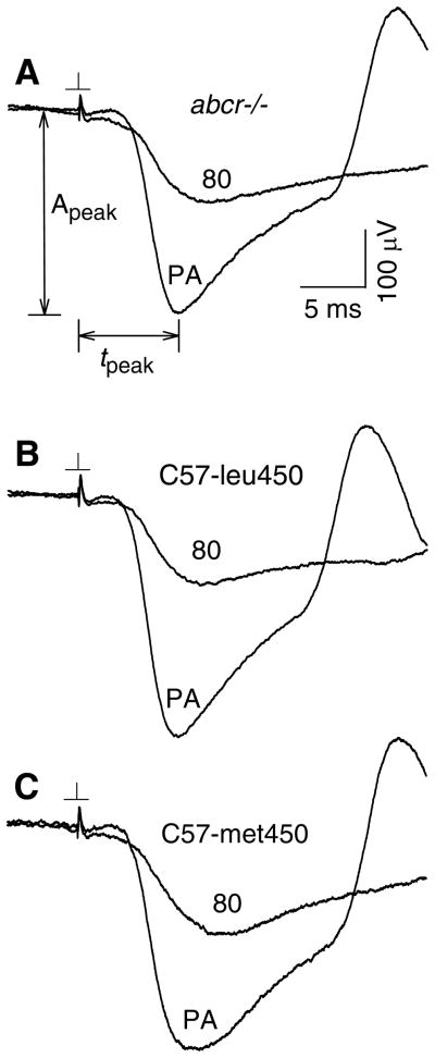
Waveforms labeled PA in Figs. 1A–C show, respectively, dark-adapted responses recorded from abcr−/−, C57-leu450 and C57-met450 mice in “probe-alone” trials, i.e., on presentation of only the bright probe flash (773 sc cd s m−2). These probe-alone responses were analyzed for the peak amplitude Apeak and time-to-peak tpeak. Sections A–C of Table 1 show, for each of the three investigated strains, the number of experiments and ages of the mice for which determinations of Apeak (column 1 in the Table) and tpeak (column 2) were made. Columns 1–2 in Table 1 sections D–F show data (means ± SDs) obtained for Apeak and tpeak in abcr−/−, C57-leu450 and C57-met450 mice. Peak amplitude of the probe-alone response ranged, on average, from 315 to 369 μV. Between-groups ANOVA showed a significant difference among the three investigated strains for Apeak [F(2,52) = 3.183; P = 0.050]; in addition, values of tpeak among the strains differed significantly [F(2,52) = 6.974; P = 0.002]. Post-hoc comparisons of Apeak and tpeak values for abcr−/− and C57-leu450 mice indicated a significant difference only for tpeak (P = 0.001). For abcr−/−vs. C57-met450 mice, there were significant differences in both Apeak and tpeak (P = 0.015 and P = 0.008, respectively). Between C57-leu450 and C57-met450 mice, Apeak and tpeak values did not differ significantly. Average values of tpeak ranged from 7.10–7.64 ms among strains.
Fig. 1.
Dark-adapted responses recorded from abcr−/− (panel A), C57-leu450 (B) and C57-met450 (C) mice in single-flash and paired-flash trials. Each illustrated waveform is a single response. Traces PA: responses to the 773 sc cd s m−2 probe flash presented alone. The peak amplitude Apeak and time-to-peak tpeak of the probe response were determined as shown in the upper panel. Traces labeled 80: probe responses recorded in paired-flash trials with an 80-ms test probe interval.
Table 1.
Dark-adapted characterization and recovery parameters.
| Approximate bleach ** | |||||||||
|---|---|---|---|---|---|---|---|---|---|
| 1 | 2 | 3 | 4 | 5 | 6 | 7 | |||
| Type of measurement | Dark-adapted characteristics * | ~10−6 τr |
~3×10⁻⁵ τω |
~3% τ |
~30–40% Slope (σ) |
||||
| Apeak | tpeak | Norm DR | |||||||
| A | abcr−/− | no. of mice age (days) |
18 55 ± 30 |
18 55 ± 30 |
15 57 ± 32 |
3 40 ± 6 |
5 43 ± 6 |
8 63 ± 40 |
4 58 ± 27 |
| B | C57-leu450 | no. of mice age (days) |
17 58 ± 26 |
17 58 ± 26 |
17 58 ± 26 |
4 53 ± 26 |
3 43 ± 24 |
8 61 ± 24 |
4 63 ± 36 |
| C | C57-met450 | no. of mice age (days) |
20 56 ± 19 |
20 56 ± 19 |
17 58 ± 19 |
3 62 ± 3 |
5 51 ± 13 |
9 50 ± 21 |
5 72 ± 14 |
| Determined parameters | Dark-adapted characteristics * | ~10−6 τr (ms) |
~3×10⁻⁵ τω (ms) |
~3% τ (min) |
~30–40% σ (min−1) *** |
|||
|---|---|---|---|---|---|---|---|---|
| Apeak (μV) | tpeak (ms) | Norm DR | ||||||
| D | abcr−/− | 315 ± 60 | 7.10 ± 0.37 | 0.65 ± 0.07 | 319 ± 24 320 (R2=.95) |
321 ± 39 315 (R2=.99) |
2.34 ± 0.74 2.45 (R2=.96) |
−0.0281 ± 0.0079 −0.0280 (R2=.95) |
| E | C57-leu450 | 340 ± 56 | 7.64 ± 0.36 | 0.67 ± 0.04 | 171 ± 26 181 (R2 = .98) |
362 ± 29 362 (R2= .98) |
5.36 ± 2.20 4.95 (R2=.99) |
−0.0099 ± 0.0068 −0.0079 (R2=.87) |
| F | C57-met450 | 369 ± 79 | 7.50 ± 0.56 | 0.65 ± 0.08 | 213 ± 41 211 (R2=.99) |
395 ± 39 392 (R2=.96) |
5.92 ± 2.44 4.93 (R2=.97) |
−0.0200 ± 0.0127 −0.0225 (R2=.98) |
Apeak and tpeak: peak amplitude and time-to-peak, respectively, of the dark-adapted response to the 773 sc cd s m−2 probe flash; Norm DR: normalized derived response to a 0.3 sc cd s m−2 test flash at t = 86 ms. Determinations of Apeak and tpeak within a given experiment are based on data obtained in 3 presentations of the probe flash. Determinations of Norm DR within a given experiment are based on data obtained in 3 paired-flash trials.
Columns 4–7 indicate, for the four investigated bleaching conditions, the numbers of mice investigated (sections A–C) and the determinations of recovery parameters (sections D–F). In sections D–F, recovery kinetics determined with fractional bleaches of ~10−6, ~3×10⁻⁵ and ~0.03 are described in relation to the time constants τr, τω and τ, respectively. These time constants are defined by eqs. 2, 3 and 4. Recovery kinetics determined with the ~30–40% bleach are described in relation to the slope σ as defined by eq. 5. Values of R2 in sections D–F denote the goodness of fit of equation 2, 3, 4, or 5 (see text) to the aggregate data.
Determinations of slope σ within a given experiment are based on fitting a straight line to ≥3 data points obtained within ~12–20 min after bleaching.
The dark-adapted characterization conducted in the initial phase of each experiment typically also included paired-flash determinations of the normalized derived response to a weak test flash (0.3 sc cd s m−2). Waveforms labeled “80” in Figs. 1A–C show probe responses obtained in paired-flash trials with use of an 80-ms test-probe interval. As indicated in column 3 of Table 1 sections D–F, the average, normalized derived response amplitudes determined for abcr−/−, C57-leu450 and C57-met450 mice were within a narrow range (0.65 – 0.67). Between-groups ANOVA showed no significant difference among strains for the dark-adapted normalized derived response [F(2,46) = 0.387; P = 0.681]. Post-hoc pair-wise comparisons showed no significant difference between values for abcr−/−vs. C57-leu450 mice (P=0.472). In addition, there was no significant difference for abcr−/−vs. C57-met450 mice (P = 0.972) or for C57-leu450 vs. C57-met450 mice (P = 0.436).
~10−6 and ~3×10⁻⁵ fractional bleaches
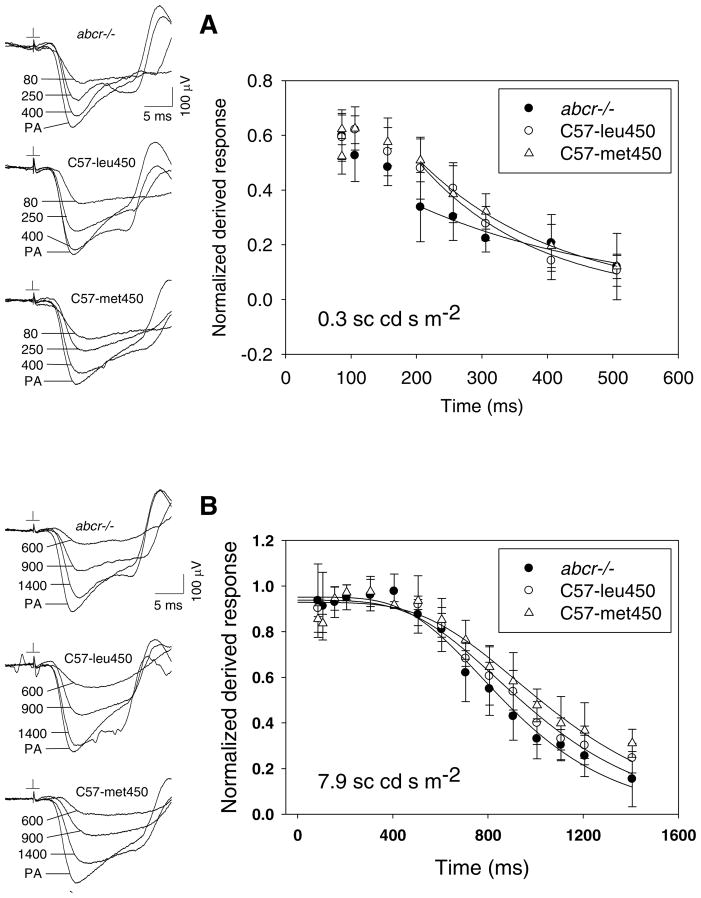
Fig. 2A shows the recovery time course of the derived response to a brief flash (0.3 sc cd s m−2) estimated to produce a fractional bleach of ~10−6. The strength of this flash was identical to that used to measure weak-flash sensitivity under dark-adapted conditions (see above). Waveforms at the left in Fig. 2A show probe responses obtained in paired-flash experiments on abcr−/−, C57-leu450 and C57-met450 mice. To quantify the time course of recovery of the paired-flash-derived response, determinations of the normalized response A(t)/AmoD beginning at tprobe = 200 ms (i.e., t = 206 ms) were analyzed in relation to the exponential decay function (curves in Fig. 2A)
Fig. 2.
Recovery following relatively weak bleaching illumination. A: Bleaching flash of 0.3 sc cd s m−2 (~10−6 fractional bleach). Left: paired-flash data obtained in single representative experiments. Labels indicate values of tprobe; PA is the dark-adapted probe-alone response. Right: aggregate results obtained from 3 abcr−/− mice, 4 C57-leu450 mice, and 3 C57-met450 mice. The curves illustrate the fitting of eq. 2 to data obtained with tprobe ≥ 200 ms. B: Bleaching flash of 7.9 sc cd s m−2 (~3×10⁻⁵ fractional bleach): Left: data obtained in single representative experiments. Right: Aggregate results obtained from 5 abcr−/−, 3 C57-leu450 and 5 C57-met450 mice. The curves illustrate the fitting of eq. 3 to the data.
| (2) |
where the dimensionless parameter η and the exponential time constant τr are free parameters. Determinations of the recovery time constant τr in these ~10−6 bleach experiments (numbers of experiments and animal ages shown in column 4 of Table 1 sections A–C) are summarized in column 4 of Table 1 sections D–F. These data are organized to indicate results obtained from the fitting of eq. 2, both to data from individual experiments and to the aggregate data set obtained from a given strain. Thus, for example, the column 4 data in Table 1 section D indicate, for abcr−/− mice, the mean ± SD of τr values obtained in individual experiments (upper entry) and the single τr value determined from eq. 2 fitting to the aggregate data set (lower entry). Accompanying the aggregate best-fit value is the corresponding goodness of fit (R2) value. Among the investigated strains, average values of τr determined from the individual fits ranged from 171 to 319 ms and corresponded closely with the aggregate fitted values of τr. Between-samples ANOVA of τr values indicated a significant difference [F(2,7) = 20.517; P = 0.001]. Post-hoc pair-wise comparisons of the data showed that τr for abcr−/− mice significantly exceeded those for both C57-leu450 mice (P <0.001) and C57-met450 mice (P = 0.004). There was no significant difference between τr values for C57-leu450 vs. C57-met450 mice. For the three investigated strains, values of η (eq. 2) were 0.34 ± 0.07 (abcr−/−), 0.51 ± 0.07 (C57-leu450), and 0.50 ± 0.04 (C57-met450).
Fig. 2B shows recovery results obtained with the 7.9 sc cd s m−2 flash. This flash, of strength sufficient to saturate the rod response (38), produced a fractional bleach of ~3×10− 5. Recovery data obtained in these experiments were analyzed in relation to a nested exponential function similar to those used previously (38,39):
| (3) |
where the dimensionless parameters θ1and θ2, and the recovery time constant τω are free parameters. Column 5 of Table 1 sections D–F summarizes the results obtained. Here, the upper entry in a given row is the mean ± SD for the recovery time constant τω based on the fitting of eq. 3 to individual data sets; the lower entry indicates the value of τω obtained by fitting eq. 3 to the aggregate data set for a given strain. As illustrated in Fig. 2B, aggregate recovery data obtained from the three investigated strains exhibited a generally similar pattern, although recovery in C57-met450 mice was on average somewhat slower than that in abcr−/− and C57-leu450 mice. ANOVA of the values of τω (column 5 of Table 1 sections D–F) indicated a significant difference [F(2,10) = 4.808; P = 0.034]. Post-hoc pair-wise comparisons showed a significant difference between abcr−/− and C57-met450 (P = 0.011); no significant difference between abcr−/− and C57-leu450; and no significant difference between C57-leu450 and C57-met450. Values of θ1 and θ2 (eq. 3) for the three strains were, respectively, 0.96 ± 0.03 and 11.76 ± 4.28 (abcr−/−); 0.93 ± 0.02 and 10.67 ± 0.51 (C57-leu450); and 0.92 ± 0.02 and 11.07 ± 3.38 (C57-met450).
~3% bleach
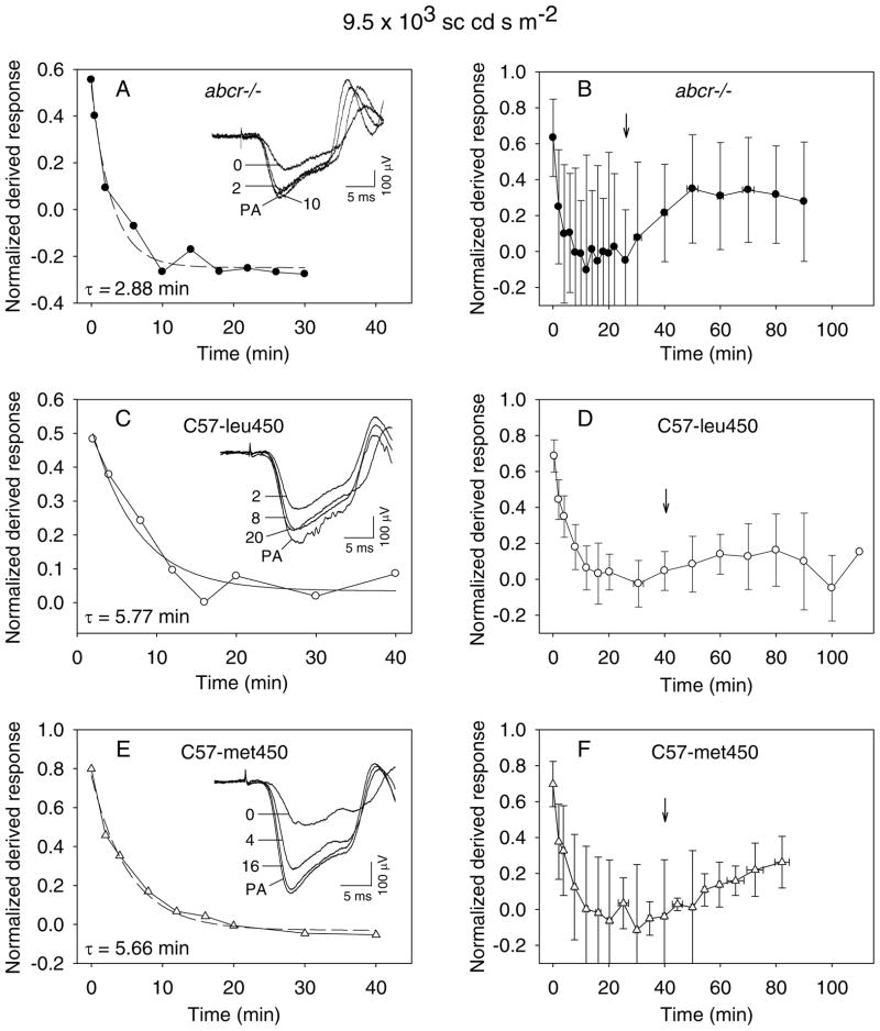
Figs. 3A, C and E show results from single representative experiments on abcr−/−, C57-leu450 and C57-met450 mice, respectively, that involved a cumulative luminance of 9.5×10³ sc cd s m−2 (series of bright flashes delivered over an approximately 100-s period; see Methods) and produced a fractional bleach of ~0.03. Figs. 3B, D and F show aggregate results obtained in a total of 8 experiments on abcr−/− mice, 8 on C57-leu450 mice, and 9 on C57-met450 mice (column 6 of Table 1 sections A–C). In these experiments, the normalized derived response determined after the bleaching illumination declined toward pre-bleach baseline, and in a number of cases exhibited an overshoot, i.e., an amplitude that exceeded the dark-adapted amplitude AmoD. A similar overshoot in the post-bleach recovery of wildtype rods has previously been reported (43; also cf. 44). To quantify the results obtained, data collected in each experiment (which typically differed in post-bleach times of determination of the derived response) were linearly interpolated to yield values of A(t)/AmoD at post-bleach times of 2, 3…30 min and (for a subset of the data set) at times 31, 32…60 min. Repeated-measures ANOVA demonstrated a significant interaction between strains as a function of time for the 2–30 min interval [F(56,616) = 3.934; P < 0.001]. Post-hoc tests showed a significant difference between abcr−/− and C57-leu450 and between abcr−/− and C57-met450, but no significant difference between C57-leu450 and C57-met450. Unlike the case of the 2–30 min interval, repeated-measures ANOVA indicated no significant difference among the investigated strains for the 31–60 min interval.
Fig. 3.
Recovery following ~3% bleach. A, C and E: Recovery data obtained in single experiments on abcr−/−, C57-leu450 and C57-met450 mice, respectively. Insets: Representative responses to the probe flash. Response PA: dark-adapted probe-alone response. Dashed curve: simple exponential function fitted to the data obtained from the time of conclusion of the bleaching light (time zero to through the apparent plateau of recovery (see text). B, D and F: Aggregate data obtained in groups of experiments on abcr−/−, C57-leu450 and C57-met450 mice respectively. In cases where data obtained at different values of tprobe were binned, the abscissa value and horizontal error bar of the illustrated data point represents the mean ± SD of the binned tprobe values. The vertical arrow in each panel identifies the conclusion of the period over which data were analyzed in relation to the exponential function.
The recovery time course in these ~3% bleach experiments was further characterized by determinations of a characteristic time constant, τ. In the first of two methods used, data obtained in a single given experiment were analyzed by fitting a simple exponential function,
| (4) |
where α, β and τ are free parameters, to recovery data obtained between time zero (i.e., the time of conclusion of the bleaching illumination) and post-bleach time T. The value of T was chosen based on visual inspection of the data, and corresponded with the time of completion of a visually apparent plateau in the derived response amplitude. Among experiments, values of the selected period T were, respectively, 29.75 ± 10.28 min (abcr−/−), 42.50 ± 8.86 min (C57-leu450), and 40.78 ± 14.65 min (C57-met450). Overall results obtained for the recovery time constant τ are shown by the upper entries in column 6 of Table 1 sections D–F. The average value of τ determined for abcr−/− mice (2.34 min) was substantially less than that determined for C57-leu450 and C57-met450 mice (5.36 min and 5.92 min, respectively). ANOVA of these determinations of τ indicated a significant difference between strains [F(2,22) = 7.144; P = 0.004]. Furthermore, post-hoc comparisons between the strains indicated a significant difference between the abcr−/− and C57-leu450 mice (P = 0.008), and between abcr−/− and C57-met450 mice (P=0.002). There was no significant difference between C57-leu450 and C57-met450 mice. The second of the two analysis methods involved the fitting of eq. (4) to aggregate data obtained from a given strain (Figs. 3B, D and F). Vertical arrows in panels B, D and F indicate the conclusions of the post-bleach periods used for this analysis of aggregate data, and lower entries in column 6 of Table 1 sections D–F show the resulting values of τ obtained. These aggregate-fit values of τ corresponded closely with the average values determined by fitting to the individual data sets.
Aggregate data obtained from all three strains (Figs. 3B, D and F) exhibited an upward trend of the derived response in the later phase of the experiments, i.e., a positive-directed trend that opposed the downward-directed recovery process. This upward-directed process, if of sufficient magnitude, might be anticipated to skew determination of (i.e., lead to underestimation of) the recovery time constant τ. However, ANOVA of the values of the excursion β of the simple exponential function fitted to individual data sets (eq. 4) (values of β: 0.68 ± 0.26 for abcr−/−; 0.74 ± 0.17 for C57-leu450; and 0.77 ± 0.27 for C57-met450) showed no significant differences [F(2,22) = 0.252; P = 0.779], and post-hoc pair-wise comparisons indicated no significant differences. In addition, aggregate data for the y-intercept of this fitted exponential function [i.e., for the time zero value, given by the sum (α + β)] were similar among strains [0.66 ± 0.22 (abcr−/−), 0.74 ± 0.10 (C57-leu450), and 0.68 ± 0.12 (C57-met450)]. Thus, although the basis of the opposing process remains unclear, the similarities of the excursion β and of the y-intercept (α + β) among strains suggest that this process operates in similar fashion among strains and does not account for the relatively fast recovery determined for the abcr−/−.
~30–40% bleach
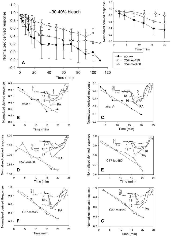
Fig. 4 shows overall results obtained in experiments with the 2-min illumination that bleached ~30–40% of the rhodopsin. Panel A shows aggregate results obtained from the investigated strains over periods that ranged up to about 100–110 min after the bleach. As illustrated by Fig. 4A, recovery in abcr−/− mice was on average faster than those in C57-leu450 and C57-met450 mice. The initial ~12–20 min of recovery determined in the investigated strains was further analyzed by determining the slope of a straight line
Fig. 4.
Recovery following ~30–40% bleach. A: Aggregate data obtained from 4 abcr−/−, 4 C57-leu450, and 5 C57-met450 mice. Inset: Panel A data illustrated on a faster time scale. B–G: Data obtained in representative single experiments on abcr−/− (panels B–C), C57-leu450 (D–E) and C57-met450 (F–G) mice over the first ~12–20 min after conclusion of the bleaching illumination. Each panel illustrates the fitting of a straight line to the data. Insets: paired-flash data obtained in representative single experiments. Labels indicate the post-bleach time, in min. PA: dark-adapted probe-alone response.
| (5) |
where the slope σ (in units of inverse time) and the dimensionless intercept ψ are free parameters fitted to the data obtained over the initial ~12–20 min post-bleach period. Figs. 4B–G illustrate results obtained in representative single experiments on abcr−/− (panels B-C), C57-leu450 (D-E) and C57-met450 (F-G) mice, and data for the determinations of slope are summarized in column 7 of Table 1 sections D–F. ANOVA of the values of the slope σ indicated no significant differences among the investigated strains [F(2,10) = 3.420; P = 0.074]. However, post-hoc pair-wise comparisons showed that slopes determined for abcr−/− differed significantly from those of C57-leu450 mice (P = 0.026). There was no significant difference between either abcr−/− and C57-met450 mice, or between C57-leu450 and C57-met450 mice. For the three investigated strains, values of the y-intercept Ψ of the fitted linear functions were 0.88 ± 0.12 (abcr−/−), 0.92 ± 0.08 (C57-leu450), and 0.97 ± 0.04 (C57-met450). ANOVA of the values of Ψ yielded no significant differences among strains [F(2,10) = 1.516; P = 0.266], and post-hoc pair-wise comparisons also indicated no significant differences between strains.
Age dependence
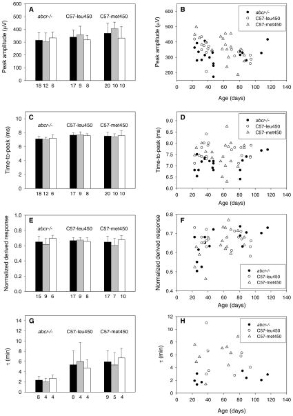
The preceding sections have considered rod response properties of abcr−/−, C57-leu450 and C57-met450 mice, independent of the age of the animals. To investigate the possibility of a difference in properties exhibited by older vs. younger mice, we separately grouped and analyzed data obtained from mice <2 months of age (1–59 days) and ≥2 months (60 days and greater) of age. The histograms of Figs. 5A, C, E and G summarize data for these two sub-groups with respect to Apeak (panel A) and tpeak (panel C) of the dark-adapted response to the probe flash; to the normalized weak-flash response at t = 86 ms (panel E); and to values of the recovery time parameter τ determined with ~3% bleaching (panel G). The triplets of histograms within each of these panels describe results obtained from abcr−/−, C57-leu450, and C57-met450 mice. The filled bar within each triplet indicates the overall (i.e., age-independent) result obtained and is identical to that described in the corresponding column of Table 1 sections D–F. The shaded and open bars of each triplet indicate results for mice of age <2 months and ≥2 months, respectively. Beneath each histogram bar is the number of mice from which the data were obtained. The scatter plots of Figs. 5B, D, F and H provide further description of the data sets summarized in the histograms. These scatter plots show, as a function of age, and for each of the investigated mice, the value of the parameter considered in the accompanying left-hand histogram. Two-way ANOVA with age (<2 months vs. ≥2 months) and strain as between-sample factors indicated no significant differences for either Apeak or tpeak. There was a significant difference for the normalized derived response [F(2,49) = 3.446; P = 0.041]. Among abcr−/− mice, ANOVA for <2-month vs. ≥2-month animals yielded a significant difference only for the normalized derived response [F(1,13) = 5.841; P = 0.031) and for τ [F(1,6) = 18.273; P. = 0.005]. For C57-leu450 mice, there was no significant effect of age on any of the parameters described in Fig. 5. For C57-met450 mice, there was a significant effect of age only for Apeak [F(1,18) = 5.340; P = 0.033). Across strains, ANOVA of data obtained from a given age group yielded, for <2-month mice, a significant difference only for Apeak [F(2,28) = 7.399; P = 0.003] and tpeak [F(2,28) = 8.236; P = 0.002]. For ≥2-month mice, there was no significant effect for any of the investigated parameters. Post-hoc pair-wise comparisons indicated, for <2 month abcr−/− vs. C57-leu450 mice, a significant difference in tpeak (P = 0.001) and a marginal effect with respect to normalized dark-adapted derived response (P = 0.051). For ≥2-month abcr−/−vs. C57-leu450 mice, there were no significant differences. For ≥2-month abcr−/−vs. C57-met450 mice, there was a significant effect only with respect to Apeak (P = 0.001) and tpeak (P =0.016). For ≥2-month abcr−/−vs. C57-met450 mice, there were no significant differences. For <2-month C57-leu450 vs. C57-met450 mice, the only significant difference was with respect to the normalized derived response (P = 0.050). For ≥2-month C57-leu450 vs. C57-met450 mice, there were no significant differences.
Fig. 5.
Analysis of data in relation to animal age. Parameters described are: the peak amplitude Apeak of the dark-adapted response to the 773 sc cd s m−2 probe flash (panels A–B); the time-to-peak tpeak of this response (C–D); the normalized derived response to a 0.3 sc cd s m−2 test flash at t = 86 ms (E–F); and the recovery time constant τ determined with ~3% bleaching (G–H). Filled bars in the histograms of A, C, E and G replicate aggregate data reported in Table 1. Shaded and open bars represent, respectively, results obtained from animals <2 months of age and ≥2 months of age. The number beneath each histogram bar indicates the number of mice from which the data were collected. See text for further details.
DISCUSSION
The present study addresses the kinetics of rod recovery in abcr−/− vs. control mice following bleaching stimuli that correspond with fractional rhodopsin bleaches of ~10−6 to ~30–40%. The most striking difference in recovery kinetics concerns the exponential time constant that describes recovery following ~3% bleaching. As shown by Table 1 and Fig. 3, the exponential time constant that describes recovery in abcr−/− rods under this condition is about half that exhibited by C57-leu450 and C57-met450 controls. In addition, the rate of initial recovery in abcr−/− mice following ~30–40% bleaching significantly exceeds that for C57-leu450 mice and is on average faster than that in C57-met450 mice (Table 1; and Fig. 4 and accompanying text). The relatively fast recovery time course in abcr−/− mice cannot be attributed to differences among the investigated strains in pupil size, other pre-retinal factors, or absorptivity (i.e., amount) of rhodopsin in the rods. That is, such differences would be expected to have produced differences in the dark-adapted, derived rod response to a weak test flash, yet these determinations of weak-flash sensitivity were similar among abcr−/− mice and controls (Table 1). Accordingly, we interpret the significantly faster recovery kinetics observed in abcr−/− mice under the above-summarized conditions to reflect an intrinsically rapid process of post-bleach recovery in abcr−/− rods.
The relationship observed here between abcr−/− mice and wildtype controls following significant bleaching differs from that reported by Weng et al. (1). That is, the absolute time course of recovery reported by Weng et al. (1) for abcr−/− mice after a 45% bleach is comparable with that reported here following a roughly similar (~30–40%) bleach. However, by contrast with the Weng et al. (1) study, we find post-bleach rod recovery in the wildtype strains to be (for C57-met450) on average slower than, or (for C57-leu450) significantly slower than, that in abcr−/− mice after ~30–40% bleaching (present Fig. 4). In addition, the presently observed rod recoveries in both wildtype strains were substantially slower than that in abcr−/− after ~3% bleaching (see above Discussion). Conceivably, differences in the specific wildtype strains used, and perhaps also the ages of the investigated mice, could be the basis of the contrasting findings of the present study and that by Weng et al. (1).
The wildtype mice used here were pigmented C57-derived lines possessing the leu450 and met450 variants of RPE65, the isomerohydrolase of the retinoid visual cycle that promotes the conversion of all-trans retinyl ester to 11-cis retinol in the RPE (36,37). The leu450 and met450 variants of RPE65 mice are known to differ with respect to the rate of isomerohydrolase activity (35,45,46). Specifically, leu450, the RPE65 variant expressed by the presently studied abcr−/− mice, has greater catalytic activity than the met450 variant. In light of evidence that the isomerohydrolase reaction is a critical step in the retinoid visual cycle (36,37), and that rhodopsin regeneration is critical for full dark adaptation of the rods (47,48), the comparison of rod recovery in the abcr−/− with that in controls possessing both the leu450 and met450 variants of RPE65 was important to the present study. Interestingly, these two wildtype strains exhibited remarkably similar rod recovery properties under the presently investigated experimental conditions. In particular, there were no significant differences among data obtained from C57-leu450 and C57-met450 following a ~3% bleach, or during the initial ~12–20-min period following ~30–40% bleaching. These findings imply that occurrence of the leu450 variant of RPE65 in abcr−/− mice does not underlie the observed relatively rapid recovery of the abcr−/− rod response following the larger bleaches used here. Significant differences in rod recovery kinetics in albino mice possessing the leu450 vs. met450 variant of RPE65 have been reported by Nusinowitz et al. (34). Specifically, these investigators compared ERG a-wave recovery in BALB/c mice, which possess the leu450 variant, with that in c2J mice, which possess the met450 variant. For bleaching illuminations of 3.61 and 3.97 log scotopic Troland-s, the exponential time constant describing recovery in the BALB/c was, respectively, about 48% and 21% faster than in c2J mice. Furthermore, following ~80–90% bleaching, recovery of the ERG b-wave proceeded more rapidly in the BALB/c mice.
The observation of relatively fast post-bleach rod recovery in abcr−/− mice raises several interesting issues relevant to the processing of all-trans retinal in abcr−/− mice and to the activity of ABCR in wildtype mice. Previous biochemical experiments indicate that ABCR’s facilitation of all-trans retinal movement across the disk membrane contributes to the post-bleach processing of all-trans retinal in the visual cycle. For example, Weng et al. (1) found that all-trans retinal per eye in abcr−/− mice at up to 1 hr after a 45% bleach significantly exceeds that in wildtype controls. However, the magnitude of this increase, on average up to ~30 pmol per eye (Fig. 3C of 1) represents only a small fraction of the decrease in rhodopsin produced by the 45% bleach (Figs. 3A–B of 1). Thus, ABCR deficiency under the conditions investigated by Weng et al. (1) corresponds with a relatively small albeit significant prolongation of all-trans retinal clearance. Interestingly, the rod outer segments of abcr−/− mice contain an abnormally high level of phosphatidylethanolamine (PE), the lipid that combines with all-trans retinal to form N-ret-PE. A high PE level in (presumably, the disk membranes of) abcr−/− rods conceivably could underlie the present observation of relatively fast recovery kinetics in the abcr−/−. For example, the abnormally large amount of PE in abcr−/− disk membranes (either the luminal surface, the cytosolic surface, or both; cf. 49) might promote, by mass action, the sequestering of all-trans retinal as N-ret-PE at a rate considerably exceeding that in wildtype rods, thereby accelerating response recovery relative to the wildtype. Alternatively, the high level of PE in the abcr−/− rod, by binding 11-cis retinal arriving from the RPE through operation of the visual cycle, might localize 11-cis retinal to the vicinity of opsin and thereby promote rhodopsin regeneration (50). A further possibility derives from the finding that ABCR itself can bind 11-cis retinal (50). That is, the ABCR of wildtype rods, a major protein of the disk rims, might delayrecovery relative to abcr−/− by competing with opsin for 11-cis retinal and thereby slowing rhodopsin regeneration.
In summary, the present results suggest that ABCR in normally functioning rods may directly or indirectly prolong, rather than accelerate, post-bleach recovery of the rod photoresponse over much of its excursion following substantial bleaching. This notion might seem puzzling in view of the likely disadvantage, in photoreceptor evolution, of processes that slow dark adaptation. The seeming paradox is resolved, however, if the primary physiological role of the ABCR-mediated reaction is to promote clearance, from the disk lumen, of minute, residual amounts of all-trans retinal that other mechanisms, such as thermal diffusion across the disk membrane, cannot achieve. That is, the ABCR-mediated reaction may have little if any accelerating effect on removal of the major portion of bleach-generated all-trans retinal and, thus, on the bulk removal of MII-like complexes of all-trans retinal and opsin. Rather, ABCR’s essential function may be to eliminate trace amounts of all-trans retinal from the disk lumen and thereby oppose the build-up, over the lifetime of the rod disk (51), of retinoid-based compounds that otherwise would be transferred to the RPE and there accumulate as A2E and other retinoid-based components of lipofuscin (18). On this hypothesis, the slower rod recovery observed in normal rods upon the bleach-induced elevation of all-trans retinal in the disk lumen represents a cost, or trade-off, associated with the presence of a system (ABCR) that can clear tiny remaining amounts of all-trans retinal. Beyond its consistency with the observed modest difference in post-bleach all-trans retinal levels in ABCR-deficient vs. wildtype rods (1), this hypothesis is consistent with the finding that abnormal A2E build-up in abcr−/− mice amounts, on average, to only several tens of pmol per eye [about 21 pmol per eye over 4–5 months (1); about 30 pmol per eye over 8–9 months (45)], a molar quantity small by comparison with, e.g., the amount of 11-cis retinal present as rhodopsin chromophore in fully dark-adapted rods (42,52). The hypothesis is also consistent with the near-normal course of rod recovery frequently observed in human subjects with Stargardt disease and an ABCA4 (i.e., ABCR) mutation (25,26), and with the prolongation, in these subjects, of primarily the final, tail phase of psychophysically measured dark adaptation following major bleaching of the rhodopsin (Fig. 9 of 25).
Acknowledgments
The authors thank Drs. Gabriel H. Travis, Michael Danciger, Theodore G. Wensel, Steven Nusinowitz, David G. Birch and Gerald A. Fishman for helpful discussions during the course of this study. Supported by NIH grants EY005494, EY016094, EY001792 and AG028662; by grants from the Daniel F. and Ada L. Rice Foundation (Skokie, IL), the Macular Degeneration Program of the American Health Assistance Foundation (Clarksburg, MD), and the CINN Foundation (Chicago, IL); and by an unrestricted departmental award from Research to Prevent Blindness, Inc (New York, NY). D.R.P. is a Senior Scientific Investigator of Research to Prevent Blindness.
Footnotes
Commercial Relationships: None.
References
- 1.Weng J, Mata NL, Azarian SM, Tzekov RT, Birch DG, Travis GH. Insights into the function of Rim protein in photoreceptors and etiology of Stargardt’s disease from the phenotype in abcr knockout mice. Cell. 1999;98:13–23. doi: 10.1016/S0092-8674(00)80602-9. [DOI] [PubMed] [Google Scholar]
- 2.Ahn J, Wong JT, Molday RS. The effect of lipid environment and retinoids on the ATPase activity of ABCR, the photoreceptor ABC transporter responsible for Stargardt macular dystrophy. J Biol Chem. 2000;275:20399–20405. doi: 10.1074/jbc.M000555200. [DOI] [PubMed] [Google Scholar]
- 3.Mata NL, Weng J, Travis GH. Biosynthesis of a major lipofuscin fluorophore in mice and humans with ABCR-mediated retinal and macular degeneration. Proc Natl Acad Sci USA. 2000;97:7154–7159. doi: 10.1073/pnas.130110497. [DOI] [PMC free article] [PubMed] [Google Scholar]
- 4.Sun H, Nathans J. Mechanistic studies of ABCR, the ABC transporter in photoreceptor outer segments responsible for autosomal recessive Stargardt disease. J Bioenerg Biomembr. 2001;33:523–530. doi: 10.1023/a:1012883306823. [DOI] [PubMed] [Google Scholar]
- 5.Ahn J, Beharry S, Molday LL, Molday RS. Functional interaction between the two halves of the photoreceptor-specific ATP binding cassette protein ABCR (ABCA4): evidence for a non-exchangeable ADP in the first nucleotide binding domain. J Biol Chem. 2003;278:39600–39608. doi: 10.1074/jbc.M304236200. [DOI] [PubMed] [Google Scholar]
- 6.Biswas-Fiss EE. Interaction of the nucleotide binding domains and regulation of the ATPase activity of the human retina specific ABC transporter, ABCR. Biochemistry. 2006;45:3813–3823. doi: 10.1021/bi052059u. [DOI] [PubMed] [Google Scholar]
- 7.Molday RS. ATP-binding cassette transporter ABCA4: Molecular properties and role in vision and macular degeneration. J Bioenerg Biomembr. 2007 Nov 10; doi: 10.1007/s10863-007-9118-6. [Epub ahead of print] [DOI] [PubMed] [Google Scholar]
- 8.Saari JC. Biochemistry of visual pigment regeneration: the Friedenwald lecture. Invest Ophthalmol Vis Sci. 2000;41:337–348. [PubMed] [Google Scholar]
- 9.Rando RR. The biochemistry of the visual cycle. Chem Rev. 2001;101:1881–1896. doi: 10.1021/cr960141c. [DOI] [PubMed] [Google Scholar]
- 10.McBee JK, Palczewski K, Baehr W, Pepperberg DR. Confronting complexity; the interlink of phototransduction and retinoid metabolism in the vertebrate retina. Prog Retinal Eye Res. 2001;20:469–529. doi: 10.1016/s1350-9462(01)00002-7. [DOI] [PubMed] [Google Scholar]
- 11.Lamb TD, Pugh EN., Jr Dark adaptation and the retinoid cycle of vision. Progr Retinal Eye Res. 2004;23:307–380. doi: 10.1016/j.preteyeres.2004.03.001. [DOI] [PubMed] [Google Scholar]
- 12.Hofmann KP, Pulvermuller A, Buczylko J, Van Hooser P, Palczewski K. The role of arrestin and retinoids in the regeneration pathway of rhodopsin. J Biol Chem. 1992;267:15701–15706. [PubMed] [Google Scholar]
- 13.Jager S, Palczewski K, Hofmann KP. Opsin/all-trans-retinal complex activates transducin by different mechanisms than photolyzed rhodopsin. Biochemistry. 1996;35:2901–2908. doi: 10.1021/bi9524068. [DOI] [PubMed] [Google Scholar]
- 14.Surya A, Knox BE. Enhancement of opsin activity by all-trans-retinal. Exper Eye Res. 1998;66:599–603. doi: 10.1006/exer.1997.0453. [DOI] [PubMed] [Google Scholar]
- 15.Sachs K, Maretzki D, Meyer CK, Hofmann KP. Diffusible ligand all-trans-retinal activates opsin via a palmitoylation-dependent mechanism. J Biol Chem. 2000;275:6189–6194. doi: 10.1074/jbc.275.9.6189. [DOI] [PubMed] [Google Scholar]
- 16.Ben-Shabat S, Parish CA, Vollmer HR, et al. Biosynthetic studies of A2E, a major fluorophore of retinal pigment epithelial lipofuscin. J Biol Chem. 2002;222:7183–7190. doi: 10.1074/jbc.M108981200. [DOI] [PubMed] [Google Scholar]
- 17.Fishkin NE, Sparrow JR, Allikmets R, Nakanishi K. Isolation and characterization of a retinal pigment epithelial cell fluorophore: an all-trans-retinal dimer conjugate. Proc Natl Acad Sci USA. 2005;102:7091–7096. doi: 10.1073/pnas.0501266102. [DOI] [PMC free article] [PubMed] [Google Scholar]
- 18.Sparrow JR, Boulton M. RPE lipofuscin and its role in retinal pathobiology. Exper Eye Res. 2005;80:595–606. doi: 10.1016/j.exer.2005.01.007. [DOI] [PubMed] [Google Scholar]
- 19.Allikmets R, Shroyer NF, Singh N, et al. Mutation of the Stargardt disease gene (ABCR) in age-related macular degeneration. Science. 1997;277:1805–1807. doi: 10.1126/science.277.5333.1805. [DOI] [PubMed] [Google Scholar]
- 20.Cremers FP, van de Pol DJ, van Driel M, et al. Autosomal recessive retinitis pigmentosa and cone-rod dystrophy caused by splice site mutations in the Stargardt’s disease gene ABCR. Hum Mol Genet. 1998;7:355–362. doi: 10.1093/hmg/7.3.355. [DOI] [PubMed] [Google Scholar]
- 21.Allikmets R. Simple and complex ABCR: genetic predisposition to retinal disease. Am J Hum Genet. 2000;67:793–799. doi: 10.1086/303100. [DOI] [PMC free article] [PubMed] [Google Scholar]
- 22.Birch DG, Peters AY, Locke KL, Spencer R, Megarity CF, Travis GH. Visual function in patients with cone-rod dystrophy (CRD) associated with mutations in the ABCA4 (ABCR) gene. Exper Eye Res. 2001;73:877–886. doi: 10.1006/exer.2001.1093. [DOI] [PubMed] [Google Scholar]
- 23.Shroyer NF, Lewis RA, Yatsenko AN, Wensel TG, Lupski JR. Cosegregation and functional analysis of mutant ABCR (ABCA4) alleles in families that manifest both Stargardt disease and age-related macular degeneration. Human Molec Genet. 2001;10:2671–2678. doi: 10.1093/hmg/10.23.2671. [DOI] [PubMed] [Google Scholar]
- 24.Fishman GA, Stone EM, Eliason DA, Taylor CM, Lindeman M, Derlacki DJ. ABCA4 gene sequence variations in patients with autosomal recessive cone-rod dystrophy. Arch Ophthalmol. 2003;121:851–855. doi: 10.1001/archopht.121.6.851. [DOI] [PubMed] [Google Scholar]
- 25.Kang Derwent JJ, Derlacki DJ, Hetling JR, et al. Dark adaptation of rod photoreceptors in normal subjects, and in patients with Stargardt disease and an ABCA4 mutation. Invest Ophthalmol Vis Sci. 2004;45:2447–2456. doi: 10.1167/iovs.03-1178. [DOI] [PubMed] [Google Scholar]
- 26.Cideciyan AV, Aleman TS, Swider M, et al. Mutations in ABCA4 result in accumulation of lipofuscin before slowing of the retinoid cycle: a reappraisal of the human disease sequence. Human Molec Genet. 2004;13:525–534. doi: 10.1093/hmg/ddh048. [DOI] [PubMed] [Google Scholar]
- 27.Melia TJ, Jr, Cowan CW, Angleson JK, Wensel TG. A comparison of the efficiency of G protein activation by ligand-free and light-activated forms of rhodopsin. Biophys J. 1997;73:3182–3191. doi: 10.1016/S0006-3495(97)78344-9. [DOI] [PMC free article] [PubMed] [Google Scholar]
- 28.Firsov ML, Kolesnikov AV, Golobokova EY, Govardovskii VI. Two realms of dark adaptation. Vision Res. 2005;45:147–151. doi: 10.1016/j.visres.2004.08.005. [DOI] [PubMed] [Google Scholar]
- 29.Lyubarsky AL, Daniele LL, Pugh EN., Jr From candelas to photoisomerizations in the mouse eye by rhodopsin bleaching in situ and the light-rearing dependence of the major components of the mouse ERG. Vision Res. 2004;44:3235–3251. doi: 10.1016/j.visres.2004.09.019. [DOI] [PubMed] [Google Scholar]
- 30.Calvert PD, Govardovskii VI, Krasnoperova N, Anderson RE, Lem J, Makino CL. Membrane protein diffusion sets the speed of rod phototransduction. Nature. 2001;411:90–94. doi: 10.1038/35075083. [DOI] [PubMed] [Google Scholar]
- 31.Pawar AS, Qtaishat NM, Pepperberg DR. Photoreceptor recovery in abcr−/− and wildtype mice following weak bleaching of rhodopsin. Invest Ophthalmol Vis Sci. 2006;47 ARVO E-Abstract 4736. [Google Scholar]
- 32.Pawar AS, Qtaishat NM, Pepperberg DR. Initial recovery kinetics of post-bleach photoreceptor responses in abcr−/− mice. Invest Ophthalmol Vis Sci. 2007;48 ARVO E-Abstract 608. [Google Scholar]
- 33.Qtaishat NM, Pepperberg DR. Preservation of retinoid influx into eye tissues of ABCR- deficient mice. Current Eye Res. 2007;32:1073–1082. doi: 10.1080/02713680701715933. [DOI] [PubMed] [Google Scholar]
- 34.Nusinowitz S, Nguyen L, Radu R, Kashani Z, Farber D, Danciger M. Electroretinographic evidence for altered phototransduction gain and slowed recovery from photobleaches in albino mice with a MET450 variant in RPE65. Exper Eye Res. 2003;77:627–638. doi: 10.1016/s0014-4835(03)00217-3. [DOI] [PubMed] [Google Scholar]
- 35.Wenzel A, Reme CE, Williams TP, Hafezi F, Grimm C. The Rpe65 Leu450Met variation increases retinal resistance against light-induced degeneration by slowing rhodopsin regeneration. J Neurosci. 2001;21:53–58. doi: 10.1523/JNEUROSCI.21-01-00053.2001. [DOI] [PMC free article] [PubMed] [Google Scholar]
- 36.Moiseyev G, Chen Y, Takahashi Y, Wu BX, Ma J. RPE65 is the isomerohydrolase in the retinoid visual cycle. Proc Natl Acad Sci USA. 2005;102:12413–12418. doi: 10.1073/pnas.0503460102. [DOI] [PMC free article] [PubMed] [Google Scholar]
- 37.Redmond TM, Poliakov E, Yu S, Tsai J-Y, Lu Z, Gentleman S. Mutation of key residues of RPE65 abolishes its enzymatic role as isomerohydrolase in the visual cycle. Proc Natl Acad Sci USA. 2005;102:13658–13663. doi: 10.1073/pnas.0504167102. [DOI] [PMC free article] [PubMed] [Google Scholar]
- 38.Hetling JR, Pepperberg DR. Sensitivity and kinetics of mouse rod flash responses determined in vivo from paired-flash electroretinograms. J Physiol. 1999;516:593–609. doi: 10.1111/j.1469-7793.1999.0593v.x. [DOI] [PMC free article] [PubMed] [Google Scholar]
- 39.Kang Derwent JJ, Qtaishat NM, Pepperberg DR. Excitation and desensitization of mouse rod photoreceptors in vivo following bright adapting light. J Physiol. 2002;541:201–218. doi: 10.1113/jphysiol.2001.013227. [DOI] [PMC free article] [PubMed] [Google Scholar]
- 40.Kang Derwent JJ, Saszik SJ, Maeda H, et al. Test of the paired-flash electroretinographic method in mice lacking b-waves. Visual Neurosci. 2007;24:141–149. doi: 10.1017/S0952523807070162. [DOI] [PubMed] [Google Scholar]
- 41.Pepperberg DR. Paired-flash ERG analysis of rod phototransduction and adaptation. In: Heckenlively JR, Arden GB, editors. Principles and Practice of Clinical Electrophysiology of Vision. 2. MIT Press; Cambridge, MA: 2006. pp. 519–532. [Google Scholar]
- 42.Qtaishat NM, Okajima T-IL, Li S, Naash MI, Pepperberg DR. Retinoid kinetics in eye tissues of VPP transgenic mice and their normal littermates. Invest Ophthalmol Vis Sci. 1999;40:1040–1049. [PubMed] [Google Scholar]
- 43.Mata NL, Tzekov RT, Liu X, Weng J, Birch DG, Travis GH. Delayed dark-adaptation and lipofuscin accumulation in abcr+/− mice: implications for involvement of ABCR in age- related macular degeneration. Invest Ophthalmol Vis Sci. 2001;42:1685–1690. [PubMed] [Google Scholar]
- 44.Xu JW, Hou M, Slaughter MM. Photoreceptor encoding of supersaturating light stimuli in salamander retina. J Physiol. 2005;569:575–585. doi: 10.1113/jphysiol.2005.092239. [DOI] [PMC free article] [PubMed] [Google Scholar]
- 45.Kim SR, Fishkin N, Kong J, Nakanishi K, Allikmets R, Sparrow JR. Rpe65 Leu450Met variant is associated with reduced levels of the retinal pigment epithelium lipofuscin fluorophores A2E and iso-A2E. Proc Natl Acad Sci USA. 2004;101:11668–11672. doi: 10.1073/pnas.0403499101. [DOI] [PMC free article] [PubMed] [Google Scholar]
- 46.Lyubarsky AL, Savchenko AB, Morocco SB, Daniele LL, Redmond TM, Pugh EN., Jr Mole quantity of RPE65 and its productivity in the generation of 11-cis-retinal from retinyl esters in the living mouse eye. Biochemistry. 2005;44:9880–9888. doi: 10.1021/bi0505363. [DOI] [PubMed] [Google Scholar]
- 47.Pepperberg DR, Brown PK, Lurie M, Dowling JE. Visual pigment and photoreceptor sensitivity in the isolated skate retina. J Gen Physiol. 1978;71:369–396. doi: 10.1085/jgp.71.4.369. [DOI] [PMC free article] [PubMed] [Google Scholar]
- 48.Cornwall MC, Fain GL. Bleached pigment activates transduction in isolated rods of the salamander retina. J Physiol. 1994;480:261–79. doi: 10.1113/jphysiol.1994.sp020358. [DOI] [PMC free article] [PubMed] [Google Scholar]
- 49.Wu G, Hubbell WL. Phospholipid asymmetry and transmembrane diffusion in photoreceptor disc membranes. Biochemistry. 1993;32:879–888. doi: 10.1021/bi00054a020. [DOI] [PubMed] [Google Scholar]
- 50.Sun H, Molday RS, Nathans J. Retinal stimulates ATP hydrolysis by purified and reconstituted ABCR, the photoreceptor-specific ATP-binding cassette transporter responsible for Stargardt disease. J Biol Chem. 1999;274:8269–8281. doi: 10.1074/jbc.274.12.8269. [DOI] [PubMed] [Google Scholar]
- 51.Young RW, Bok D. Participation of the retinal pigment epithelium in the rod outer segment renewal process. J Cell Biology. 1969;42:392–403. doi: 10.1083/jcb.42.2.392. [DOI] [PMC free article] [PubMed] [Google Scholar]
- 52.Saari JC, Garwin GG, Van Hooser JP, Palczewski K. Reduction of all-trans-retinal limits regeneration of visual pigment in mice. Vision Res. 1998;38:1325–1333. doi: 10.1016/s0042-6989(97)00198-3. [DOI] [PubMed] [Google Scholar]


