Abstract
Purpose
The objective of this study was to determine if biomechanical and neuromuscular risk factors related to abnormal movement patterns increased in females, but not males, during the adolescent growth spurt.
Methods
315 subjects participated in two testing sessions approximately one year apart. Male and female subjects were classified based on their maturation status as pubertal or post-pubertal. Three trials of a drop vertical jump (DVJ) were collected. Maximum knee abduction angle and external moments were calculated during the DVJ deceleration phase using a 3D motion analysis system. Changes in knee abduction from the first to second year were compared among four subject groups (female pubertal, female post-pubertal, male pubertal and male post-pubertal).
Results
There were no sex differences in peak knee abduction angle or moment during DVJ between pubertal males and females (p>0.05). However, pubertal females increased peak abduction angle from the first to second year (p<0.001), while males demonstrated no similar change (p=0.90) in the matched developmental stages. Following puberty, the peak abduction angle and moment were greater in females relative to males (angle: female -9.3±5.7°, male -3.6±4.6°, p<0.001; moment: female:-21.9±13.5 Nm, male:-13.0±12.0 Nm, p=0.017).
Conclusion
This study identified, through longitudinal analyses, that knee abduction angle was significantly increased in pubertal females during rapid adolescent growth, while males showed no similar change. In addition, knee abduction motion and moments were significantly greater for subsequent year in young female athletes, following rapid adolescent growth, compared to males. The combination of longitudinal, sex and maturational group differences indicate that early puberty appears to be a critical phase related to the divergence of increased ACL injury risk factors.
Keywords: anterior cruciate ligament injury, female, drop vertical jump landing, ACL injury risk factor
Introduction
Biomechanical and neuromuscular risk factors may be critical components of the mechanisms which underlie the higher rate of ACL injuries in female compared to male athletes. Specifically, altered movement patterns such as knee abduction (valgus) may relate to an increase ACL injury risk.(18) Several studies have shown increased knee abduction motion and load in females compared to males during a variety of landing and pivoting movements.(7, 12-14, 16-17, 22, 27) Similar lower extremity abduction postures have been reported in females at the time of injury.(24, 30) In addition, a prospective combined biomechanical-epidemiologic study showed that knee abduction moments (valgus torques) and angles were significant predictors of future ACL injury risk. Knee abduction moments predicted ACL injury risk with 73% sensitivity and 78% specificity.(18) Knee abduction angles were 8 degrees greater in the ACL-injured than the uninjured groups. It is therefore likely that the increases in knee abduction moment and motion in the injured group were a significant risk factor that may have predisposed these athletes to ACL injury.
The onset of these neuromuscular risk factors may coincide with rapid adolescent growth that results in the divergence of a multitude of biomechanical and neuromuscular parameters between sexes. There is little if any evidence which demonstrates a sex difference in ACL injury rates in pre-pubescent athletes, which is in direct contrast to injury rates in adolescent and adult populations.(1) Although ACL injuries increase with age in both males and females, females have higher rates immediately following the growth spurt.(37) Following an adolescent growth spurt, increased body mass and height of the center of mass may lead to altered movement patterns.(17) Musculoskeletal growth during puberty, in the absence of corresponding neuromuscular adaptation, may facilitate the development of certain intrinsic risk factors.(17) These intrinsic risk factors, if not addressed at the proper time period, may continue through adolescence into maturity and predispose athletes to ACL injuries. The differences in neuromuscular performance between the sexes during and following puberty may contribute to altered biomechanics and resultant forces on the knee. These excessive knee loads, especially in the frontal plane may explain the increased risk of ACL injury in females following puberty and may help identify the optimal time to implement injury prevention programs. Therefore, the timing of adolescent growth may be the critical phase of growth and development related to sex differences in ACL injury risk. However, the effects of rapid growth and development in females on ACL risk factors is currently unknown.
The purpose of this study was to determine if neuromuscular risk factors related to abnormal movement patterns increase in females, but not males, during the adolescent growth spurt. The hypothesis was that during adolescent growth, pubertal females would demonstrate longitudinal increases in knee abduction moments and motion compared to pubertal males. In addition, we hypothesized that following the adolescent growth spurt, post-pubertal females would have significantly greater knee abduction moments and motion than post-pubertal males.
Methods
Subjects
A nested cohort design (total sample female n = 709; total sample male n = 250) was used to identify 401 subjects that had consecutive test session approximately one year apart. The final study population consisted of 315 pubertal and post-pubertal subjects (pre-pubertal subjects were excluded). The maturational status, either pubertal (n = 182) or post-pubertal (n = 133), was based on the modified Pubertal Maturational Observational Scale (PMOS).(9, 32) The modified PMOS incorporates both parental questionnaires and investigator observations to classify subjects into the pubertal categories. The PMOS scale has shown high reliability and can be used to differentiate between pubertal stages.(9, 17, 31) The maturation observation checklist specifically included items related to acne, recent growth spurt, sweating after physical activities, evidence of muscular development and darkened underarm hair (or shaves). Female specific criteria consisted of menarche, breast development and darkened leg hair (or shaves) while male criteria were evidence of facial hair (or shaves) and deepening voice (or currently breaking). If the subject scored a 1 or less on the checklist they were excluded from the current study. If the subject scored a two through five in either the first or second year of testing they were operationally categorized as pubertal. Subjects classified as post-pubertal (at least six characteristics) had to meet the post-pubertal criteria on the first year of testing. The subjects in this study involved one school district with a matched school for male and female athletics in order to control for potential differences in sport exposure and level of play. The level of sports play was similar across the male and female teams (i.e. Varsity, Junior Varsity etc.).
Height was measured with a stadiometer with the subject in bare feet. Body mass was collected on a calibrated physician scale. Detailed demographics of both the male and female subjects are included in Table 1. Each subject participated in the first testing session immediately prior to their basketball or soccer season. Approximately one year after the initial testing session (mean 365.7 ± 14.7 days) the subjects were retested using the same methods. Subjects were excluded from this study if they had previous history of knee or ankle surgery.. Regression equations were used to estimate adult stature for female and males.(23)
Table 1.
Mean (±SD) subject demographics
| Pubertal | Post-Pubertal | ||||
|---|---|---|---|---|---|
| Variable | Test Session | Female | Male | Female | Male |
| Age (yrs) | Year 1 | 12.3 ± 0.8 | 13.0 ± 1.1 | 14.4 ± 1.4 | 15.1 ± 1.1 |
| Year 2 | 13.3 ± 0.8 | 14.0 ± 1.1 | 15.4 ± 1.4 | 16.1 ± 1.1 | |
| Height (cm) | Year 1 | 155.9 ± 6.8 | 165.2 ± 10.2 | 164.4 ± 5.8 | 180.8 ± 7.9 |
| Year 2 | 160.7 ± 5.9 | 171.8 ± 9.2 | 165.2 ± 5.8 | 182.5 ± 7.6 | |
| % Change | 3.1 ± 1.6 | 4.1 ± 1.8 | 0.5 ± 0.9 | 0.9 ± 1.2 | |
| Mass (kg) | Year 1 | 47.8 ± 10.2 | 54.5 ± 10.2 | 59.0 ± 8.5 | 70.1 ± 8.4 |
| Year 2 | 52.7 ± 9.9 | 61.1 ± 10.0 | 60.9 ± 8.7 | 74.9 ± 7.6 | |
| % Change | 11.1 ± 7.3 | 12.8 ± 7.2 | 3.4 ± 5.7 | 7.2 ± 3.0 | |
The data collection procedures were approved by Cincinnati Children's Hospital Institutional Review Board. Each parent or guardian reviewed and signed the Institutional Review Board approved written informed consent to participate form prior to data collection. Child assent was also obtained from each subject prior to study participation.
Procedures
Each subject was instrumented with 37 retroreflective markers placed bilaterally on the shoulder, elbow, wrist, ASIS, greater trochanter, thigh, medial and lateral knee, tibial tubercle, shank, distal shank, medial and lateral ankle, heel, dorsal surface of the midfoot, lateral foot (5th metarsal) and toe (between 2nd and 3rd metatarsals) in addition to the sacrum, left PSIS and sternum. A static trial was collected in which the subject was instructed to stand still in the anatomical position with foot placement standardized. This static measurement was used as each subject's neutral (zero) alignment. Each subject performed three trials of the drop vertical jump (DVJ). The DVJ consisted of the subject starting on top of a 31 cm box with their feet positioned 35 cm apart and arms held comfortably at their side. They were instructed to drop directly down off the box and immediately perform a maximum vertical jump, raising both arms as if they were jumping for a basketball rebound.(12)
Trials were collected with EVaRT (Version 4, Motion Analysis Corporation, Santa Rosa, CA) using a motion analysis system consisting of eight digital cameras (Eagle cameras, Motion Analysis Corporation, Santa Rosa, CA) positioned in the laboratory and sampled at 240 Hz. Prior to data collection the motion analysis system was calibrated with a two-step process, first using a static calibration frame to orient the cameras with respect to the laboratory coordinate system and second using dynamic wand data to fine tune camera positions, calculate the lens distortion maps and calculate the lens focal length. Two force platforms (AMTI, Watertown, MA) were sampled at 1200 Hz and time synchronized with the motion analysis system. The force platforms were embedded into the floor and positioned 8 cm apart so that each foot would contact a different platform during the stance phase of the DVJ.
Data Analysis
Following data collection, 3D marker trajectories were examined for accurate marker identification within EVaRT (Version 4, Motion Analysis Corporation, Santa Rosa, CA) and exported to a C3D formatted file. The C3D files were then further analyzed in Visual3d (Version 4.0, C-Motion, Inc., Germantown, MD). The procedures within Visual3D first consisted of the development of a static model customized for each subject. The model's coordinate system convention was +X towards the subject's right (medial-lateral), +Y forward (posterior-anterior) and +Z up (distal-proximal).
The pelvis, thigh, shank and foot segments were generated based on reflective markers from the static trial. Pelvis proximal joint center was assumed to be located at 50% of the distance between left and right ASIS markers (mid-ASIS) (X direction) and 50% of the distance from marker on the sacrum to the mid-ASIS (Y direction). The sacrum, right ASIS and left ASIS markers were used as tracking markers for the pelvis segment. The hip joint center was aligned in the anterior direction (Y) with greater trochanter marker. The hip joint center in the Y direction was positioned medially from the ASIS 14% of the inter-ASIS distance.(2) In the Z direction, the hip joint center was located distally from the ASIS 30% of the inter-ASIS distance.(2) Tracking markers for the thigh segment were placed on the greater trochanter, middle of the thigh and lateral knee. The knee and ankle joint centers were positioned 50% of the distance between the medial and lateral knee and ankle markers, respectively. Markers placed on the tibial tubercle and middle and distal portions of the shank were used as tracking markers for the shank. The foot segment tracking markers were the heel, instep, lateral foot and toe.
Segment lengths were estimated as the distance between the proximal and distal joint center (e.g. thigh segment distance was equal to the distance between the hip joint center to knee joint center). The subjects were positioned for the static trial in a standardized position in order to align the global coordinate system with each segment coordinate system. The pelvis coordinate system was considered aligned with the global lab. The Z axis for thigh segment coordinate system was calculated as the unit vector from the hip to knee joint center. The Y axis was then defined as the unit vector perpendicular to the Z axis and the anatomical plane formed based on the hip and knee joint centers and lab projected lateral hip marker. The X axis was orthogonal to the Y and Z axes based on the right hand rule. The local shank segment coordinate system Z axis was the unit vector from the knee to ankle joint center. The unit vector perpendicular to the shank Z axis and frontal plane (knee and ankle joint centers and lab projected lateral knee marker) formed the shank Y axis. The shank X axis was then defined orthogonal to the Y and Z axes. The foot local Y axis was calculated as the unit vector from the ankle joint center to the toe. The local Z axis was then defined as perpendicular to the Y axis and plane formed based on projected lateral ankle marker and toe and ankle center. The mass and inertial properties for each segment were based on sex-specific parameters from de Leva.(10) The subject's height and body mass were included in each model.
Custom MATLAB code was used to process each subject through the Visual3D pipeline. The code generated a text file that the Visual3D pipeline engine could read and process the subject-specific model and kinematic and kinetic analyses. 3D marker trajectories from each trial were filtered at a cutoff frequency of 12 Hz (low-pass fourth order Butterworth filter) determined based on residual analysis techniques. 3D knee joint angles were calculated according to a cardan rotation sequence (i.e. flexion/extension, abduction-adduction and internal-external rotation). Kinematic data were combined with force data to calculate knee joint moments using inverse dynamics. The ground reaction force was filtered through a low-pass fourth order Butterworth filter at a cutoff frequency of 12 Hz in order to minimize possible impact peak errors.(6) Net external knee moments are described in this paper and represent the external load on the joint. Knee abduction angle and knee external abduction moment are represented as negative values based on the analysis convention. Knee abduction moment was analyzed un-normalized (Nm) as well as normalized to body mass (Nm/kg), height (Nm/m) and body mass times height [(Nm/(kg*m)]. The kinematic and kinetic data were exported to MATLAB (mat file) and the peak knee abduction angle and moment (negative) were calculated during the deceleration phase of the DVJ stance phase (ground contact). Data from initial contact (vertical ground reaction force first exceeded 10 N) to toe off (vertical ground reaction force dropped below 10 N) was operationally defined as the stance phase. The deceleration phase was defined as being from initial contact to the lowest vertical position of the body center of mass during the stance phase. The right side data were used for statistical analysis.
Statistical Analysis
Two between group independent variables of sex (female, male) and maturation level (pubertal, post-pubertal) in addition to the within subject independent variable (repeated measure) of two consecutive year screening sessions were used in the statistical design. The dependent variables were peak knee abduction angle and moment. A 2X2X2 ANOVA (maturation, sex, session) was used to test each hypothesis. It is important to clarify that longitudinal analyses were used for the within group comparison of the change from the first to second year of testing while cross-sectional comparisons were made between maturational levels. Post-hoc analyses with simple mean comparisons (T-test) were used if significant interactions were observed between factors. An α ≤ 0.05 was used to indicate statistical significance. Analyses were conducted using SPSS v16.0 (SPSS Inc., Chicago, IL).
RESULTS
Stature Change
Percent of adult stature was estimated in order to present a related measure of somatic maturation. The pubertal subjects (female year 1: 87.8 ± 3.4%, female year 2: 91.8 ± 2.7%; male year 1: 87.7 ± 4.5%, male year 2: 91.7 ± 3.9%) and post-pubertal subjects (female year 1: 94.7 ± 3.4%, female year 2: 96.9 ± 3.0%; male year 1: 96.7 ± 2.3%, male year 2: 98.4 ± 1.5%) was similar (p = 0.3) between sexes. The within subject change in stature in the pubertal group was 4.8 ± 2.4 cm in females and 6.6 ± 2.8 cm in males (within subject p < 0.001). The increase in stature from the first to second year was less than 2 cm in post-pubertal subjects (post-pubertal female 0.8 ± 1.5 cm, post-pubertal male 1.6 ± 2.2 cm) (within subject p > 0.05). The percentage change in height was significantly greater in pubertal but not post-pubertal athletes (p < 0.001, Table 1). Pubertal subjects increased in height on average 3.3 ± 1.7% compared to 0.5 ± 0.9% for post-pubertal subjects.
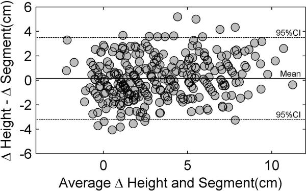
The changes from year one to year two in subject height measured with a standard stadiometer were compared to yearly changes in length of the segments calculated from the motion analysis system (summed tibia, femur, and trunk). The average difference in stature over all subjects measured with the stadiometer 3.4 ± 3.1 cm was not different compared to 3.2 ± 2.8 cm for the change in summed segment length (p > 0.05). Figure 1 shows the agreement between the change in length measured with the stadiometer and segment motion analysis. The two measures demonstrated high reliability (ICC(3,1) = 0.83).
Figure 1.
Bland-Altman plot demonstrating the agreement between the change in length measured with the stadiometer and segment motion analysis. Solid line represents the mean difference between the change in height – change in segment lengths. Less than five percent of the 315 cases fall outside the 95% confidence interval.
Knee Abduction Angle
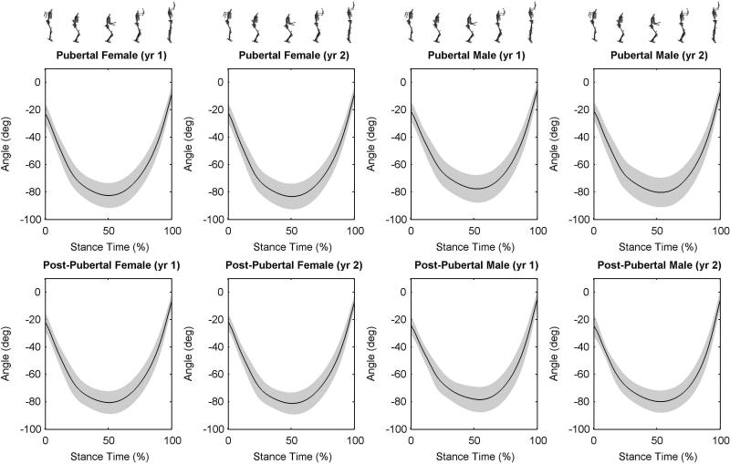
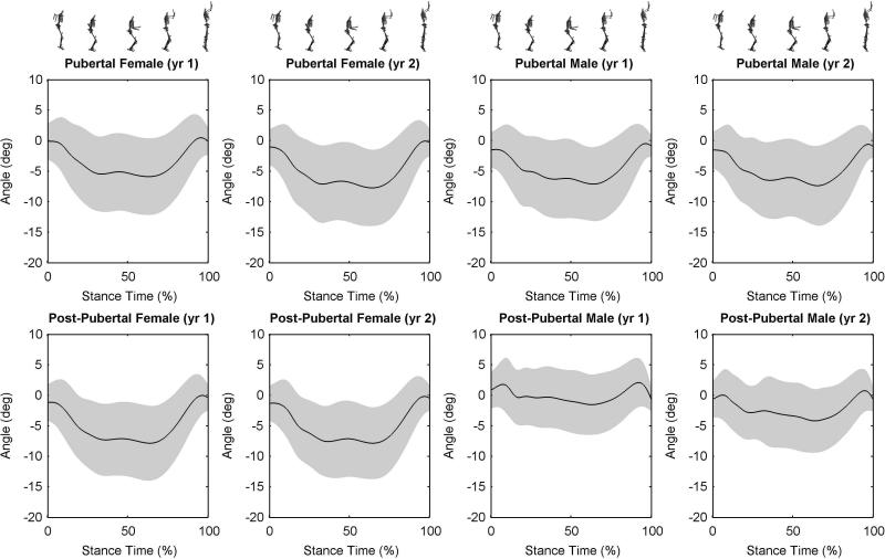
Mean knee flexion and abduction angle during the DVJ stance phase are presented in Figures 2 and 3, respectively. There was a significant three-way interaction with peak knee abduction angle (p = 0.029). Post hoc analysis identified, within the pubertal group, a significant longitudinal increase in peak abduction angle in females (p < 0.001) with no change in males (p = 0.90). Table 2 shows the longitudinal increase in knee abduction angle during one year of adolescent growth in pubertal females compared to pubertal males.
Figure 2.
Ensemble average plot of knee flexion angle (+/- one standard deviation, gray shaded area) throughout the stance phase of the drop vertical jump. The stance phase begins with initial ground contact (0% stance) and ends with toe off (100% stance).
Figure 3.
Ensemble average plot of knee abduction angle (+/- one standard deviation, gray shaded area) throughout the stance phase of the drop vertical jump. The stance phase begins with initial ground contact (0% stance) and ends with toe off (100% stance).
Table 2.
Mean (±SD) peak knee abduction angle and moment for pubertal and post-pubertal subjects
| Pubertal | Post-Pubertal | ||||
|---|---|---|---|---|---|
| Variable | Test Session | Female | Male | Female | Male |
| Knee Abduction Angle (deg)a,b | Year 1 | -7.7 ± 6.1 | -8.2 ± 5.8 | -9.3 ± 5.7 | -2.6 ± 4.4 |
| Year 2 | -9.3 ± 6.2 | -8.3 ± 6.2 | -9.4 ± 5.6 | -4.6 ± 4.8 | |
| Knee Abduction Moment (Nm)b | Year 1 | -14.4 ± 10.9 | -16.4 ± 12.3 | -20.6 ± 13.7 | -9.6 ± 9.4 |
| Year 2 | -19.2 ± 12.1 | -19.4 ± 15.0 | -23.2 ± 13.4 | -16.3 ± 14.7 | |
Denotes interaction of year, sex, maturation
Denotes interaction of sex and maturation. (p<0.05)
Within this cohort of post-pubertal athletes, females and males did not show significant longitudinal changes (p > 0.05) in peak knee abduction angle (Table 2). However, post-pubertal females had significantly greater overall peak abduction angle following adolescent growth compared to males (female -9.3 ± 5.7°; male -3.6 ± 4.6°; p < 0.001).
Knee Abduction Moment
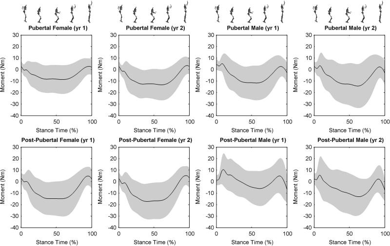
Figure 4 details the mean knee abduction moment for maturation and sex groups over each year. There was a significant main effect of year (p <0.001) indicative of longitudinal increases in peak knee abduction moment in the subjects overall. A two-way interaction between sex and maturation group was identified (p = 0.013). Post-hoc analysis indicated that post-pubertal females had significantly greater peak knee abduction moment compared to post-pubertal males (female -21.9 ± 13.5 Nm; male -13.0 ± 12.0 Nm; p = 0.017). Sex differences in knee abduction moment were not found in pubertal subjects (p > 0.05).
Figure 4.
Ensemble average plot of knee abduction moment (+/- one standard deviation, gray shaded area) throughout the stance phase of the drop vertical jump. The stance phase begins with initial ground contact (0% stance) and ends with toe off (100% stance).
Similar findings were observed when knee abduction moment was normalized to body mass (main effect of year p = 0.001), height (main effect of year p < 0.001) and body mass times height (main effect of year p = 0.005)(Table 3). Regardless of how the moment was normalized there were within-subject increases in the magnitude of knee abduction moment from the first to second year of testing. In addition, a two-way interaction between sex and maturation group was consistent with the un-normalized moment analysis (p = 0.016). Post-hoc analysis indicated that post-pubertal females landed with significantly greater, approximately double, body mass normalized (female -0.37 ± 0.22 Nm/kg; male -0.18 ± 0.16 Nm/kg; p = 0.002), height normalized (female -13.2 ± 8.0 Nm/m; male -7.1 ± 6.6 Nm/m; p = 0.006) and body mass times height normalized (female -0.22 ± 0.14 Nm/(kg*m); male -0.10 ± 0.09 Nm/(kg*m); p = 0.001) knee abduction moment. Sex differences in normalized knee abduction moment were not found in pubertal subjects (p > 0.05).
Table 3.
Normalized mean (±SD) peak knee abduction moment for pubertal and post-pubertal subjects
| Pubertal | Post-Pubertal | ||||
|---|---|---|---|---|---|
| Variable | Test Session | Female | Male | Female | Male |
| Mass Normalized | |||||
| Knee Abduction Moment (Nm/kg)b,c | Year 1 | -0.30 ± 0.22 | -0.32 ± 0.24 | -0.35 ± 0.23 | -0.14 ± 0.13 |
| Year 2 | -0.37 ± 0.23 | -0.34 ± 0.28 | -0.38 ± 0.22 | -0.22 ± 0.20 | |
| Height Normalized | |||||
| Knee Abduction Moment (Nm/m)b,c | Year 1 | -9.12 ± 6.74 | -9.94 ± 7.27 | -12.45 ± 8.15 | -5.32 ± 5.30 |
| Year 2 | -11.86 ± 7.31 | -11.34 ± 8.68 | -13.96 ± 7.93 | -8.91 ± 7.85 | |
| Mass*Height Normalized | |||||
| Knee Abduction Moment (Nm/(kg*m))b,c | Year 1 | -0.19 ± 0.14 | -0.19 ± 0.15 | -0.21 ± 0.14 | -0.08 ± 0.07 |
| Year 2 | -0.23 ± 0.14 | -0.20 ± 0.16 | -0.23 ± 0.13 | -0.12 ± 0.11 | |
Denotes interaction of sex and maturation. (p<0.05)
Denotes interaction of sex and maturation. (p<0.05)
DISCUSSION
The purpose of this investigation was to determine if a neuromuscular risk factor related to abnormal movement patterns increase in females, but not males, during the adolescent growth spurt. The pubertal group of male and female athletes showed an increase in stature that indicated they were going through rapid adolescent growth compared to the post-pubertal group. Knee abduction angle was not changed in post-pubertal females from the first to second year testing session. However, the post-pubertal females exhibited significantly greater knee abduction angle than post-pubertal males.
The findings from the current study support the first hypothesis that through longitudinal analyses pubertal female athletes increased peak knee abduction angle during a year of rapid adolescent growth. These combined results have not been previously identified and are likely important consequences of maturation which increase risk of injury following puberty. The pubertal group was estimated at approximately 88% to 92% of adult stature during the first and second year of testing, respectively. These percent adult stature values indicate that the male and female subjects were at similar stages of somatic growth. Peak height velocity (greatest increase in height during adolescence) occurs at approximately 91% of adult stature in both male and females, with the onset of the adolescent growth spurt near 81 - 84%.(36) Therefore, this further supports that the pubertal group appeared to be within the phase of rapid growth.
Both male and female athletes had increased knee abduction moments from the first to second testing session. The within-subject increases in moments were consistent regardless of the normalization method (body mass, height or body mass times height). The increases in knee abduction, a potentially modifiable risk factor, may coincide with the increased risk of ACL injury in female athletes compared to males.(17) While several studies have demonstrated that females land with greater knee abduction angles, the current study identifies, through longitudinal measures, a possible relationship between maturational group and increases in ACL injury risk factors. The combination of increased motion and torque in female athletes may predispose them to increased risk of injury as they develop and mature.(17-18)
Cross-sectional differences in knee abduction angle among maturational groups has been previously reported.(17) Similar to our current study, Hewett et al. found significant differences in knee abduction angle in post pubertal but not pubertal athletes.(17) The post-pubertal females also had a significantly greater knee abduction angle than pubertal females.(17) In addition, a recent study showed similar results with maturing females exhibiting greater knee valgus posture during DVJ.(33) Schmitz et al.(33) found in a cross-sectional study that females classified as Tanner Stage 1 or 2 had less knee valgus posture compared to females classified as Tanner Stage 4 or 5. The measure of knee valgus in the Schmitz et al.(33) study incorporated a two-dimensional frontal plane angle calculated from digital video. While this single plane measure may involve confounding additional rotations of the hip, knee and ankle, these authors did find similar results based on maturation within female subjects. In the current study, we chose to examine pubertal compared to post-pubertal athletes with the hypothesis that increased knee abduction would be observed during puberty in females, but not in males. Yu et al.(39) investigated the effects of age and gender on lower extremity movement in male and female soccer players between 11 and 16 years as they performed a stop jump task. They found that knee abduction angle was greater in female subjects that were older compared to the younger subjects.(39)
Within subject increases in knee abduction motion and moments are of particular concern given previous cadaveric and modeling work that have linked these measures to increased ACL strain.(5, 11, 20, 26, 35, 38) In a recent cadaveric study, Withrow et al. simulated landings in ten specimens with knee abduction angles at 15° and 0°(neutral).(38) The impact with the knee in abduction led to a primary abduction (valgus) moment that increased ACL strain compared to the neutral landings. Shin et al. used a similar single leg landing impact that resulted in a knee abduction (valgus) moment to a three-dimensional dynamic knee model and found a 35% increase in ACL strain compared to a “neutral-lander” (zero knee abduction moment).(35) Based on incremental increase of the knee abduction moment, these authors showed that peak ACL strain increased rapidly between approximately 20 and 50 Nm.(35) While direct comparisons are difficult between modeling and in vivo data, post-pubertal females (21.9 Nm) were within the steepest region of ACL strain curve,(35) which indicates that slight increases in abduction moments would result in large increases in ligament strain. It is important to point out that all the knee loading conditions in this study were non-injurious (i.e. no injuries occurred during DVJ testing). However, it is likely that repeatedly performing a task with increased knee abduction moment and motion may place females at risk of an ACL injury when an unanticipated or unbalanced landing occurs. For, example, during unanticipated cutting, knee abduction-adduction and internal-external moments can be twice as high compared to pre-planned directional cutting.(3)
Mechanisms of growth and development in females may underlie the increased risk of ACL injury through altered neuromuscular control and movement biomechanics such as knee abduction.(8, 17) ACL injuries do not appear to be different between male and female athletes prior to the onset of puberty and the adolescent growth spurt.(1, 15) However, following rapid pubertal growth, females have higher rates of knee sprains compared to males.(37) The lower extremity segments also go through rapid increases in length during adolescent growth and may potentially lead to increases in knee moments. Increased inertial properties of the segment could partially explain the higher joint moments. Peak body mass velocity, greatest increase in body mass during adolescence, for both sexes generally occurs after peak height velocity.(4) It is interesting that both male and females had increases in knee abduction moment, but knee abduction motion changes were found only in pubertal females. Therefore, growth alone is not likely to be responsible for the increased abduction motion in females. An additional mechanism that differs between sexes as they mature likely plays a role in increased risk of ACL injury in females compared to males. Fat mass remains relatively consistent in males, with skeletal tissue and muscle mass gains being primarily responsible for the observed mass increase.(4, 25) In contrast, females have less overall gains in skeletal tissue and muscle mass compared to males, in addition to a continuous rise in fat mass throughout puberty.(4, 25) The higher center of mass, that results from skeletal growth, and subsequent mass gain during adolescence, makes muscular control of body position more difficult and may translate into larger joint forces at the knee.(17) Males naturally demonstrate a “neuromuscular spurt” (increased strength and power during maturational growth and development) to match the increased demands of growth and development.(17, 19, 21, 25, 31) A recent longitudinal study concluded that males demonstrated a neuromuscular spurt as evidenced by increased vertical jump height and increased ability to attenuate landing force.(31) The absence of similar adaptations in females during maturation may facilitate the mechanisms that lead to increased risk of ACL injury.(34)
A limitation in our study was that group differences between pubertal groups were cross-sectional in nature. We did not follow the subjects through pubertal development and into post-pubertal status. Knee abduction angle was decreased (closer to neutral) in the post-pubertal males compared to pubertal males. The possible protective mechanism in post-pubertal males should be further investigated which may limit excessive knee abduction.
The timing of rapid growth may be the critical phase of development related to sex differences in ACL injury risk. As females reach maturity a variety of discrete sex differences in lower extremity measures have been shown in females compared to males performing landing and cutting maneuvers.(7, 12-13, 16-17, 22, 27) Sex differences between post-pubertal athletes, combined with the timing of the onset of these variables, indicates that an appropriate time to implement an intervention program would be during early puberty. A prospective study that will investigate the timing of intervention is warranted. Prevention programs that incorporate plyometrics, technique biofeedback, and dynamic balance components appear to be effective and reducing measures of knee abduction and the occurrence of ACL injuries.(18, 28-29)
Conclusions
Knee abduction angle was significantly increased in pubertal females during rapid adolescent growth compared to males. In addition, important reported risk factors of knee abduction motion and torque were significantly greater across consecutive years in young female athletes, following rapid adolescent growth, compared to males. An important maturational time period related to potential increase risk of ACL injury appears to be early within pubertal development. The current investigation may also help explain the mechanisms which underlie this phenomenon of sex related injury risk divergence. Future studies should focus on specific mechanisms that may limit or regulate dynamic abduction motion and torque, such as lower extremity strength, muscular co-contraction and joint stiffness parameters in young females that are at increased risk of ACL injury.
ACKNOWLEDGEMENTS
This work was supported by NIH/NIAMS Grants R01-AR049735, R01-AR055563, and R01-AR056259.
Disclosure of Funding: NIH/NIAMS Grants R01-AR049735, R01-AR055563, and R01-AR056259.
Footnotes
CONFLICT OF INTEREST: The authors have no conflicts of interest related to the results of this study. The results of this study do not constitute endorsement by ACSM.
Publisher's Disclaimer: This is a PDF file of an unedited manuscript that has been accepted for publication. As a service to our customers we are providing this early version of the manuscript. The manuscript will undergo copyediting, typesetting, and review of the resulting proof before it is published in its final citable form. Please note that during the production process errors may be discovered which could affect the content, and all legal disclaimers that apply to the journal pertain.
REFERENCES
- 1.Andrish JT. Anterior cruciate ligament injuries in the skeletally immature patient. Am J Orthop. 2001;30(2):103–10. [PubMed] [Google Scholar]
- 2.Bell AL, Pedersen DR, Brand RA. A comparison of the accuracy of several hip center location prediction methods. J Biomech. 1990;23(6):617–21. doi: 10.1016/0021-9290(90)90054-7. [DOI] [PubMed] [Google Scholar]
- 3.Besier TF, Lloyd DG, Ackland TR, Cochrane JL. Anticipatory effects on knee joint loading during running and cutting maneuvers. Med Sci Sports Exerc. 2001;33(7):1176–81. doi: 10.1097/00005768-200107000-00015. [DOI] [PubMed] [Google Scholar]
- 4.Beunen G, Malina RM. Growth and physical performance relative to the timing of the adolescent spurt. Exerc Sport Sci Rev. 1988;16:503–40. [PubMed] [Google Scholar]
- 5.Beynnon BD, Fleming BC. Anterior cruciate ligament strain in-vivo: a review of previous work. J Biomech. 1998;31(6):519–25. doi: 10.1016/s0021-9290(98)00044-x. [DOI] [PubMed] [Google Scholar]
- 6.Bisseling RW, Hof AL. Handling of impact forces in inverse dynamics. J Biomech. 2006;39(13):2438–44. doi: 10.1016/j.jbiomech.2005.07.021. [DOI] [PubMed] [Google Scholar]
- 7.Chappell JD, Yu B, Kirkendall DT, Garrett WE. A comparison of knee kinetics between male and female recreational athletes in stop-jump tasks. Am J Sports Med. 2002;30(2):261–7. doi: 10.1177/03635465020300021901. [DOI] [PubMed] [Google Scholar]
- 8.Croce RV, Russell PJ, Swartz EE, Decoster LC. Knee muscular response strategies differ by developmental level but not gender during jump landing. Electromyogr Clin Neurophysiol. 2004;44(6):339–48. [PubMed] [Google Scholar]
- 9.Davies PL, Rose JD. Motor skills of typically developing adolescents: awkwardness or improvement? Phys Occup Ther Pediatr. 2000;20(1):19–42. [PubMed] [Google Scholar]
- 10.de Leva P. Joint center longitudinal positions computed from a selected subset of Chandler's data. J Biomech. 1996;29(9):1231–3. doi: 10.1016/0021-9290(96)00021-8. [DOI] [PubMed] [Google Scholar]
- 11.Fleming BC, Ohlen G, Renstrom PA, Peura GD, Beynnon BD, Badger GJ. The effects of compressive load and knee joint torque on peak anterior cruciate ligament strains. Am J Sports Med. 2003;31(5):701–7. doi: 10.1177/03635465030310051101. [DOI] [PubMed] [Google Scholar]
- 12.Ford KR, Myer GD, Hewett TE. Valgus knee motion during landing in high school female and male basketball players. Med Sci Sports Exerc. 2003;35(10):1745–50. doi: 10.1249/01.MSS.0000089346.85744.D9. [DOI] [PubMed] [Google Scholar]
- 13.Ford KR, Myer GD, Smith RL, Vianello RM, Seiwert SL, Hewett TE. A comparison of dynamic coronal plane excursion between matched male and female athletes when performing single leg landings. Clinical Biomechanics. 2006;21(1):33–40. doi: 10.1016/j.clinbiomech.2005.08.010. [DOI] [PubMed] [Google Scholar]
- 14.Ford KR, Myer GD, Toms HE, Hewett TE. Gender differences in the kinematics of unanticipated cutting in young athletes. Med Sci Sports Exerc. 2005;37(1):124–9. [PubMed] [Google Scholar]
- 15.Gallagher SS, Finison K, Guyer B, Goodenough S. The incidence of injuries among 87,000 Massachusetts children and adolescents: results of the 1980-81 Statewide Childhood Injury Prevention Program Surveillance System. Am J Public Health. 1984;74(12):1340–7. doi: 10.2105/ajph.74.12.1340. [DOI] [PMC free article] [PubMed] [Google Scholar]
- 16.Hewett TE, Ford KR, Myer GD, Wanstrath K, Scheper M. Gender Differences in Hip Adduction Motion and Torque During a Single Leg Agility Maneuver. J Orthop Res. 2006;24(3):416–421. doi: 10.1002/jor.20056. [DOI] [PubMed] [Google Scholar]
- 17.Hewett TE, Myer GD, Ford KR. Decrease in neuromuscular control about the knee with maturation in female athletes. J Bone Joint Surg Am. 2004;86-A(8):1601–1608. doi: 10.2106/00004623-200408000-00001. [DOI] [PubMed] [Google Scholar]
- 18.Hewett TE, Myer GD, Ford KR, et al. Biomechanical Measures of Neuromuscular Control and Valgus Loading of the Knee Predict Anterior Cruciate Ligament Injury Risk in Female Athletes: A Prospective Study. Am J Sports Med. 2005;33(4):492–501. doi: 10.1177/0363546504269591. [DOI] [PubMed] [Google Scholar]
- 19.Hewett TE, Myer GD, Ford KR, Slauterbeck JL. Preparticipation Physical Exam Using a Box Drop Vertical Jump Test in Young Athletes: The Effects of Puberty and Sex. Clin J Sport Med. 2006;16(4):298–304. doi: 10.1097/00042752-200607000-00003. [DOI] [PubMed] [Google Scholar]
- 20.Kanamori A, Woo SL, Ma CB, et al. The forces in the anterior cruciate ligament and knee kinematics during a simulated pivot shift test: A human cadaveric study using robotic technology. Arthroscopy. 2000;16(6):633–9. doi: 10.1053/jars.2000.7682. [DOI] [PubMed] [Google Scholar]
- 21.Kellis E, Tsitskaris GK, Nikopoulou MD, Moiusikou KC. The evaluation of jumping ability of male and female basketball players according to their chronological age and major leagues. J Strength Cond Res. 1999;13(1):40–46. [Google Scholar]
- 22.Kernozek TW, Torry MR, VH H, Cowley H, Tanner S. Gender differences in frontal and sagittal plane biomechanics during drop landings. Med Sci Sports Exerc. 2005;37(6):1003–12. discussion 1013. [PubMed] [Google Scholar]
- 23.Khamis HJ, Roche AF. Predicting adult stature without using skeletal age: the Khamis-Roche method. Pediatrics. 1994;94(4 Pt 1):504–7. [PubMed] [Google Scholar]
- 24.Krosshaug T, Nakamae A, Boden BP, et al. Mechanisms of anterior cruciate ligament injury in basketball: video analysis of 39 cases. Am J Sports Med. 2007;35(3):359–67. doi: 10.1177/0363546506293899. [DOI] [PubMed] [Google Scholar]
- 25.Malina RM, Bouchard C. Growth, maturation, and physical activity. Human Kinetics; Champaign, Il: 1991. p. 501. [Google Scholar]
- 26.Markolf KL, Burchfield DM, Shapiro MM, Shepard MF, Finerman GA, Slauterbeck JL. Combined knee loading states that generate high anterior cruciate ligament forces. J Orthop Res. 1995;13(6):930–5. doi: 10.1002/jor.1100130618. [DOI] [PubMed] [Google Scholar]
- 27.McLean SG, Huang X, Su A, van den Bogert AJ. Sagittal plane biomechanics cannot injure the ACL during sidestep cutting. Clin Biomech. 2004;19:828–838. doi: 10.1016/j.clinbiomech.2004.06.006. [DOI] [PubMed] [Google Scholar]
- 28.Myer GD, Ford KR, McLean SG, Hewett TE. The Effects of Plyometric Versus Dynamic Stabilization and Balance Training on Lower Extremity Biomechanics. Am J Sports Med. 2006;34(3):490–8. doi: 10.1177/0363546505281241. [DOI] [PubMed] [Google Scholar]
- 29.Myer GD, Ford KR, Palumbo JP, Hewett TE. Neuromuscular training improves performance and lower-extremity biomechanics in female athletes. J Strength Cond Res. 2005;19(1):51–60. doi: 10.1519/13643.1. [DOI] [PubMed] [Google Scholar]
- 30.Olsen OE, Myklebust G, Engebretsen L, Bahr R. Injury mechanisms for anterior cruciate ligament injuries in team handball: a systematic video analysis. Am J Sports Med. 2004;32(4):1002–12. doi: 10.1177/0363546503261724. [DOI] [PubMed] [Google Scholar]
- 31.Quatman CE, Ford KR, Myer GD, Hewett TE. Maturation Leads to Gender Differences in Landing Force and Vertical Jump Performance: A Longitudinal Study. Am J Sports Med. 2006;34(5):806–13. doi: 10.1177/0363546505281916. [DOI] [PubMed] [Google Scholar]
- 32.Quatman CE, Ford KR, Myer GD, Paterno MV, Hewett TE. The effects of gender and pubertal status on generalized joint laxity in young athletes. J Sci Med Sport. 2008;11(3):257–263. doi: 10.1016/j.jsams.2007.05.005. [DOI] [PMC free article] [PubMed] [Google Scholar]
- 33.Schmitz RJ, Shultz SJ, Nguyen AD. Dynamic valgus alignment and functional strength in males and females during maturation. J Athl Train. 2009;44(1):26–32. doi: 10.4085/1062-6050-44.1.26. [DOI] [PMC free article] [PubMed] [Google Scholar]
- 34.Shea KG, Pfeiffer R, Wang JH, Curtin M, Apel PJ. Anterior cruciate ligament injury in pediatric and adolescent soccer players: an analysis of insurance data. J Pediatr Orthop. 2004;24(6):623–8. doi: 10.1097/00004694-200411000-00005. [DOI] [PubMed] [Google Scholar]
- 35.Shin CS, Chaudhari AM, Andriacchi TP. The effect of isolated valgus moments on ACL strain during single-leg landing: A simulation study. J Biomech. 2009;42(3):280–285. doi: 10.1016/j.jbiomech.2008.10.031. [DOI] [PMC free article] [PubMed] [Google Scholar]
- 36.Tanner JM, Whitehouse RH, Marubini E, Resele LF. The adolescent growth spurt of boys and girls of the Harpenden Growth Study. Annals of Human Biology. 1976;3(2):109–126. doi: 10.1080/03014467600001231. [DOI] [PubMed] [Google Scholar]
- 37.Tursz A, Crost M. Sports-related injuries in children. A study of their characteristics, frequency, and severity, with comparison to other types of accidental injuries. Am J Sports Med. 1986;14(4):294–299. doi: 10.1177/036354658601400409. [DOI] [PubMed] [Google Scholar]
- 38.Withrow TJ, Huston LJ, Wojtys EM, Ashton-Miller JA. The effect of an impulsive knee valgus moment on in vitro relative ACL strain during a simulated jump landing. Clin Biomech (Bristol, Avon) 2006;21(9):977–83. doi: 10.1016/j.clinbiomech.2006.05.001. [DOI] [PubMed] [Google Scholar]
- 39.Yu B, McClure SB, Onate JA, Guskiewicz KM, Kirkendall DT, Garrett WE. Age and gender effects on lower extremity kinematics of youth soccer players in a stop-jump task. Am J Sports Med. 2005;33(9):1356–64. doi: 10.1177/0363546504273049. [DOI] [PubMed] [Google Scholar]


