The early growth response gene product (Egr-1) is a zinc finger transcription factor (1, 2) first identified because of its characteristic pattern of expression following exposure of cells to mediators associated with growth and differentiation. Egr-1 (also called Zif268, NGF1-A, Krox24, or TIS8) has been termed an immediate-early response protein, based on the brisk kinetics of its induction, within minutes of a stimulus, and its rapid decay, often within hours.
The initial association of Egr-1 with growth and development suggested that it might act in cell differentiation, and cell culture studies indicated a crucial role for Egr-1 in promoting differentiation along a macrophage lineage (3). However, the generation of Egr-1–null mice by S. Lee and colleagues provided a new perspective on Egr-1 biology (4). Monocyte differentiation, they found, is unaffected by the deletion of the Egr-1 gene, and knockout animals appear normal with the exception of infertility in homozygous null females (4). These observations indicated that physiologic roles of Egr-1 might only become manifest in response to environmental challenge. Consistent with this possibility, in vitro studies identified a number of gene products — TNFα, ICAM-1, CD44, PDGF A/B chain, TGFβ, M-CSF, among others (5) — that are induced by Egr-1 and that participate in the physiological response to various kinds of stress.
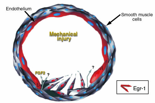
Recent work from Khachigian and colleagues has provided an important step forward in our understanding of Egr-1 biology (6, 7). This group has found that the promoter from the gene for PDGF A chain contains a GC-rich element with overlapping binding sites for Egr-1 and Sp1. Under quiescent conditions, these sites are occupied by Sp1, which is believed to be required for basal expression of this gene. However, following stimulation of cells with phorbol ester, levels of Egr-1 rise, allowing Egr-1 to displace Sp1 from this region (6). Support for the in vivo relevance of Egr-1–mediated gene expression comes from experiments with denuding injury to the rat aorta; Egr-1 and a variety of Egr-1 target genes are induced in endothelium at the wound margins (7). Cultured endothelial monolayers subjected to an analogous mechanical injury release fibroblast growth factor 2 (FGF2), which stimulates Egr-1 expression in this system (8). Furthermore, a DNA-enzyme that specifically cleaves Egr-1 mRNA blocks arterial neointima formation in a rat carotid angioplasty model (9). Although experiments with the DNA enzyme approach can be difficult to interpret and the relevance of the rat angioplasty model to human neointimal disease is controversial, these observations clearly support a role for Egr-1 in the response to vascular injury (Figure 1).
Figure 1.
Schematic depiction of Egr-1 induction in response to mechanical injury to the vessel wall. Egr-1 expression is especially evident in endothelial cells at the wound margin, as well as in the migrating and proliferating smooth muscle cells in the forming neointima. FGF2, as well as other factors yet to be identified, are likely to be involved in Egr-1 induction in this setting.
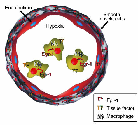
The pathological role of Egr-1 in blood vessels is not limited to its promotion of neointimal formation following mechanical injury. There is an unexpected role for Egr-1 during hypoxemia, when it triggers deposition of fibrin in the vasculature. Recent studies have established cause-effect relationships between hypoxemia, Egr-1 expression, and fibrin deposition in this system (10–13) (Figure 2). Mice subjected to normobaric hypoxia rapidly induce expression in the lung of active Egr-1, which drives expression of tissue factor (TF), the cell-surface cofactor responsible for initiation of coagulation. Increased TF, which promotes intravascular fibrin accumulation (10), is observed in mononuclear phagocytes and smooth muscle cells, the same cells exhibiting increased Egr-1 in response to oxygen deprivation (11). The central role of Egr-1 in this pathway is clear from observations in Egr-1–null mice, which do not induce TF and which, as a consequence, remain free of vascular fibrin deposits under these conditions.
Figure 2.
Schematic depiction of Egr-1 induction in the vessel wall and mononuclear phagocytes/macrophages in response to hypoxemia. Oxygen deprivation causes expression of Egr-1 in mononuclear phagocytes and vascular smooth muscle cells thereby activating downstream target genes such as TF. Expression of TF in hypoxemic/ischemic vasculature provides a mechanism triggering the procoagulant pathway and, potentially, resulting in fibrin deposition.
The best-characterized pathway for biosynthetic adaptation to oxygen deprivation involves hypoxia-inducible factor-1 (HIF-1), which promotes expression of genes critical for survival in the oxygen-scarce environment (14). However, the hypoxia-triggered pathway involving Egr-1 is independent of HIF-1 and appears to act through the rapid activation of protein kinase C isoform βII (PKCβII) when oxygen levels decline. This kinase initiates a signaling cascade that activates the transcription factor Elk-1 and thereby initiates transcription of Egr-1 (11, 12). Consistent with this model, Egr-1 and TF are not expressed in hypoxic mice that carry a homozygous deletion in PKCβ, although these animals retain HIF-1–dependent responses (13). This novel pathway for hypoxia-inducible responses may well be relevant to the pathobiology of ischemic injury.
In each of these studies, Egr-1 appears to participate in the acute response to physical injury or hypoxia, but, in the current issue of the JCI, McCaffrey and colleagues demonstrate sustained Egr-1 expression in atherosclerosis, a chronic condition (15). These investigators harvested RNA from the fibrous cap of carotid endarterectomy samples and used a cDNA expression array to analyze changes in gene expression associated with the disease. They demonstrated an approximately 5-fold increase in Egr-1 mRNA in lesions, compared with adjacent media. Crucially, these lesions also showed an increase in known Egr-1 target transcripts, such as TNFα, ICAM-1, M-CSF, that are believed to contribute to this disease process. In addition, they confirmed that the Egr-1 protein accumulates during postnatal development in atherosclerosis-prone mice and is enriched in atherosclerotic lesions, especially in smooth muscle cells.
At this juncture, these observations represent nothing more than an association, although a provocative one, between Egr-1 and the atherogenic process. The challenge remains to establish cause-effect relationships pertinent to outcome, and the availability of Egr-1–null mice is likely to be critical for this purpose, as well as in the identification of physiologically relevant targets of Egr-1. It will also be important to dissect the regulation Egr-1 expression and activity, including the role of the inducible corepressor NAB2 (16), and to determine if other members of the Egr-1 family contribute to atherogenesis or other conditions, such as neointimal hyperplasia or thrombosis. If such studies implicate Egr-1 as an important pathogenetic factor, the apparent well-being of Egr-1–null mice (4) suggests that Egr-1 expression or function could be inhibited for therapeutic purposes with little ill effect.
References
- 1.Milbrandt J. A nerve growth factor induced gene encodes a possible transcriptional regulatory factor. Science. 1988;238:797–799. doi: 10.1126/science.3672127. [DOI] [PubMed] [Google Scholar]
- 2.Gashler A, Sukhatme V. Egr-1: prototype of a zinc finger family of transcription factors. Prog Nucleic Acid Res Mol Biol. 1995;50:191–224. doi: 10.1016/s0079-6603(08)60815-6. [DOI] [PubMed] [Google Scholar]
- 3.Nguyen H, Hoffman-Liebermann B, Liebermann D. Egr-1 is essential for and restricts differentiation along the macrophage cell lineage. Cell. 1993;72:197–209. doi: 10.1016/0092-8674(93)90660-i. [DOI] [PubMed] [Google Scholar]
- 4.Lee S, et al. LH deficiency and female infertility in mice lacking the transcription factor Egr-1. Science. 1996;273:1219–1221. doi: 10.1126/science.273.5279.1219. [DOI] [PubMed] [Google Scholar]
- 5.Khachigian L, Collins T. Inducible expression of Egr-1-dependent genes. A paradigm of transcriptional activation in vascular endothelium. Circ Res. 1997;81:457–461. doi: 10.1161/01.res.81.4.457. [DOI] [PubMed] [Google Scholar]
- 6.Khachigian L, Williams A, Collins T. Interplay of Sp1 and Egr-1 in the proximal PDGF A-chain promoter in cultured vascular endothelial cells. J Biol Chem. 1995;270:27679–27686. doi: 10.1074/jbc.270.46.27679. [DOI] [PubMed] [Google Scholar]
- 7.Khachigian L, Lindner V, Williams A, Collins T. Egr-1-induced endothelial gene expression: a common theme in vascular injury. Science. 1996;271:1427–1431. doi: 10.1126/science.271.5254.1427. [DOI] [PubMed] [Google Scholar]
- 8.Santiago F, Lowe H, Day F, Chesterman C, Khachigian L. EGR-1 induction by injury is triggered by release and paracrine activation by FGF-2. Am J Pathol. 1999;154:937–944. doi: 10.1016/S0002-9440(10)65341-2. [DOI] [PMC free article] [PubMed] [Google Scholar]
- 9.Santiago F, et al. New DNA enzyme targeting Egr-1 mRNA inhibits vascular smooth muscle proliferation and regrowth after injury. Nat Med. 1999;5:1264–1269. doi: 10.1038/15215. [DOI] [PubMed] [Google Scholar]
- 10.Lawson C, et al. Monocytes and tissue factor promote thrombosis in a murine model of oxygen deprivation. J Clin Invest. 1997;99:1729–1738. doi: 10.1172/JCI119337. [DOI] [PMC free article] [PubMed] [Google Scholar]
- 11.Yan S-F, et al. Tissue factor transcription driven by Egr-1 is a critical mechanism of murine pulmonary fibrin deposition in hypoxia. Proc Natl Acad Sci USA. 1998;95:8298–8303. doi: 10.1073/pnas.95.14.8298. [DOI] [PMC free article] [PubMed] [Google Scholar]
- 12.Yan S-D, et al. Hypoxia-associated induction of EGR-1 gene expression. J Biol Chem. 1999;274:15030–15040. doi: 10.1074/jbc.274.21.15030. [DOI] [PubMed] [Google Scholar]
- 13.Yan, S.-F., et al. Protein Kinase C-β and oxygen deprivation: a novel Egr-1-dependent pathway for fibrin deposition in hypoxemic vasculature. J. Biol. Chem. In press. [DOI] [PubMed]
- 14.Semenza G. Regulation of mammalian oxygen homeostasis by HIF-1. Annu Rev Cell Dev Biol. 1999;15:551–578. doi: 10.1146/annurev.cellbio.15.1.551. [DOI] [PubMed] [Google Scholar]
- 15.McCaffrey T, et al. High-level expression of Egr-1, and Egr-1-inducible genes in mouse and human atherosclerosis. J Clin Invest. 2000;105:653–662 (2000). doi: 10.1172/JCI8592. [DOI] [PMC free article] [PubMed] [Google Scholar]
- 16.Svaren J, et al. NAB2, a corepressor of EGR-1, is induced by proliferative and differentiative stimuli. Mol Cell Biol. 1996;16:3545–3553. doi: 10.1128/mcb.16.7.3545. [DOI] [PMC free article] [PubMed] [Google Scholar]


