

Abstract
The occupational chemical 4-vinylcyclohexene diepoxide (VCD) selectively destroys ovarian small pre-antral follicles in rats and mice via apoptosis. Detoxification of VCD can occur through glutathione conjugation, catalyzed by glutathione S-transferase (GST) enzymes. Further, GST class pi (GSTp) can negatively regulate JNK activity through protein:protein interactions in extra-ovarian tissues. Dissociation of this protein complex in the face of chemical exposure releases the inhibition of pro-apoptotic JNK. Increased JNK activity during VCD-induced ovotoxicity has been shown in isolated ovarian small pre-antral follicles following in vivo dosing of rats (80mg/Kg/d; 15d, i.p). The present study investigated the pattern of ovarian GSTp expression during VCD exposure. Additionally, the effect of VCD on an ovarian GSTp:JNK protein complex was investigated. PND4 F344 rat ovaries were incubated in control medium ± VCD (30 μM) for 2-8d. VCD increased ovarian GSTp mRNA (P <0.05) relative to control on d4-d8; whereas GSTp protein was increased (P < 0.05) on d6-d8. A GSTp:JNK protein complex was detected by immunoprecipitation and Western blotting in ovarian tissues. Relative to control, the amount of GSTp-bound JNK was increased (P = 0.09), while unbound JNK was decreased (P < 0.05) on d6 of VCD exposure. The VCD-induced decrease in unbound JNK was preceded by a decrease in phosphorylated c-Jun which occurred on d4. These findings are in support of a possible dual protective role for GSTp in the rat ovary, consisting of metabolism of VCD and inhibition of JNK-initiated apoptosis.
Keywords: 4-vinylcyclohexene diepoxide, ovotoxicity, glutathione S-transferase pi
Introduction
4-vinylcyclohexene (VCH) is an occupational chemical formed by dimerization of 1,3-butadiene and is produced as a by-product in the pesticide, rubber, plastic and flame retardant industries (Rappaport and Fraser, 1977). A metabolite of VCH, 4-vinylcyclohexene diepoxide (VCD) is used as an industrial diluent for epoxides (IARC, 1976). VCD is ovotoxic and specifically destroys small pre-antral follicles (primordial and primary) in ovaries of rats and mice (Kao et al., 1999; Smith et al., 1990) by enhancing the natural process of atresia in a time- and dose-dependent manner (apoptosis; Devine et al., 2002a; Springer et al., 1996a,b,c; Hu et al., 2001a,b; 2002). In the ovary the cytochrome P450 enzyme isoform CYP2E1 may play a role in converting VCH to the ovotoxic form, VCD (Cannady et al., 2003; Rajapaska et al., 2007a). Ovarian detoxification of VCD to inactive metabolites is catalyzed through the action of microsomal epoxide hydrolase (mEH) and the glutathione S-transferase (GST) family of enzymes.
GST comprise a large family of enzymes that catalyze the conjugation of glutathione (GSH) to a variety of xenobiotics, allowing for their inactivation and more rapid excretion. There are a number of classes of GST enzymes, alpha, mu, omega, pi, sigma and theta, and each of these in turn contains a number of isoforms. The mouse ovary has been shown to synthesize GSH (Luderer et al., 2001), and form VCD-GSH adducts (Rajapaska et al., 2007b). A previous study in cultured postnatal day (PND) 4 B6C3F1 mouse ovaries showed no effect of VCD on mRNA encoding GST classes alpha, omega or theta (Keating et al., 2008). However, VCD exposure increased (P < 0.05) mRNA encoding GST classes pi (GSTp) and mu (GSTm) on d4 by 1.55- and 1.7-fold, respectively. Yet this effect was reversed on d6 and d8. GSTp, but not GSTm protein was elevated by 47% after 8d of VCD exposure. It was hypothesized that in mice, despite early up-regulation of GST, repeated VCD exposure eventually overwhelmed the induction of GST enzymes, thereby reducing its detoxification capacity during the onset of ovotoxicity (Keating et al., 2008).
In addition to its role in glutathione conjugation, the GST family has another possible indirect role in regulation of cell signaling. GSTp has been shown to form a protein complex with JNK and c-Jun (Adler et al., 1999), keeping JNK in an inactive state. This interaction prevents JNK mediated phosphorylation and activation of c-Jun (phosphorylated c-Jun; p-c-Jun). Following chemical exposure, this complex dissociates, relieving the inhibitory action of GSTp on JNK (Adler et al., 1999). A role for both JNK and p-c-Jun during VCD-induced ovotoxicity has been demonstrated. Following 15d of daily dosing of rats with VCD (80mg/Kg; i.p.), relative to controls, both JNK (protein and activity) and p-c-Jun (protein) increased in isolated small pre-antral follicles (targeted by VCD; Hu et al., 2002).
An in vitro culture system has been developed using ovaries from PND4 rats (enriched in small pre-antral follicles) to examine in vitro effects of ovotoxicants without a metabolic influence from the liver (Devine et al., 2002b; 2004). Whether the GST and JNK pathways interact in the ovary as in other tissues is unknown. Therefore, the present study was designed to characterize in rats the association between VCD and GSTp mRNA and protein as relates to VCD-induced follicle loss using the in vitro ovary culture system. The formation of an ovarian GSTp and JNK-containing protein complex was also investigated.
Materials and Methods
Reagents
VCD (mixture of isomers, >99% purity), 2-β-mercaptoethanol, 30% acrylamide/0.8% bis-acrylamide, ammonium persulfate, glycerol, N’,N’,N’,N’-Tetramethyl-ethylenediamine (TEMED), Tris base, Tris HCl, sodium chloride, Tween-20, bovine serum albumin (BSA), ascorbic acid (Vitamin C), phosphatase inhibitor, protease inhibitor and transferrin were purchased from Sigma-Aldrich Inc. (St Louis, MO). Dulbecco’s Modified Eagle Medium: nutrient mixture F-12 (Ham) 1X (DMEM/Ham’s F12), albumax, penicillin/streptomycin (5000U/ml, 5000 μg/ml, respectively), Hanks’ Balanced Salt Solution (without CaCl2, MgCl2, or MgSO4), custom designed primers, and superscript III one-step RT-PCR System were obtained from Invitrogen Co. (Carlsbad, CA). Millicell-CM filter inserts, anti-p-c-Jun and anti-GSTp ant ibodies were purchased from Millipore (Bedford, MA). 48 well cell culture plates were obtained from Corning Inc. (Corning, NY). RNeasy Mini kit, QIAshredder kit, RNeasy MinElute kit, and Quantitect™ SYBR Green PCR kit were purchased from Qiagen Inc. (Valencia, CA). Anti-JNK antibody was purchased from Santa Cruz Biotechnology (Santa Cruz, CA). Anti-ACTB antibody and agarose G beads were purchased from Santa Cruz Biotechnology (Santa Cruz, CA). Goat anti-rabbit and goat anti-mouse secondary antibodies were purchased from Pierce Biotechnology (Rockford, IL).
Animals
A breeding colony was established from Fischer 344 (F344) rats that were originally purchased from Harlan Laboratories (Indianapolis, IN) to use as a source of PND4 female rat pup ovaries for culture. All pregnant animals were housed singly in plastic cages, and maintained in a controlled environment (22 ± 2°C; 12h light/ 12h dark cycles). Animals were provided a standard diet with ad libidum access to food and water, and allowed to give birth. All animal experiments were approved by the University of Arizona’s Institutional Animal Care and Use Committee.
In vitro ovarian culture
Ovaries from PND4 F344 rats were cultured as described by Devine et al. (2002a). Briefly, PND4 female F344 rats were euthanized by CO2 inhalation followed by decapitation. Each ovary was removed, the oviduct and excess tissue were trimmed, and it was placed on a piece of Millicell-CM membrane floating on 250 μl of DMEM/Ham’s F12 medium containing 1 mg/ml BSA, 1 mg/ml Albumax, 50 μg/ml ascorbic acid, 5 U/ml penicillin/5 μg/ml streptomycin, and 27.5 μg/ml transferrin per well in a 48 well plate previously equilibrated to 37°C. Using fine forceps, a drop of medium was placed to cover the top of the ovary to prevent drying. Plates containing ovaries were cultured at 37°C and 5% CO2 in air. Culture media was used as a solvent for VCD. The concentration of VCD used (30 μM) was determined previously to cause loss of primordial and small primary follicles following 6 days of exposure using the in vitro rat ovary culture system (Keating et al., 2009). For those cultures lasting more than 2d, media were removed and fresh media and treatment were replaced every 2d.
RNA isolation
Following 2, 4, 6 or 8d of in vitro culture, ovaries treated with control or VCD (30 μM) were stored in RNAlater at −80°C. Total RNA was isolated (n=3; 10 ovaries per pool) using an RNeasy Mini kit. Briefly, ovaries were lysed and homogenized using a motor pestle followed by applying the mixture onto a QIAshredder column. The QIAshredder column containing ovarian tissue sample was then centrifuged at 14,000 rpm for 2 min. The resulting flow-through was applied to an RNeasy mini column, allowing RNA to bind to the filter cartridge. Following washing, RNA was eluted from the filter, and concentrated using an RNeasy MinElute kit. Briefly, isolated RNA was applied to an RNeasy MinElute spin column, and after washing, RNA was eluted using 14 μL of RNase-free water. RNA concentration was determined using an ND-1000 Spectrophotometer (λ = 260/280nm; NanoDrop technologies, Inc., Wilmington, DE).
First strand cDNA synthesis and real-time polymerase chain reaction (PCR)
Total RNA (0.5 μg) was reverse transcribed into cDNA utilizing the Superscript III One-Step RT-PCR System. cDNA was diluted (1:25) in RNase-free water. Two microliters of diluted cDNA were amplified on a Rotor-Gene 3000 using Quantitect™ SYBR Green PCR kit and custom designed primers as described in Keating et al. (2008). The cycling program consisted of a 15 min hold at 95°C and 45 cycles of: denaturing at 95°C for 15s, annealing at 58°C for 15s, and extension at 72°C for 20s at which point data were acquired. Product melt conditions were determined using a temperature gradient from 72°C to 99°C with a 1°C increase at each step. There was no difference in β-actin (Actb) mRNA between control and VCD treated ovaries. Therefore, each sample was normalized to ACTB before quantification using the 2−ΔΔCt method.
Protein Isolation
Pools of whole ovarian protein (10-20 ovaries/pool) homogenates were prepared from cultured ovaries via homogenization in tissue lysis buffer containing protease and phosphatase inhibitors as previously described (Thompson et al., 2005). For immunoprecipitation protein isolation, SDS was omitted from the tissue lysis buffer. Briefly, homogenized samples were placed on ice for 30 min, followed by two rounds of centrifugation at 10,000 rpm for 15 min. Supernatant was aliquoted and stored at −80°C until further use. Protein was quantified using a standard BCA protocol on a 96-well assay plate. Emission absorbance was detected with a λ= 540nm excitation on a Synergy™ HT Multi-Detection Microplate Reader using KC4™ software (Bio-Tek® Instruments Inc., Winooski, VT). Protein concentrations were calculated from a BSA protein standard curve.
Protein Immunoprecipitation
Ovarian protein (100 μg) was incubated overnight at 4°C with 20 μl of anti-GSTp antibody. Protein G agarose beads were washed, added to the protein-antibody mixture and incubated at 4°C for 3 hours with turning. This mixture was centrifuged at 10,000rpm, and the supernatant (unbound fraction) was removed for Western blotting. The beads were washed three times with tissue lysis buffer (500 μl). Laemmli sample buffer (20 μl) was added and beads incubated at 95°C for 10 mins. The beads were centrifuged and 10 μl supernatant (bound fraction) was used for Western blotting.
Western Blot Analysis
SDS-PAGE (10%) was used to separate protein homogenates (n=3; 10 μg total protein or 10 μl unbound or bound immunoprecipitation fractions) and subsequently transferred onto nitrocellulose membranes as previously described (Thompson et al., 2005). Briefly, membranes were blocked for 1 h with shaking at 4°C in 5% milk in Tris-buffered saline with Tween-20 (TTBS). Membranes were incubated with primary antibody in 5% milk in TTBS overnight at 4°C. Antibody dilutions used were anti-GSTp (1:200), anti-p-c-Jun (1:500), anti-JNK (1:500) and anti-ACTB (1:5000). Membranes were washed three times for 10 min each with TTBS. HRP-conjugated secondary antibody (1:2000 dilution) was added for 1 h at room temperature. Membranes were washed three times for 10 min each in TTBS, followed by a single wash for 10 min in Tris Buffered Saline (TBS). Western blots were detected by chemiluminescence (using ECL plus chemiluminescence detection substrate) and exposed to X-ray film. Densitometry of the appropriate bands was perfomed using ImageJ software (NCBI). Individual treatment values were normalized to ACTB for total protein and immunoprecipitated unbound protein and expressed as a percentage of the control mean. Bound immunoprecipitated JNK was normalized to GSTp protein and expressed as a percentage of the control mean.
Statistical analysis
Comparisons were made between treatments using Statview software Fisher’s protected least significant difference (PLSD) multiple range test. Statistical analysis was carried out on raw data and, for graphical purposes, values were expressed as a percentage of control (n = 3), and one control value of 100% presented per graph. The assigned level of significance for all tests was P < 0.05, with P < 0.1 considered as a trend for a difference.
Results
Temporal effect of VCD on GSTp mRNA in F344 rat ovaries
Previous studies have determined that VCD-induced follicle loss in PND4 rat ovaries begins on d6 of culture (Keating et al., 2009). In order to compare changes in GSTp mRNA expression over the time-course of VCD-induced ovotoxicity, PND4 F344 rat ovaries were cultured in VCD (30 μM) for 2, 4, 6, or 8d. Relative to control ovaries, GSTp mRNA was increased (P < 0.05) on d4-d8 (Figure 1 - d2: CT = 1, VCD = 0.92 ± 0.33; d4: CT = 1, VCD = 1.91 ± 0.19 - 91% increase; d6: CT = 1, VCD = 2.19 ± 0.16 - 119% increase; d8: CT = 1, VCD = 1.74 ± 0.08 - 74% increase).
Figure 1. Temporal effect of VCD on GSTp mRNA.
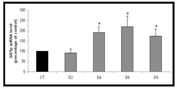
Ovaries from PND4 F344 rats were cultured with control (CT) medium or medium containing 30 μM VCD for 2-8d. Following incubation, total RNA was isolated, reverse transcribed to cDNA and analyzed for GSTp or Actb mRNA expression by RT-PCR. Values are percentage of control ± SE; n=3 (8-10 ovaries per pool). *P < 0.05; different from control.
Temporal effect of VCD on GSTp protein in F344 rat ovaries
The effect of VCD on GSTp protein level was evaluated by Western blotting. PND4 F344 rat ovaries were cultured in control media or media containing VCD (30 μM) for 4, 6, or 8d. There was no effect of VCD on GSTp protein on d4. Relative to control, GSTp protein levels were increased (P < 0.05) on d6 and d8 (Figure 2 - d4: CT = 4.41 ± 0.33, VCD = 4.1 ± 0.12; d6: CT = 3.55 ± 0.26, VCD = 5.49 ± 0.39 - 54% increase; d8: CT = 4.37 ± 0.18, VCD = 5.55 ± 0.19 - 27% increase).
Figure 2. Temporal effect of VCD on GSTp protein.

Ovaries from PND4 F344 rats were cultured with control (CT) medium or medium containing 30 μM VCD for 4-8d. Following incubation, total protein was isolated and Western blotting carried out for GSTp and ACTB. (A) Values are normalized to ACTB protein and expressed as a percentage of control mean ± S.E.; n=3; * P <0.05; Different from control. (B) Representative Western blot is shown on d6; control = c; VCD = v.
Presence of and effect of VCD on GSTp:JNK protein complex formation in F344 rat ovaries
In order to investigate the existence of ovarian protein complex formation between GSTp, and JNK at a time prior to (d4) and at the time of (d6) VCD-induced follicle loss, PND4 F344 rat ovaries were cultured for 4d and 6d in control media. Immunoprecipitation was performed using an anti-GSTp antibody followed by Western blotting to detect JNK. The presence of an ovarian GSTp:JNK protein complex was detected on d4 and d6 of culture (Figure 3A). The presence of an ovarian GSTp:p-c-Jun protein complex was also detected (data not shown).
Figure 3. Effect of VCD on GSTp:JNK protein complex formation.

Ovaries from PND4 F344 rats were cultured with control (CT) medium or medium containing 30 μM VCD for 4d (D4) or 6d (D6). Following incubation, total protein was isolated and immunoprecipitation carried out using an anti-GSTp antibody as described in materials and methods. (A) Representative Western blot of GSTp immunoprecipitation, followed by detection of JNK protein (control ovary; d6). (B) Ratio of JNK protein bound to GSTp on D4 and D6. (C) JNK protein relative to total GSTp. Values are percentage of control ± SE. * P <0.05; # P < 0.1; Different from control.
The effect of VCD (30 μM) on the amount of JNK complexed with GSTp at a time prior to (d4) and at the time of (d6) VCD-induced follicle loss was assessed in F344 PND4 rat ovaries. There was no effect of VCD on d4, but there was a trend for an increase (P = 0.09) in the ratio of JNK to GSTp in the protein complex on d6 (Figure 3B - d4: CT = 3.22 ± 0.29, VCD = 3.33 ± 0.12; d6: CT = 2.46 ± 0.04, VCD = 2.85 ± 0.31 - 15.8% increase). The amount of GSTp-bound JNK protein was calculated by normalization to the total GSTp protein present at both timepoints (d4 and d6). Relative to control, there was no effect of VCD on JNK protein levels after 4d. There was a 74% increase (P < 0.05) in JNK protein due to VCD exposure on d6 (Figure 3C - d4: CT = 3.21 ± 0.17, VCD = 3.33 ± 0.07; d6: CT = 2.46 ± 0.02, VCD = 4.38 ± 0.27 - 74% increase). In addition, there was no effect of VCD on the amount of p-c-Jun protein bound to GSTp (data not shown).
Effect of VCD on unbound JNK and p-c-Jun protein levels
The effect of VCD exposure on the amount of unbound JNK and its downstream phosphorylation target, c-Jun, at a time prior to (d4) and at the time of (d6) VCD-induced follicle loss was assessed in F344 PND4 rat ovaries cultured in control media or media containing VCD (30 μM). Western blotting was performed on non-immunoprecipitated protein to detect unbound JNK and p-c-Jun. ACTB protein was monitored to assess gel loading efficiency (Figure 4A). The amount of unbound JNK was not changed on d4 in response to VCD exposure, but decreased (P < 0.05) on d6 (Figure 4B - d4: CT = 0.52 ± 0.05, VCD = 0.44 ± 0.03; d6: CT = 2.0 ± 0.05, VCD = 1.71 ± 0.05 – 15.2% decrease). There was a decrease (P < 0.05) in the amount of unbound p-c-Jun (Figure 4C - d4: CT = 1.16 ± 0.008, VCD = 1.08 ± 0.023 – 6.5% decrease; d6: CT = 1.31 ± 0.03, VCD = 1.15 ± 0.02 – 11.7% decrease).
Figure 4. Effect of VCD on unbound JNK and p-c-Jun protein levels.

Ovaries from PND4 F344 rats were cultured with control (CT) medium or medium containing 30 μM VCD for 4d (D4) or 6d (D6). Following incubation, total protein was isolated and immunoprecipitation carried out using an anti-GSTp antibody as described in materials and methods. JNK or p-c-Jun proteins were detected by Western blotting. Image J software was used to measure integrated density of protein bands. (A) Representative Western blot for JNK, p-c-Jun and ACTB on d6; control = c; VCD = v. (B) Unbound JNK protein level, normalized to ACTB. (C) Unbound p-c-Jun protein level, normalized to ACTB. Values are expressed as percentage of control ± SE; n=3; *P <0.05; different from control.
Discussion
The ovary has the potential to detoxify VCD by VCD-glutathione (GSH) conjugate formation through the action of the GST family of enzymes. VCD-GSH conjugates can then be further processed and excreted from the body. VCD (30 μM) exposure in PND4 rat ovaries results in loss of primordial and small primary follicles after 6d in culture (Keating et al., 2009). Thus, an in vitro rat whole ovarian culture system (Devine et al., 2002a,b; 2004) was used in the present study to investigate ovarian expression of GSTp mRNA and protein in response to VCD in rats. An increase in GSTp mRNA was first detected after d4 of VCD exposure and was sustained on d6 and d8. In addition, VCD increased GSTp protein levels on d6 and d8. Thus, the increase in GSTp mRNA preceded the increase in GSTp protein.
In neonatal mouse ovaries maintained in culture with VCD, GSTp mRNA was increased only on d4 but returned to control levels on d6 and d8 (Keating et al., 2008). However, the mouse studies used 15 μM VCD whereas 30 μM VCD was used in incubations of rat ovaries in the present study. Thus, whether there are species differences between rats and mice cannot be concluded from these findings. At any rate, these results collectively support a role for GSTp in the response of ovaries to VCD in rats as well as mice.
In extra-ovarian tissues, GSTp has been shown to play a role in inhibiting the action of pro-apoptotic JNK (Adler et al., 1999; Yu et al., 2009). GSTp forms a protein complex with both JNK and c-Jun, preventing phosphorylation of c-Jun by JNK, thus acting as a negative regulator of JNK-induced apoptosis (Adler et al., 1999). It is proposed that this protein complex can become disrupted following an environmental stress exposure. This disruption results in the release of GSTp for it to catalyze GSH conjugation to xenobiotics (metabolism). As a result, there is relief of the inhibitory effect afforded by the protein complex on JNK activity (Adler et al., 1999). This event then favors apoptosis. The involvement of JNK and its downstream substrate, c-Jun, in VCD-induc ed ovotoxicity has been shown following in vivo dosing of rats with VCD (80mg/Kg/day; i.p; Hu et al., 2002). At the time of impending follicle loss, there was an increase in JNK activity and protein (d10 and d15) and an increase in phosphorylated c-Jun (d15; protein; p-c-Jun - active state) in small pre-antral (primordial and small primary) follicles isolated from dosed animals, with no effect in the VCD non-target follicle pool (secondary follicles). Thus, a role for both JNK and p-c-Jun was supported during VCD-induced ovotoxicity, however, an involvement of GSTp protein was not evaluated in that study.
In the present study, GSTp and JNK were identified in an ovarian protein complex in ovarian tissue suggesting a possible link between GSTp and the previously reported involvement of both JNK and p-c-Jun in VCD-induced follicle loss (Hu et al., 2002). The amount of GSTp protein as well as JNK protein bound to GSTp was increased in response to VCD (d6). This increase in GSTp-bound JNK at the onset of follicle loss in cultured ovaries corresponds to the increase in total JNK protein and activity in ovarian small follicles isolated from VCD-dosed rats (Hu et al., 2002). In the present study, there was also a concomitant decrease in the amount of unbound JNK protein on d6, indicating that the level of free JNK that is available for initiating apoptosis has become limited. The VCD-induced reduction in unbound JNK (d6) was preceded by a decrease in unbound p-c-Jun after 4d of VCD exposure. Thus, although the decrease in unbound JNK was not seen until d6 of exposure, it is likely that JNK activity was affected prior to this time. This observation supports that, in response to VCD, JNK pro-apoptotic activity (phosphorylation of c-Jun) has become reduced prior to the onset of follicle loss (d6).
Ovotoxicity induced by VCD requires repeated daily dosing in vivo, or continuous exposure in vitro. Interestingly, a single dose of VCD in rats was associated with decreased JNK protein and activity in the follicles targeted by VCD (Hu et al., 2002). Furthermore, the single dose of VCD protected against follicle loss 15d later (Borman et al., 1999). Therefore, results of the present study show that the requirement for continuous exposure to VCD may partially relate to an early induction of ovarian GSTp. Because JNK was found to be complexed with GSTp, it is possible that the increase in GSTp serves to inhibit JNK activity. Thus, initially, increased levels of GSTp may be sufficient to play a dual role in protecting the ovary from VCD-induced ovotoxicity: (1) metabolism of VCD, and (2) negative regulation of JNK activity.
In summary, the increase in expression of mRNA encoding GSTp in cultured neonatal rat ovaries is maintained over the time-course of VCD-induced follicle loss. The increase in GSTp protein induced by VCD is highest at the time that follicle loss is first seen (d6). The interaction of GSTp with JNK corresponds to inhibition of JNK activity, as demonstrated by the reduction in its downstream target, p-c-Jun. Taken together, these findings are in support of a protective role for GSTp in the ovary, by catalyzing VCD-GSH adduct formation and by inhibiting the action of JNK. Ultimately, however, it is possible that the repeated chemical assault may compromise the capacity of GSTp to carry out both roles and ovotoxicity commences. These findings raise concerns about low-dose repeated exposure of women to ovotoxic chemicals which may pose a risk equal to or greater than a single exposure to a higher dose (Borman et al., 2000).
Acknowledgments
The project described was supported by award number K99ES016818 to AFK from the National Institutes of Environmental Health Sciences. The content is solely the responsibility of the authors and does not necessarily represent the official views of the National Institute of Environmental Health Sciences or the National Institutes of Health.
Footnotes
This work was supported by National Institutes of Health grant ES09246, Center Grant 06694, and K99 ES016818 (to AFK).
Publisher's Disclaimer: This is a PDF file of an unedited manuscript that has been accepted for publication. As a service to our customers we are providing this early version of the manuscript. The manuscript will undergo copyediting, typesetting, and review of the resulting proof before it is published in its final citable form. Please note that during the production process errors may be discovered which could affect the content, and all legal disclaimers that apply to the journal pertain.
Conflicts of Interest Statement: The authors declare that there are no conflicts of interest.
Summary sentence: GSTp is activated in the ovary during VCD-induced ovotoxicity in rats.
Reference List
- Adler V, Yin Z, Fuch SY, Benezra M, Rosario L, Tew KD, Pincus MR, Sardana M, Henderson CJ, Wolf CR, Davis RJ, Ronai Z. Regulation of JNK signaling by GSTp. EMBO. J. 1999;18:1321–1334. doi: 10.1093/emboj/18.5.1321. [DOI] [PMC free article] [PubMed] [Google Scholar]
- Borman SM, VanDePol BJ, Kao S, Thompson KE, Sipes IG, Hoyer PB. A single dose of the ovotoxicant 4-vinylcyclohexene diepoxide is protective in rat primary ovarian follicles. Toxicol. Appl. Pharmacol. 1999;158:244–252. doi: 10.1006/taap.1999.8702. [DOI] [PubMed] [Google Scholar]
- Borman SM, Christian PJ, Sipes IG, Hoyer PB. Ovotoxicity in female fischer rats and B6 mice induced by low-dose exposure to three polycyclic aromatic hydrocarbons: comparison through calculation of an ovotoxic index. Toxicol. Appl. Pharmacol. 2000;167:191–198. doi: 10.1006/taap.2000.9006. [DOI] [PubMed] [Google Scholar]
- Cannady EA, Dyer CA, Christian PJ, Sipes IG, Hoyer PB. Expression and activity of cytochrome P450 2E1, 2A, and 2B in the mouse ovary: The effect of 4- vinylcyclohexene and its diepoxide metabolite. Toxicol. Sci. 2003;73:423–430. doi: 10.1093/toxsci/kfg077. [DOI] [PubMed] [Google Scholar]
- Devine PJ, Sipes IG, Skinner MK, Hoyer PB. Characterization of a rat in vitro ovarian culture system to study the ovarian toxicant 4-vinylcyclohexene diepoxide. Toxicol. Appl. Pharmacol. 2002a;184:107–115. [PubMed] [Google Scholar]
- Devine PJ, Rajapaska KS, Hoyer PB. In vitro ovarian tissue and organ culture: a review. Front. Biosci. 2002b;7:1979–1989. doi: 10.2741/devine. [DOI] [PubMed] [Google Scholar]
- Devine PJ, Sipes IG, Hoyer PB. Initiation of delayed ovotoxicity by in vitro and in vivo exposure of rat ovaries to 4-vinylcyclohexene diepoxide. Reprod. Toxicol. 2004;19:71–77. doi: 10.1016/j.reprotox.2004.06.002. [DOI] [PubMed] [Google Scholar]
- Hu X, Christian PJ, Thompson KE, Sipes IG, Hoyer PB. Apoptosis induced in rats by 4-vinylcyclohexene diepoxide is associated with activation of the caspase cascades. Biol. Reprod. 2001a;65:87–93. doi: 10.1095/biolreprod65.1.87. [DOI] [PubMed] [Google Scholar]
- Hu X, Christian PJ, Sipes IG, Hoyer PB. Expression and redistribution of cellular Bad, Bax, and Bcl-X(L) protein is associated with VCD-induced ovotoxicity in rats. Biol. Reprod. 2001b;65:1489–1495. doi: 10.1095/biolreprod65.5.1489. [DOI] [PubMed] [Google Scholar]
- Hu X, Flaws JA, Sipes IG, Hoyer PB. Activation of mitogen-activated protein kinases and AP-1 transcription factor in ovotoxicity induced by 4-vinylcyclohexene diepoxide in rats. Biol. Reprod. 2002;67:718–724. doi: 10.1095/biolreprod.102.004259. [DOI] [PubMed] [Google Scholar]
- IARC . IARC Monograph on the evaluation of carcinogenic risk of chemicals to humans. Vol. 11. International Agency for Research on Cancer; Lyon, France: 1976. Cadmium, nickel, some epoxides, miscellaneous industrial chemicals and general considerations on volatile anaesthetics; pp. 141–145. [PubMed] [Google Scholar]
- Kao SW, Sipes IG, Hoyer PB. Early effects of ovotoxicity induced by 4-vinylcyclohexene diepoxide in rats and mice. Reprod. Toxicol. 1999;13:67–75. doi: 10.1016/s0890-6238(98)00061-6. [DOI] [PubMed] [Google Scholar]
- Keating AF, Sipes IG, Hoyer PB. Expression of ovarian microsomal epoxide hydrolase and glutathione S-transferase during onset of VCD-induced ovotoxicity in B6C3F1 mice. Toxicol. Appl. Pharmacol. 2008;230:109–116. doi: 10.1016/j.taap.2008.02.016. [DOI] [PMC free article] [PubMed] [Google Scholar]
- Keating AF, Mark CJ, Sen N, Sipes IG, Hoyer PB. Effect of phosphatidylinositol-3 kinase inhibition on ovotoxicity caused by 4-vinylcyclohexene diepoxide and 7,12-dimethylbenz[a]anthracene in neonatal rat ovaries. Toxicol. Appl. Pharmacol. 241:127–34. doi: 10.1016/j.taap.2009.08.012. [DOI] [PMC free article] [PubMed] [Google Scholar]
- Luderer U, Kavanagh TJ, White CC, Faustman EM. Gonadotropin regulation of glutathione synthesis in the rat ovary. Reprod. Toxicol. 2001;15:495–504. doi: 10.1016/s0890-6238(01)00157-5. [DOI] [PubMed] [Google Scholar]
- Rajapaska KS, Cannady EA, Sipes IG, Hoyer PB. Involvement of CYP 2E1 enzyme in ovotoxicity caused by 4-vinylcyclohexene and its metabolites. Toxicol. Appl. Pharmacol. 2007a;221:215–221. doi: 10.1016/j.taap.2007.03.009. [DOI] [PMC free article] [PubMed] [Google Scholar]
- Rajapaska KS. Thesis. The University of Arizona; Tucson, U.S.A.: 2007b. The role of ovarian metabolism in 4-vinylcyclohexene metabolites and 7, 12-dimethylbenz[a]anthracene-induced ovotoxicity in mice. [Google Scholar]
- Rappaport SM, Fraser DA. Air sampling and analysis in a rubber vulcanization area. Am. Ind. Hyd. Assoc. J. 1977;38:205–210. doi: 10.1080/0002889778507601. [DOI] [PubMed] [Google Scholar]
- Smith BJ, Mattison DR, Sipes IG. The role of epoxidation in 4-vinylcyclohexene-induced ovarian toxicity. Toxicol. Appl. Pharmacol. 1990;105:372–381. doi: 10.1016/0041-008x(90)90141-g. [DOI] [PubMed] [Google Scholar]
- Springer LN, McAsey ME, Flaws JA, Tilly JL, Sipes IG, Hoyer PB. Involvement of apoptosis in 4-vinylcyclohexene diepoxide-induced ovotoxicity in rats. Toxicol. Appl. Pharmacol. 1996a;139:394–401. doi: 10.1006/taap.1996.0180. [DOI] [PubMed] [Google Scholar]
- Springer LA, Tilly JL, Sipes IG, Hoyer PB. Enhanced expression of bax in small preantral follicles during 4-vinylcyclohexene diepoxide-induced ovotoxicity in the rat. Toxicol. Appl. Pharmacol. 1996b;139:402–410. doi: 10.1006/taap.1996.0181. [DOI] [PubMed] [Google Scholar]
- Springer LN, McAsey ME, Flaws JA, Sipes IG, Hoyer PB. Involvement of apoptosis in 4-vinylcyclohexene diepoxide-induced ovotoxicity in rats. Toxicol. Appl. Pharmacol. 1996c;139:394–401. doi: 10.1006/taap.1996.0180. [DOI] [PubMed] [Google Scholar]
- Thompson KE, Bourguet SM, Christian PJ, Benedict JC, Sipes IG, Flaws JA, Hoyer PB. Differences between rats and mice in the involvement of the aryl hydrocarbon receptor in 4-vinylcyclohexene diepoxide-induced ovarian follicle loss. Toxicol. Appl. Pharmacol. 2005;203:114–123. doi: 10.1016/j.taap.2004.07.010. [DOI] [PubMed] [Google Scholar]
- Yu ST, Chen TM, Chern JW, Tseng SY, Chen YH. Downregulation of GST expression by tryptanthrin contributing to sensitization of doxorubicin-resistant MCF-7 cells through c-jun NH2-terminal kinase mediated apoptosis. Anticancer Drug. 2009;20:382–8. doi: 10.1097/CAD.0b013e32832a2cd4. [DOI] [PubMed] [Google Scholar]