Abstract
Purpose
To describe the treatment of chronic stromal Acanthamoeba keratitis with oral voriconazole monotherapy.
Methods
All cases of chronic stromal Acanthamoeba keratitis recalcitrant to traditional therapy subsequently treated with systemic voriconazole seen at the University of Illinois Eye and Ear Infirmary between June, 2003 and July, 2009 were reviewed for clinical presentation, clinical course and outcome.
Results
Three eyes of two patients were identified with culture-confirmed chronic stromal Acanthamoeba keratitis unresponsive to traditional anti-acanthamoebal therapies, requiring topical corticosteroids to maintain corneal clarity. Oral voriconazole 200mg twice daily achieved a rapid but transient reduction of inflammation and elimination of corticosteroid dependency, but, in both patients, recrudesced approximately 6 weeks after its discontinuation. Subsequent repeated and/or extended use of oral voriconazole alone resulted in complete resolution ranging from 7-11 months off all medications with final BCVA ranging from 20/20-20/25.
Conclusions
Recalcitrant, chronic Acanthamoeba stromal keratitis was successfully treated with extended systemic voriconazole administration with good preservation of vision. The clinical resolution of chronic stromal keratitis in our two cases suggests that voriconazole may have a larger role in the treatment of Acanthamoeba keratitis.
Introduction
The prognosis for patients with Acanthamoeba keratitis (AK) has improved significantly over the last two decades with the introduction of topical biguanides, but medical options remain limited for cases failing current medical therapy especially those with deeper corneal involvement sometimes requiring surgical intervention.1 One potential class of drugs, the azoles which act to interrupt the synthesis of ergosterol, an integral component of fungal cell walls which has also been identified in the plasma membrane of Acanthamoeba sp, have been suggested in the treatment of AK.2 The successful use of earlier generation azoles, such as miconazole, clotrimazole and itraconazole, for Acanthamoeba keratitis has been reported previously.3-6 Unfortunately, these cases almost always involved their adjunctive use with other established anti-acanthamoebal drugs or, rarely, surgery, thereby, making it difficult to determine the actual contribution of each individual agent.
Newer triazoles, specifically voriconazole (Vfend, Roerig/Pfizer Inc., New York, NY) have been shown to have significantly improved in vitro activity and tissue penetration, but with the additional presumptive evidence of efficacy in systemic Acanthamoeba infection.2, 7 However, success in systemic disease is preliminary and not universal.8 Herein, we report 2 cases of steroid-dependent stromal Acanthamoeba keratitis refractory to traditional therapy treated with oral voriconazole monotherapy.
Report of Cases
Case 1
A seventeen year old girl was referred to the University of Illinois Eye and Ear Infirmary (IEEI) with a 1 month history of bilateral contact lens related keratitis complaining of blurred vision and ocular discomfort. She reported using AMO Complete MoisturePlus and having poor lens hygiene habits. Prior treatment with topical gatifloxacin and ciprofloxacin ointment was unsuccessful, experiencing symptomatic improvement only after topical corticosteroids were started two weeks prior. Visual acuity was 20/40 and 20/30 in the right and left eyes, respectively. She exhibited a bilateral epitheliitis with relative peripheral corneal clearing accompanied by a radial keratoneuritis in the left eye. Confocal microscopy and a smear, utilizing the Diff-Quik (Difco, Detroit, Mi) modified Romanowski stain, both demonstrated Acanthamoeba cysts with positive cultures reported the next day. The patient was treated with topical propamidine isethionate (Brolene, Sanofi-Aventis, Paris, France) and chlorhexidine gluconate 0.02%, tapered over the next 5 months by her private ophthalmologist. Her vision returned to 20/20 OU when she independently elected to return to contact lens wear.
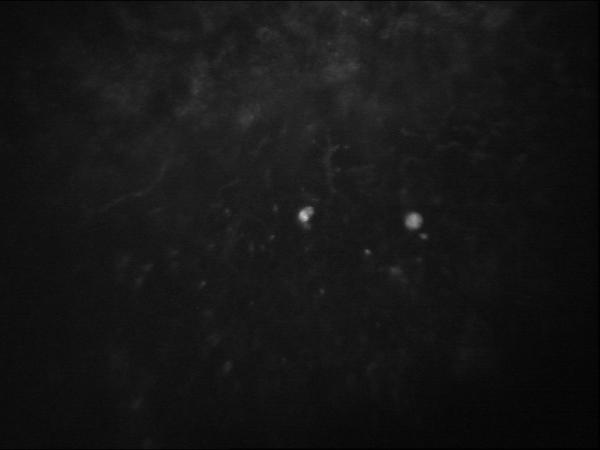
She again presented to her ophthalmologist complaining of blurred vision and photophobia 6 months later and was treated with low dose corticosteroids for bilateral stromal keratitis. After 5 months of therapy, she was referred to the IEEI for a persistently symptomatic and slowly progressive steroid dependent, multifocal stromal keratitis without epithelial, endothelial involvement or neovascularization. Confocal microscopy was positive for Acanthamoeba in the left eye (Figure 1) and equivocal in the right. Scrapings were not performed at this visit because her anterior cornea appeared uninvolved. Propamidine and polyhexamethylene biguanide (PHMB) 0.02% were prescribed hourly with the addition of voriconazole 200mg twice daily by mouth because of the deep keratitis. Propamidine was discontinued after one day because of intolerance, but, despite this, the patient was able to eliminate steroid use over the next three weeks. PHMB was continued for a total of 4 months, tapered to 4 times daily after 6 weeks. The voriconazole was discontinued at the same time. Inflammation returned 6 weeks later and voriconazole 200mg twice daily without corticosteroids was again introduced for two additional months with good subjective and objective response. Eleven months later, she remained asymptomatic with a complete resolution of her corneal inflammation. Liver function tests were monitored throughout her course of therapy and remained normal. Final spectacle corrected visual acuity was 20/25 and 20/20 in the right and left eyes, respectively.
Figure 1.

Case 1. Confocal microscopy showing a dense circular bright white opacity characteristic of the cyst form of Acanthamoeba after 5 months of steroid therapy for inflammatory stromal keratitis.
Case 2
A 14 year old boy presented July, 2006 to the IEEI with an 8 month history of stromal keratitis of the left eye with significant redness, blurred vision and photophobia. He initially had symptoms in November of 2005 after swimming in his contact lenses while vacationing in St. Lucia. The patient had failed treatment with commercially available topical antibacterials and antivirals and was now reliant on topical corticosteroids for comfort. Visual acuity was 20/30, best corrected in the affected eye. Deep and superficial stromal inflammatory lesions found both in the peripheral and central cornea occasionally associated with deep neovascularization extending from the limbus. Minimal epithelial or endothelial changes were noted and no ring-type infiltrate was present. Confocal microscopy was equivocal likely because of the density and depth of the infiltrates. Cultures were initially negative. Immune and infectious serologic tests including syphilis, HSV, EBV, and Lyme disease were also negative. Stromal lesions slowly progressed and the patient remained symptomatic despite empiric chlorhexidine 0.02% for 10 weeks and courses of systemic itraconazole and valacyclovir. (Figure 2) A corneal biopsy was then performed which showed chronic non-granulomatous inflammation and no organisms. The patient remained dependent on a minimum of 1-2 times per week topical corticosteroids and Restasis (Allergan, Irvine, CA) twice a day with occasional but significant exacerbations.
Figure 2.
Case 2. Slit lamp photo demonstrating central stromal keratitis and inferior corneal vascularization related to Acanthamoeba.
Six months after initial presentation, the original specimen was reprocessed utilizing methods appropriate for naked amoeba, now yielding a positive culture for Acanthamoeba. After extensive discussion and informed consent, the patient was given a course of oral voriconazole 200 mg twice daily for two months without concomitant topical treatment. The patient was able to discontinue low dose topical steroids 6 weeks later. 6 weeks after discontinuing voriconazole, mild recurrent inflammation ensued. Further anti-acanthamoebal therapy was offered, but the family elected to continue use of periodic topical corticosteroids only. Episodes continued for the following 14 months until a severe episode of stromal keratitis occurred in October, 2008. A 6 month course of oral voriconazole was administered with rapid and sustained resolution of corneal inflammation without the need for corticosteroids. The eye remained completely free of inflammation 7 months after discontinuing the drug. Liver function tests were monitored throughout his course of therapy and remained normal. Final best corrected visual acuity was 20/25.
Discussion
Our two cases represent patients with culture-confirmed, chronic Acanthamoeba stromal keratitis resistant to treatment with traditional anti-acanthamoebal medications successfully treated with oral voriconazole monotherapy. Although voriconazole was initially used in conjunction with other anti-acanthamoebal drugs, recurrent inflammation resolved with the sole, extended or repeated use of systemic voriconazole. Each introduction of voriconazole induced a rapid reduction in inflammation and allowed for a discontinuation of chronic topical immunosuppressant use with a stable improvement of both vision and comfort.
Voriconazole exhibits better oral absorption and tissue penetration characteristics as well as significantly greater efficacy in fungal infections than previous generation azoles. We chose to use the drug systemically in these patients because of their deeper corneal involvement and to obtain more consistent drug levels. Also, since the sole use of traditional topical anti-acanthamoebal drugs did not result in a similar response, we elected to treat with systemic medications alone for subsequent courses to avoid any barrier effects of the intact epithelium present in our patients. Topical voriconazole 1% has been suggested to have good corneal penetration in the setting of active inflammation and may, in the future, be evaluated as an alternative mode of delivery in patients with active epithelial disease.9
Although primarily an antifungal agent, successful use of voriconazole has been previously reported in disseminated systemic Acanthamoeba infection but only as part of a three drug post-penetrating keratoplasty regimen for AK.7, 10 In vitro studies have shown good activity against Acanthamoeba trophozoites and rapid suppression of Acanthamoeba cysts.2 It is important to note that, in vitro, although exposure to the drug results in rapid suppression, extended exposure to the drug is required for cidal activity.2 Correspondingly, either extended or multiple courses of voriconazole were required in our patients before sustained resolution of inflammation was achieved. Recurrences, if they were to occur, presented within 6 weeks of discontinuing therapy in the form of recrudescent inflammation.
While a small proportion of chronic Acanthamoebal stromal keratitis may be immune rather than infectious,11 the resolution of inflammation in our patients with antibiotic treatment strongly suggests continued infection. It is unknown whether the subsequent infection in case 1 represents reactivation or a new infection since the disease may remain dormant for over a year.12 Regardless, confocal microscopy was positive and the patient experienced a rapid treatment response. In case 2, the patient required periodic topical corticosteroids to suppress corneal inflammation almost continuously from the time of initial presentation, but was only able to discontinue these permanently after oral voriconazole further suggesting persistent infection.
While ideally voriconazole would have been the only drug utilized during the course of therapy, the clinical response in our refractory, culture-confirmed cases presents the clearest indication to date of the efficacy of oral voriconazole as a single agent for the medical treatment of chronic stromal Acanthamoeba keratitis. Despite the availability of highly efficacious topical anti-acanthamoebal therapy, there remains a subset of patients who respond poorly and require alternative, effective compounds or surgical intervention.13, 14 Although a number of compounds have been reported as additive or adjunctive drugs for Acanthamoeba keratitis therapy, few have been demonstrated to be independently efficacious as monotherapeutic agents.15 Voriconazole, should, therefore, be considered in the treatment of chronic stromal keratitis related to Acanthamoeba infection and further studied for use in other forms of Acanthamoeba keratitis.
Footnotes
This is a PDF file of an unedited manuscript that has been accepted for publication. As a service to our customers we are providing this early version of the manuscript. The manuscript will undergo copyediting, typesetting, and review of the resulting proof before it is published in its final citable form. Please note that during the production process errors may be discovered which could affect the content, and all legal disclaimers that apply to the journal pertain.
References
- 1.Kitzmann AS, Goins KM, Sutphin JE, Wagoner MD. Keratoplasty for treatment of Acanthamoeba keratitis. Ophthalmology. 2009 May;116(5):864–869. doi: 10.1016/j.ophtha.2008.12.029. [DOI] [PubMed] [Google Scholar]
- 2.Schuster FL, Guglielmo BJ, Visvesvara GS. In-vitro activity of miltefosine and voriconazole on clinical isolates of free-living amebas: Balamuthia mandrillaris, Acanthamoeba spp., and Naegleria fowleri. J Eukaryot Microbiol. 2006 Mar-Apr;53(2):121–126. doi: 10.1111/j.1550-7408.2005.00082.x. [DOI] [PubMed] [Google Scholar]
- 3.Hirst LW, Green WR, Merz W, et al. Management of Acanthamoeba keratitis. A case report and review of the literature. Ophthalmology. 1984 Sep;91(9):1105–1111. doi: 10.1016/s0161-6420(84)34200-2. [DOI] [PubMed] [Google Scholar]
- 4.Moore MB, McCulley JP, Luckenbach M, et al. Acanthamoeba keratitis associated with soft contact lenses. Am J Ophthalmol. 1985 Sep 15;100(3):396–403. doi: 10.1016/0002-9394(85)90500-8. [DOI] [PubMed] [Google Scholar]
- 5.Driebe WT, Jr., Stern GA, Epstein RJ, Visvesvara GS, Adi M, Komadina T. Acanthamoeba keratitis. Potential role for topical clotrimazole in combination chemotherapy. Arch Ophthalmol. 1988 Sep;106(9):1196–1201. doi: 10.1001/archopht.1988.01060140356031. [DOI] [PubMed] [Google Scholar]
- 6.Ishibashi Y, Matsumoto Y, Kabata T, et al. Oral itraconazole and topical miconazole with debridement for Acanthamoeba keratitis. Am J Ophthalmol. 1990 Feb 15;109(2):121–126. doi: 10.1016/s0002-9394(14)75974-4. [DOI] [PubMed] [Google Scholar]
- 7.Walia R, Montoya JG, Visvesvera GS, Booton GC, Doyle RL. A case of successful treatment of cutaneous Acanthamoeba infection in a lung transplant recipient. Transpl Infect Dis. 2007 Mar;9(1):51–54. doi: 10.1111/j.1399-3062.2006.00159.x. [DOI] [PubMed] [Google Scholar]
- 8.Kaul DR, Lowe L, Visvesvara GS, Farmen S, Khaled YA, Yanik GA. Acanthamoeba infection in a patient with chronic graft-versus-host disease occurring during treatment with voriconazole. Transpl Infect Dis. 2008 Dec;10(6):437–441. doi: 10.1111/j.1399-3062.2008.00335.x. [DOI] [PubMed] [Google Scholar]
- 9.Vemulakonda GA, Hariprasad SM, Mieler WF, Prince RA, Shah GK, Van Gelder RN. Aqueous and vitreous concentrations following topical administration of 1% voriconazole in humans. Arch Ophthalmol. 2008 Jan;126(1):18–22. doi: 10.1001/archophthalmol.2007.8. [DOI] [PubMed] [Google Scholar]
- 10.Ben Salah S, Makni F, Cheikrouhou F, et al. Acanthamoeba keratitis: about the first two Tunisian cases. Bull Soc Pathol Exot. 2007 Feb;100(1):41–42. [PubMed] [Google Scholar]
- 11.Yang YF, Matheson M, Dart JK, Cree IA. Persistence of acanthamoeba antigen following acanthamoeba keratitis. Br J Ophthalmol. 2001 Mar;85(3):277–280. doi: 10.1136/bjo.85.3.277. [DOI] [PMC free article] [PubMed] [Google Scholar]
- 12.Gooi P, Lee-Wing M, Brownstein S, El-Defrawy S, Jackson WB, Mintsioulis G. Acanthamoeba keratitis: persistent organisms without inflammation after 1 year of topical chlorhexidine. Cornea. 2008 Feb;27(2):246–248. doi: 10.1097/ICO.0b013e31815b82a2. [DOI] [PubMed] [Google Scholar]
- 13.Mathers W. Use of higher medication concentrations in the treatment of acanthamoeba keratitis. Arch Ophthalmol. 2006 Jun;124(6):923. doi: 10.1001/archopht.124.6.923. [DOI] [PubMed] [Google Scholar]
- 14.Tu EY, Joslin CE, Sugar J, Shoff ME, Booton GC. Prognostic Factors Affecting Visual Outcome in Acanthamoeba Keratitis. Ophthalmology. 2008 Nov;115(11):1998–2003. doi: 10.1016/j.ophtha.2008.04.038. [DOI] [PMC free article] [PubMed] [Google Scholar]
- 15.Lim N, Goh D, Bunce C, et al. Comparison of polyhexamethylene biguanide and chlorhexidine as monotherapy agents in the treatment of Acanthamoeba keratitis. Am J Ophthalmol. 2008 Jan;145(1):130–135. doi: 10.1016/j.ajo.2007.08.040. [DOI] [PubMed] [Google Scholar]


