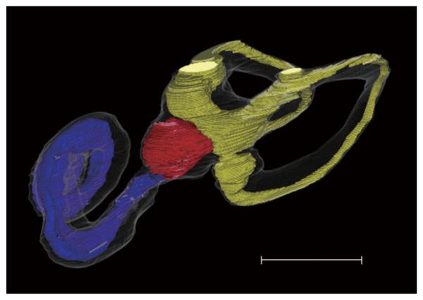
Fig. 3.
Computer-generated three-dimensional images of Ménière disease (anterior-medial view). The superior semicircular canal is not reconstructed. Blue, red, and yellow indicate cochlear duct, saccule, and utricle, respectively. Note cochlear and saccular hydrops. Scale bar indicates 5 mm. [Color figure can be viewed in the online issue, which is available at www.interscience.wiley.com.]

