Abstract
OBJECTIVE
We studied differences between patients with latent autoimmune diabetes in adults (LADA), type 2 diabetes, and classical type 1 diabetes diagnosed after age 35 years.
RESEARCH DESIGN AND METHODS
Polymorphisms in HLA-DQB1, INS, PTPN22, and CTLA4 were genotyped in patients with LADA (n = 213), type 1 diabetes diagnosed at >35 years of age (T1D>35y; n = 257) or <20 years of age (T1D<20y; n = 158), and type 2 diabetes.
RESULTS
Although patients with LADA had an increased frequency of HLA-DQB1 and PTPN22 risk genotypes and alleles compared with type 2 diabetic subjects, the frequency was significantly lower compared with T1D>35y patients. Genotype frequencies, measures of insulin secretion, and metabolic traits within LADA differed according to GAD antibody (GADA) quartiles, but even the highest quartile differed from type 1 diabetes. Having two or more risk genotypes was associated with lower C-peptide concentrations in LADA.
CONCLUSIONS
LADA patients differed genetically and phenotypically from both T1D>35y and type 2 diabetic patients in a manner dependent on GADA levels.
Patients with latent autoimmune diabetes in adults (LADA) have a progressive insulin secretion defect, have less evidence of metabolic syndrome than type 2 patients, and share a genetic predisposition with both type 1 and type 2 diabetic patients (1–5). Whether LADA merely represents older-onset type 1 diabetes or is a distinct subgroup has been debated (6), but studies comparing LADA with classical type 1 diabetes in a comparative age-range are lacking. We investigated 1) whether LADA differed genetically (with respect to genes associated with risk of type 1 diabetes: HLA-DQB1, PTPN22, INS, and CTLA4) and phenotypically (with respect to metabolic syndrome) from type 1 diabetes diagnosed after age 35 years; and 2) whether the observed clinical heterogeneity within LADA depended on GAD antibody (GADA) levels and type 1 diabetes susceptibility genotypes.
RESEARCH DESIGN AND METHODS
We included patients with age at onset >35 years with LADA (defined here as GADA-positive diabetes without insulin treatment for the first 6 months; n = 213) or with type 1 diabetes (T1D>35y; n = 35), patients with type 1 diabetes diagnosed at <20 years of age (T1D<20y; n = 158), and GADA-positive type 2 diabetic patients (n = 648) from the Botnia study (2) together with T1D>35y patients (n = 222) from the FinnDiane study (7). The non–insulin-treated subjects underwent an oral glucose tolerance test with blood samples drawn at −5, 0, 30, 60, and 120 min for measurement of plasma glucose, serum insulin, C-peptide, lipids, and GADA levels (8). The HLA-DQB1*02, *0301, *0302, *0602, and *0603 alleles were genotyped (9) and classified as risk (*0302/*02, 0302/X), protective [*0602(3)/X, *0602(3)/*0301], or neutral (all other) (X = homozygous or other allele). The INS − Hph23 A/T (rs689), CTLA4 CT60 (rs3087243), and PTPN22 1858C>T (rs2476601) variants were genotyped with TaqMan allelic discrimination (Applied Biosystems, Foster City, CA). Statistical analyses were performed with Statistical Package for Social Science software (version 13.0; SPSS, Chicago, IL) using the χ2 test, the Kruskal-Wallis test, or a general linear model. Participants gave informed consent, and the study was approved by the local ethics committee.
RESULTS
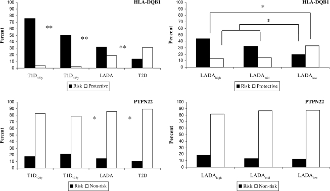
The HLA-DQB1 risk genotypes were most frequent in T1D<20y patients (75.3%), followed by T1D>35y patients (50.2%) and LADA patients (32.2%) (P < 0.00001; Fig. 1), with the opposite trend for the protective genotypes. The type 1 diabetic patients also had a higher frequency of the PTPN22 risk allele (T1D<20y 17.7% and T1D>35y 21.1%) than LADA patients (14.5%; P = 0.008). Compared with type 2 diabetes, the HLA-DQB1 (P < 0.00001) and PTPN22 risk genotypes and alleles (P = 0.04–0.008) were significantly more common in LADA. The INS and CTLA4 risk genotypes were only associated with type 1 diabetes (T1D>35y, LADA, type 2 diabetes: INS 77.4, 58.7, and 56.5%, respectively, and CTLA4 48.2, 45.0, and 41.6%, respectively).
Figure 1.
On the left, distribution of HLA-DQB1 genotypes and PTPN22 (rs2476601) alleles in type 1 diabetic patients (T1D<20y, n = 158) or after the age of 35 years (T1D>35y, n = 257), LADA patients (n = 213), and type 2 diabetic patients (T2D, n = 648). On the right, distribution of HLA-DQB1 genotypes and PTPN22 (rs2476601) alleles in LADA patients stratified according to GADA quartiles: the highest, LADAhigh (n = 52); the two middle quartiles pooled, LADAmid (n = 109); and the lowest, LADAlow (n = 52). Black columns represent risk genotypes and alleles, and white columns represent protective (HLA-DQB1) or nonrisk (PTPN22) genotypes and alleles. The χ2 test was applied to test differences between diabetic groups. Significant differences are indicated with asterisks. *P < 0.05. **P < 0.0001. HLA-DQB1: P = 0.01 across the GADA quartiles of LADA.
All risk genotypes and alleles were most prevalent in patients in the highest GADA quartile (LADAhigh, GADA >278 IU/ml) compared with the two middle (LADAmid, 44–278 IU/ml) or the lowest quartiles (LADAlow, 33–43.9 IU/ml; Fig. 1), but the difference was significant only for HLA-DQB1 (P = 0.009). However, even LADAhigh subjects differed from T1D>35y subjects (P = 0.001), and LADAlow subjects had nonsignificantly more HLA-DQB1 risk genotypes than type 2 diabetic subjects (19.6 vs. 14.0%). LADAhigh subjects also differed from the type 2 diabetic subjects with respect to the allele frequency of PTPN22 (P = 0.02) and CTLA4 (P = 0.03).
In a joint analysis of the four genes, two or more risk genotypes were most common in T1D<20y patients (82.1%), followed by T1D>35y patients (74.4%), LADA (54.1%), and type 2 diabetic (38.7%) patients (P < 0.00001). The frequency differed across the GADA quartiles (P = 0.004), and it was similar in LADAhigh and T1D>35y patients (72.5 vs. 74.4%), as well as in LADAlow and type 2 diabetic patients (46.0 vs. 38.7%) (online appendix Table A3, available at http://care.diabetesjournals.org/cgi/content/full/dc09-2188/DC1). LADA patients with an increasing number of risk genotypes had decreasing fasting serum (fS)–C-peptide concentrations (P = 0.015).
The LADA patients had higher BMI and lipid concentrations than the T1D>35y patients (online appendix Table A1). Compared with type 2 diabetic patients, LADA patients had lower insulin secretion and BMI, as well as a better lipid profile. Going from LADAlow to LADAhigh, there was a significant trend toward lower insulin secretion, lipid levels, and BMI (online appendix Table A2). However, the fS–C-peptide concentration was higher in LADAhigh patients compared with T1D>35y patients and was somewhat lower in LADAlow compared with type 2 diabetic patients. Despite the marked age difference, the T1D>35y and T1D<20y patients were metabolically similar.
CONCLUSIONS
We have shown a significant genetic difference between LADA and type 1 diabetes diagnosed after age 35 years. Although HLA-DQB1 and PTPN22 risk genotypes were increased in LADA, they were much less common than in type 1 diabetes. INS and CTLA4 were only associated with type 1 diabetes.
As suggested previously (3,5,10), genetic differences and clinical phenotype were associated with GADA levels, explaining some of the observed heterogeneity within LADA. Of note, the LADAhigh group (GADA >278 IU/ml) roughly corresponds to the high GADA group (>300 IU/ml) in the Non Insulin Requiring Autoimmune Diabetes (NIRAD) Study, in which association with PTPN22 was also found only in patients with high GADA (10).
Our study is the largest to date in adult-onset type 1 diabetic patients. In agreement with a smaller study (11), we clearly showed a lower frequency of DQB1 risk genotypes in T1D>35y compared with T1D<20y patients. However, protective HLA-DQB1 genotypes were as rare in T1D>35y as in T1D<20y patients. The prevalence of INS, PTPN22, and CTLA4 risk genotypes was similar in the two groups. As expected (2,4,10,12–14), LADA had an increased frequency of HLA-DQB1 and PTPN22 risk genotypes as well as a decreased frequency of HLA-DQB1 protective genotypes compared with type 2 diabetic subjects. Contrary to previous reports (4,14,15), the INS variant was not associated with LADA in the Finnish subjects.
In conclusion, we have shown that patients with LADA differ genetically and phenotypically from type 1 diabetic patients diagnosed after age 35 years. The LADA group was heterogeneous, and both the genotype distribution and phenotypic characteristics were associated with GADA level. A significant trend was observed toward lower insulin secretion and metabolic trait values going from the lowest GADA quartile to the highest. Thus, LADA patients with high GADA concentrations were more similar, but not identical, to type 1 diabetic patients, and those with low GADA concentrations were more similar to type 2 diabetic patients.
Acknowledgments
This study was supported by grants from the Academy of Finland, the Sigrid Juselius Foundation, the Finnish Diabetes Research Foundation, the Folkhälsan Research Foundation, the Wilhelm and Else Stockmann Foundation, the Finska Läkaresällskapet, the Novo Nordisk Foundation, the Swedish Cultural Foundation in Finland, the Ollquist Foundation, the Korsholm, Malax, Närpes, and Vasa Heath Care Centers, and the Helsinki University Central Hospital.
L.G. has been a consultant and served on advisory boards for sanofi-aventis, Glaxo- SmithKline, Novartis, Merck, Tethys Bioscience, and Xoma and has received lecture fees from Eli Lilly and Novartis. No other potential conflicts of interest relevant to this article were reported.
M.K.A., V.L., J.A.T., C.F., B.I., and T.T. researched data. M.K.A., V.L., and T.T. wrote the manuscript. J.A.T., C.F., B.I., P.-H.G., and L.G. reviewed and edited the manuscript and contributed to the discussion.
The authors acknowledge Paula Kokko, Anita Nilsson, and Barbro Gustavsson for help with the genotyping and also acknowledge the Botnia Research Group and the FinnDiane Research Group for recruiting and clinically studying the subjects.
Footnotes
The costs of publication of this article were defrayed in part by the payment of page charges. This article must therefore be hereby marked “advertisement” in accordance with 18 U.S.C. Section 1734 solely to indicate this fact.
References
- 1. Turner R, Stratton I, Horton V, Manley S, Zimmet P, Mackay IR, Shattock M, Bottazzo GF, Holman R: UKPDS 25: autoantibodies to islet-cell cytoplasm and glutamic acid decarboxylase for prediction of insulin requirement in type 2 diabetes. UK Prospective Diabetes Study Group. Lancet 1997;350:1288–1293 [DOI] [PubMed] [Google Scholar]
- 2. Tuomi T, Carlsson A, Li H, Isomaa B, Miettinen A, Nilsson A, Nissén M, Ehrnström BO, Forsén B, Snickars B, Lahti K, Forsblom C, Saloranta C, Taskinen MR, Groop LC: Clinical and genetic characteristics of type 2 diabetes with and without GAD antibodies. Diabetes 1999;48:150–157 [DOI] [PubMed] [Google Scholar]
- 3. Buzzetti R, Di Pietro S, Giaccari A, Petrone A, Locatelli M, Suraci C, Capizzi M, Arpi ML, Bazzigaluppi E, Dotta F, Bosi E: Non Insulin Requiring Autoimmune Diabetes Study Group. High titer of autoantibodies to GAD identifies a specific phenotype of adult-onset autoimmune diabetes. Diabetes Care 2007;30:932–938 [DOI] [PubMed] [Google Scholar]
- 4. Cervin C, Lyssenko V, Bakhtadze E, Lindholm E, Nilsson P, Tuomi T, Cilio CM, Groop L: Genetic similarities between latent autoimmune diabetes in adults, type 1 diabetes, and type 2 diabetes. Diabetes 2008;57:1433–1437 [DOI] [PubMed] [Google Scholar]
- 5. Pettersen E, Skorpen F, Kvaløy K, Midthjell K, Grill V: Genetic heterogeneity in latent autoimmune diabetes is linked to various degrees of autoimmune activity: results from the Nord-Trøndelag Health Study. Diabetes 2010;59:302–310 [DOI] [PMC free article] [PubMed] [Google Scholar]
- 6. Gale EA: Latent autoimmune diabetes in adults: a guide for the perplexed. Diabetologia 2005;48:2195–2199 [DOI] [PubMed] [Google Scholar]
- 7. Thorn LM, Forsblom C, Fagerudd J, Thomas MC, Pettersson-Fernholm K, Saraheimo M, Wadén J, Rönnback M, Rosengård-Bärlund M, Björkesten CG, Taskinen MR, Groop PH: FinnDiane Study Group. Metabolic syndrome in type 1 diabetes: association with diabetic nephropathy and glycemic control (the FinnDiane study). Diabetes Care 2005;28:2019–2024 [DOI] [PubMed] [Google Scholar]
- 8. Lundgren VM, Isomaa B, Lyssenko V, Laurila E, Korhonen P, Groop LC, Tuomi T: Botnia Study Group. GAD antibody positivity predicts type 2 diabetes in an adult population. Diabetes 2010;59:416–422 [DOI] [PMC free article] [PubMed] [Google Scholar]
- 9. Sjöroos M, Iitiä A, Ilonen J, Reijonen H, Lövgren T: Triple-label hybridization assay for type-1 diabetes-related HLA alleles. BioTechniques 1995;18:870–877 [PubMed] [Google Scholar]
- 10. Petrone A, Suraci C, Capizzi M, Giaccari A, Bosi E, Tiberti C, Cossu E, Pozzilli P, Falorni A, Buzzetti R: NIRAD Study Group. The protein tyrosine phosphatase nonreceptor 22 (PTPN22) is associated with high GAD antibody titer in latent autoimmune diabetes in adults: Non Insulin Requiring Autoimmune Diabetes (NIRAD) Study 3. Diabetes Care 2008;31:534–538 [DOI] [PubMed] [Google Scholar]
- 11. Caillat-Zucman S, Garchon HJ, Timsit J, Assan R, Boitard C, Djilali-Saiah I, Bougnères P, Bach JF: Age-dependent HLA genetic heterogeneity of type 1 insulin-dependent diabetes mellitus. J Clin Invest 1992;90:2242–2250 [DOI] [PMC free article] [PubMed] [Google Scholar]
- 12. Hosszúfalusi N, Vatay A, Rajczy K, Prohászka Z, Pozsonyi E, Horváth L, Grosz A, Gerõ L, Madácsy L, Romics L, Karádi I, Füst G, Pánczél P: Similar genetic features and different islet cell autoantibody pattern of latent autoimmune diabetes in adults (LADA) compared with adult-onset type 1 diabetes with rapid progression. Diabetes Care 2003;26:452–457 [DOI] [PubMed] [Google Scholar]
- 13. Desai M, Zeggini E, Horton VA, Owen KR, Hattersley AT, Levy JC, Walker M, Gillespie KM, Bingley PJ, Hitman GA, Holman RR, McCarthy MI, Clark A: An association analysis of the HLA gene region in latent autoimmune diabetes in adults. Diabetologia 2007;50:68–73 [DOI] [PMC free article] [PubMed] [Google Scholar]
- 14. Haller K, Kisand K, Pisarev H, Salur L, Laisk T, Nemvalts V, Uibo R: Insulin gene VNTR, CTLA-4 +49A/G and HLA-DQB1 alleles distinguish latent autoimmune diabetes in adults from type 1 diabetes and from type 2 diabetes group. Tissue Antigens 2007;69:121–127 [DOI] [PubMed] [Google Scholar]
- 15. Desai M, Zeggini E, Horton VA, Owen KR, Hattersley AT, Levy JC, Hitman GA, Walker M, Holman RR, McCarthy MI, Clark A: The variable number of tandem repeats upstream of the insulin gene is a susceptibility locus for latent autoimmune diabetes in adults. Diabetes 2006;55:1890–1894 [DOI] [PubMed] [Google Scholar]


