Abstract
Background
Differences in survival have been reported between pediatric and adult acute lymphoblastic leukemia. The inferior prognosis in adult acute lymphoblastic leukemia is not fully understood but could be attributed, in part, to differences in genomic alterations found in adult as compared to in pediatric acute lymphoblastic leukemia.
Design and Methods
We compared two different sets of high-density single nucleotide polymorphism array genotyping data from 75 new diagnostic adult and 399 previously published diagnostic pediatric acute lymphoblastic leukemia samples. The patients’ samples were randomly acquired from among Caucasian and Asian populations and hybridized to either Affymetrix 50K or 250K single nucleotide polymorphism arrays. The array data were investigated with Copy Number Analysis for GeneChips (CNAG) software for allele-specific copy number analysis.
Results
The high density single nucleotide polymorphism array analysis of 75 samples of adult acute lymphoblastic leukemia led to the identification of numerous cryptic and submicroscopic genomic lesions with a mean of 7.6 genomic alterations per sample. The patterns and frequencies of lesions detected in the adult samples largely reproduced known genomic hallmarks detected in previous single nucleotide polymorphism-array studies of pediatric acute lymphoblastic leukemia, such as common deletions of 3p14.2 (FHIT), 5q33.3 (EBF), 6q, 9p21.3 (CDKN2A/B), 9p13.2 (PAX5), 13q14.2 (RB1) and 17q11.2 (NF1). Some differences between adult and pediatric acute lymphoblastic leukemia were identified when the pediatric data set was partitioned into hyperdiploid and non-hyperdiploid cases and then compared to the nearly exclusively non-hyperdiploid adult samples. In this analysis, adult samples had a higher rate of deletions of chromosome 17p (TP53) and duplication of 17q.
Conclusions
Our analysis of adult acute lymphoblastic leukemia cases led to the identification of new potential target lesions relevant for the pathogenesis of acute lymphoblastic leukemia. However, no unequivocal pattern of submicroscopic genomic alterations was found to separate adult acute lymphoblastic leukemia from pediatric acute lymphoblastic leukemia. Therefore, apart from different therapy regimen, differences of prognosis between adult and pediatric acute lymphoblastic leukemia are probably based on genetic subgroups according to cytogenetically detectable lesions but not focal genomic copy number microlesions.
Keywords: SNP-array, adult ALL, pediatric ALL, comparative study
Introduction
Acute lymphoblastic leukemia (ALL) is a malignant disease resulting from the accumulation of genetic alterations of B or T lymphoid precursor cells.1,2 ALL is the most common leukemia in children and accounts for 20% of acute leukemias in adults. The 5-year event-free survival is over 80% for children but only approximately 40% for adult ALL.2
The development of ALL involves chromosomal changes including translocations causing fusion genes, as well as hyperdiploidy containing more than 50 chromosomes. The frequency of particular chromosomal alterations differs between pediatric and adult ALL. About 50% of pediatric cases of B-lineage leukemia (B-ALL) have either a t(12;21) translocation (TEL-AML1) or hyperdiploidy; only 10% of adult B-ALL cases have these abnormalities. In contrast, approximately 30% of adult B-ALL samples contain a t(9;22) translocation (BCR-ABL1), but only a few percent of pediatric B-ALL have this translocation. MLL rearrangements including t(4;11), t(11;19) and t(9;11) translocations are found in 8–13% of both adult and pediatric B-ALL.3 Over-expression of the transcription factor TAL1 is observed in about 60% of pediatric T-progenitor leukemias (T-ALL) and 30–45% of adult T-ALL.2,4,5 Although many genomic abnormalities such as translocations are known in ALL, several new cryptic abnormalities have been identified recently using new techniques such as high density single nucleotide polymorphism (SNP) arrays.1,6–8 Previously, we developed the Copy-Number Analysis for Affymetrix GeneChips (CNAG) software and a new algorithm, allele-specific copy-number analysis using anonymous references (AsCNAR).6,9 The SNP-array analysis using these tools provides a highly sensitive platform to detect large and small copy-number changes, as well as copy number neutral loss-of-heterozygosity (CNN-LOH), which is represented by one allele being deleted and the other allele duplicated. The latter cannot be detected by either karyotypic or comparative genomic hybridization analyses.
The full complement of cooperating lesions and their distribution within the known genetic subtypes of ALL remain to be defined and a detailed comparison study by SNP-array between pediatric and adult ALL has not been performed. In the present study, we investigated a cohort of 75 adult ALL samples including 61 B-ALL and 14 T-ALL samples to identify cryptic chromosomal alterations using SNP-array analysis and compared these genomic changes to those present in 399 previously analyzed and published pediatric ALL samples.7
Design and Methods
Patients’ samples
Seventy-five anonymized adult ALL samples were obtained from bone marrow or peripheral blood at initial diagnosis. Sample information included gender, immunophenotype, white blood cell count (WBC) and occurrence of BCR-ABL and MLL-AF4 fusions as examined by karyotyping, reverse transcriptase polymerase chain reaction and fluorescence in situ hybridization analysis (Online Supplementary Tables S1 and S2). The cohort contained 41 Asian (Japanese) patients who were diagnosed with ALL at the University of Tokyo between October 1994 and May 1997. These samples were acquired in line with a study involving the JALSG protocol, which was approved by the ethics boards of the University of Tokyo. The remaining 34 samples were from patients of Caucasian origin, diagnosed in the Munich Leukemia Laboratory between October 2005 and April 2006, in the context of the GMALL study protocols. Informed consent to sample acquisition and processing was obtained in accordance with the Declaration of Helsinki in all cases. All samples were acquired in the chronological order of entry without any selection or bias. In total, 61 cases of B-ALL, including two null ALL cases lacking both B and T cell markers, one mixed ALL, and 14 cases of T-ALL were examined. At diagnosis the patients were aged from 19 to 86 years old (Online Supplementary Figure S1). The karyotypes of all samples are listed in Online Supplementary Table S2.
High-density single nucleotide polymorphism-array analysis
Genomic DNA from adult ALL cells was subjected to GeneChip Human mapping processing protocols. Forty samples were processed with 50K arrays and 35 samples with 250K arrays (Affymetrix, Santa Clara, CA, USA). Detailed information about which individual samples were hybridized to specific arrays is given in the Online Supplementary Methods. Hybridization, washing and signal detection were performed on a GeneChip Fluidics Station 450 and GeneChip scanner 3000 according to the manufacturer’s protocols (Affymetrix). Microarray data were analyzed for determination of both total and allelic-specific copy numbers using the CNAG software as previously described,6,9–11 with minor modifications: 11 cases were examined with their paired normal DNA from remission samples, and 64 cases were analyzed with anonymous normal references. For clustering of ALL samples with regard to the status of copy number changes, as well as CNN-LOH, the CNAG graph software was used.7 The size, position and location of genes were identified with the UCSC Genome Browser (http://genome.ucsc.edu/). Registered copy number variants or IgG rearrangements were eliminated using either http://projects.tcag.ca/variation/orhttp://genome.ucsc.edu/.
Results
Single nucleotide polymorphism-array analysis of 75 adult acute lymphoblastic leukemia samples
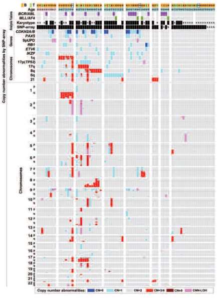
The SNP-array analysis of our 75 adult ALL samples revealed that 69/75 (92%) samples including 61 cases of BALL and 14 cases of T-ALL had one or more genomic abnormalities (Figure 1). A total of 572 genomic alterations were detected, with a mean of 7.6 genomic alterations per sample (Table 1A). Homozygous deletions (N=19) (Online Supplementary Table S3) and heterozygous deletions (N=349) (Online Supplementary Table S4) were more frequent than either duplications (N=169) (Online Supplementary Table S5) or amplifications with five or more copy number changes, which occurred in only two samples (Online Supplementary Table S6). CNN-LOH was detected in 33 regions (Online Supplementary Table S7).
Figure 1.
Summary of SNP array analysis of adult ALL. Seventy-five adult ALL samples were subjected to SNP-array analysis. The groups were separated by types of genomic abnormalities. Orange, B-cell type ALL; light green, T-cell type ALL; white, null-ALL, (cases #B-59, −60, −61); purple, BCR-ABL positive; yellow green, MLL/AF4 positive. Black box = abnormalities detected in the sample; N = no abnormalities detected in the sample. Gray = normal copy number; red = duplication; blue = heterozygous deletion; dark blue = homozygous deletion; pink = CNN-LOH; p = short arm; q = long arm; CN = copy number; 0 = 0 N (homozygous deletion); 1 = 1 N (heterozygous deletion); 2 = 2 N (normal); ¾ = 3 or 4 N (duplication); ≥5 = ≥5 N (amplification); CNN-LOH = copy number neutral loss-of-heterozygosi-ty. The groups were separated based on abnormalities and age: group 1, adolescent group (19–21 years old); group 2, 1q duplication; group 3, either 17p deletion or 17q duplication or 8q duplication; group 4, 6q deletion; group 5, one or more deletions of CDKN2A/B, PAX5, 9pUPD, RB1, ETV6 and IKZF; group 6, chromosome 21 duplication; group 7, either BCR/ABL or MLL/AF4; group 8, other than above.
Table 1A.
Number of copy number changes in adult acute lymphoblastic leukemia.
When the adult ALL cases were segregated by either age or ethnicity, no differences were found in the types of genomic lesions or their frequencies between the different groups (Online Supplementary Tables S13 and S14).
Clustering of common genetic lesions in all samples is displayed in Figure 1, in which groups were separated based on abnormalities and furthermore, adult versus adolescents (19–21 years old); 1q duplication; either 17p deletion or 17q duplication or 8q duplication; 6q deletion; one or more deletions of CDKN2A/B, PAX5, IKZF, RB1, ETV6 or CNN-LOH on chromosome 9p; chromosome 21 duplication; either BCR/ABL or MLL/AF4; and other than above.
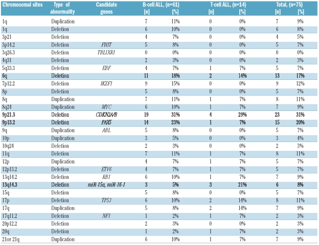
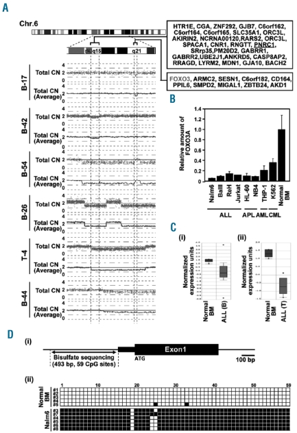
The recurrent genetic abnormalities detected by SNP-array are shown in Table 1b and Online Supplementary Figure S3. Among these, the three most common genetic abnormalities were deletions of chromosome 9p21.3 (CDKN2A/B, N=23, 31%), 9p13.2 (PAX5, N=15, 20%) and chromosome 6q (N=13, 17%). Chromosome 9p was also most frequently affected by CNN-LOH (Online Supplementary Table S7). One of the commonly deleted regions on chromosome 6 ranged from 108,940,080 to 109,998,869 and involved the FOXO3 gene as a possible candidate gene (Figure 2A). FOXO3 was also found to be frequently deleted in pediatric ALL (32/399, 8%) (Online Supplementary Figure S4).1,12 We analyzed the expression of FOXO3 in several ALL cell lines such as Nalm6, Ball, ReH and Jurkat and found markedly lower levels of FOXO3 in these cell lines than in cell lines of myeloid origin or healthy bone marrow (Figure 2B). These data, which show that deregulated FOXO3 expression in ALL may be of pathogenic relevance to this disease, were also corroborated by results found using ONCOMINE https://www.oncomine.org13 in the Andersson Leukemia study (pediatric ALL), which demonstrated that the expression of FOXO3 was lower in both B- and T-cell ALL than in normal bone marrow samples (Figure 2C). A possible reason for this differential expression of FOXO3 in ALL as compared to in healthy bone marrow could be promoter hypermethylation as we found that the promoter region of FOXO3 was more highly methylated in gDNA of the B-ALL leukemia cell line Nalm6 than in normal gDNA (Figure 2D). Another interesting common lesion was identified on chromosome 12, where BTG1, which had been found to be deleted frequently in pediatric ALL1,7 was involved in breakpoints in seven cases (9%) (Online Supplementary Table S4).
Table 1B.
Recurrent genetic abnormalities detected by SNP-array analysis in 61 B-cell type ALL and 14 T-cell type ALL.
Figure 2.
Identification of target genes by determination of commonly affected genomic regions with the CNAG (Copy Number Analyser for GeneChip®) software. (A) Deletions on chromosome 6q of a collection of adult ALL samples: candidate genes are listed on the right side of the figure. While many samples harbor large deletions containing hundreds of potential target genes of the genomic lesion, overlapping the data from other samples leads to a reduction of size of the common region. This is impressively shown here in adult ALL for heterozygous deletions pointing to the candidate region as q15 containing the FOXO3 gene, which was also involved in a breakpoint in case #B-17. (B) Expression levels of FOXO3 in various cell lines. FOXO3 expression levels were measured in ALL as well as APL, AML and CML cell lines by real-time PCR and compared with expression in normal bone marrow. (C) FOXO3 expression profiling by microarray in childhood acute leukemia by Oncomine. Gene expression analyses were performed on (i) 87 samples of B-ALL and (ii) 11 samples of T-ALL of the Andersson_Leukemia study (http://www.ncbi.nlm.nih.gov/geo/query/acc.cgi?acc=GSE7186). FOXO3 expression was lower in both B-ALL and T-ALL samples than in normal bone marrow (P= 1.6×10⁻¹¹ and 1.2×10−08, respectively). ALL(B) = B-cell type acute lymphoblastic leukemia; ALL (T) = T-cell type acute lymphoblastic leukemia; BM = bone marrow. (D) Methylation analysis of the FOXO3 promoter region (i) A scheme of the FOXO3 promoter region. (ii) Results of bisulfate sequencing are shown. At least five colonies were examined. White box = non-methylated CpG; Black box = methylated CpG.
Besides recurrent alterations, interesting submicroscopic lesions were detectable in single cases, possibly indicating new candidate genes in the pathogenesis of ALL: two focal homozygous deletions were found. In case # B-14, a region of CNN-LOH on chromosome 9p contained a homozygous deletion of PTPRD (protein tyrosine phosphatase, receptor type D) (Online Supplementary Figure S2A). Case #B-35 contained a submicroscopic homozygous deletion of chromosome 10q (98,441,682 – 98,547,438, 0.11Mb), which only contained exons 1 and 2 of PIK3AP1 (phosphoinositide-3-kinase adaptor protein 1). The remaining part of the PIK3AP1 gene was heterozygously deleted (Online Supplementary Table S3 and S4). Interestingly, this case also displayed a duplication of chromosome 14 (22,069,902 – 105,685,710, 83.616Mb), which contained AKT1 (Online Supplementary Table S5).
Of note, although 28 cases were categorized as having a normal karyotype ALL by initial karyotypic analysis, 24 of these cases (86%) had genomic alterations detected by SNP-array analysis, corroborating the power of this latter technique to reveal cryptic genomic abnormalities in hematologic malignancies.
Table 2B.
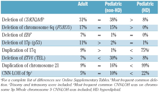
Additional differences between adult and pediatric acute lymphoblastic leukemia identified in this study.a
Identification of fusion genes in acute lymphoblastic leukemia
Genomic alterations found by SNP arrays are often involved in chromosomal translocations. Balanced translocations cannot be detected by SNP-array analysis, since they are not accompanied by copy number changes. However, unbalanced translocations associated with copy number changes are often detectable by this technique with a high resolution. An example is shown by the SNP-array tracing of case #B-20, who had a duplication in the middle of the PAX5 gene (9p13.2) and in the middle of the ETV6 gene (12p13.2) (Online Supplementary Figure S5B[i]). Initial karyotypic analysis of this case showed +8, der(9)r(9)ins(9;12), der(12)t(9;12)(?;q15). Primers, which were designed at the break-points of PAX5 and ETV6, amplified a polymerase chain reaction product; and nucleotide sequencing of this product showed that the fusion occurred between exon 4 of PAX5 and exon 3 of ETV6 (Online Supplementary Figure S5B[ii]). Apart from this fusion, PAX5 internal deletions were detected in three of our adult ALL samples underlining the important role of PAX5 gene disruptions in B-ALL.
Differences between adult and pediatric acute lymphoblastic leukemia samples
In a previous study, our group carried out a SNP array analysis of 399 pediatric ALL cases using Affymetrix 50K arrays.7 In the current study, we sought to compare the genomic changes found in our adult ALL cases with those of the pediatric cases of the previous study in order to find leads to biological differences between these two age groups. Since the analysis of the adult samples was largely carried out with higher density 250K arrays and is, therefore, only comparable to the 50K pediatric data set to a limited extent, we introduced a threshold of 141 kb per lesion for acceptance of detected genomic abnormalities from the 250K array adult data set. This is based on the technical characteristics of the 50K SNP-arrays that interrogate the genome with a total combined number of approximately 58,000 SNP and a median intermarker distance of 47 kb. Because we defined three SNP per copy number abnormality as the minimum requirement for acceptance of a genomic lesion, we defined this smallest common denominator to make the adult 250K data comparable to the 50K pediatric data. The differences detected by this comparison are summarized in Table 2, Online Supplementary Tables S8-S11 and Online Supplementary Figure S6. The data show that almost all types of recurrent genomic lesions occur at a similar rate in pediatric ALL and adult ALL.
Table 2A.
Differences between adult and pediatric acute lymphoblastic leukemia. Known differences.2
However, of note, hyperdiploidy was not detected in the 75 adult ALL samples (Online Supplementary Table S2), but was found in 116 pediatric ALL samples (29%, P=4.3×10⁻⁷).7 We, therefore, separated the pediatric ALL samples into hyperdiploid (HD) and non-hyperdiploid (non-HD) groups (Online Supplementary Table S12). Nevertheless, after subdivision of the groups, we found that deletions of 3p14.2 (FHIT), 9p21.3 (CDKN2A/B), 9p13.2 (PAX5), 13q14.2 (RB1) and 17q11.2 (NF1) occurred at a comparable rate in adult and non-HD-pediatric ALL (Online Supplementary Table S9). As expected, these deletions were rare in HD-pediatric ALL (Online Supplementary Table S10). The only two differences detected pertained to deletions encompassing ETV6 on chromosome 12 and alterations on chromosome 17: deletions of 12p13.2 (ETV6) were more common in non-HD-pediatric ALL (30%) than in adult ALL (7%, P=0.00001). Furthermore, genomic changes of both deletion of 17p (TP53) and duplication of 17q (11% and 9%, respectively) were more frequent in adult ALL than in non-HD-pediatric ALL (2% and 1%, P=0.005 and P=0.002, respectively).
Discussion
Our genome-wide SNP-array analysis of adult ALL showed that 92% of samples had one or more genomic abnormalities including deletions, duplications and/or CNN-LOH (Figure 1). As anticipated, SNP-array analysis was able to detect genomic abnormalities in numerous samples that cytogenetically appeared to be normal. Twenty-four of the 28 samples (86%) that had a normal karyotype contained genomic abnormalities detected by SNP-array (Online Supplementary Table S2).
The most common abnormalities were found on chromosome 9 in adult ALL (Online Supplementary Figure S3). Our analysis revealed that 9p CNN-LOH was frequently found in both adult and pediatric ALL (Online Supplementary Figure S6). CNN-LOH is interesting in cancer research because of the high probability of such abnormalities containing either a mutated tumor suppressor gene (TSG) or oncogene with loss of their normal allele. We have previously shown that JAK2-V617F is often duplicated by CNN-LOH in normal karyotype acute myeloid leukemia.10 Also, JAK1/2/3 have been reported to be mutated in childhood ALL.14 In our adult ALL samples, we found several recurrent CNN-LOH regions, containing genes coding for tyrosine kinases and putative tumor suppressors, which represent interesting target genes for further, functional evaluation in the context of ALL (Online Supplementary Table S7). We have previously shown in pediatric ALL that CDKN2A/B deletions are often found in 9p CNN-LOH (23 of 30, 77%), while they are less frequent in whole chromosome 9 CNN-LOH (1 of 18, 6%).7 Three of 12 samples (25%) with homozygous deletions of the CDKN2A/B genes had 9p CNN-LOH in our adult ALL samples while the rest of these focal deletions occurred without 9p CNN-LOH (Online Supplementary Figure S7 and Online Supplementary Table S3). Interestingly, in one case, 9p CNN-LOH also contained a homozygous deletion of PTPRD, suggesting that this may also be a candidate gene in this region (Online Supplementary Figure S2A). Some pre-B-cell lines and some B-cell lines express PTPRD.15 Previous studies by others and ourselves found disruption of the PTPRD gene in lung carcinoma,16,17 neuroblastoma,18 cutaneous squamous cell carcinomas19 and relapsed pediatric ALL.12
Moreover, PAX5 is located on chromosome 9p. PAX5 is critical for B-cell development; without it, cells are blocked at the pro-B stage. Lymphoid cells lacking PAX5 in an experimental model can differentiate into either T cells or myeloid cells with the appropriate signals, highlighting the important role that PAX5 plays in B-cell development.20 In our study, PAX5 deletions occurred at similar frequencies in adult and non-HD-pediatric ALL (20% and 18% of cases, respectively), but they were extremely rare in HD-pediatric ALL (3%). Therefore, 9p CNN-LOH and dysfunction of CDKN2A/B, PTPRD and PAX5 on chromosome 9p may cooperate in a common mechanism in the development of ALL.
Two other notable common lesions were deletions on chromosome 6q, which included the FOXO3 gene in five cases (Figure 2A) or BTG1 involvement in chromosomal breakpoints in seven cases (Online Supplementary Table S4) on chromosome 12. Deletions of 6q21 regions were previously reported in B-cell lymphomas and ALL21 and we observed that the FOXO3 gene was involved in a breakpoint in one case, which led us to analyze FOXO3 expression levels in several ALL cell lines. The levels of FOXO3 expression were markedly lower in the tested ALL cell lines than in healthy control samples (Figure 2B). This might be explained by promoter hypermethylation as suggested by our results of promoter hypermethylation of FOXO3 in the B-lymphoblastic cell line Nalm6 (Figure 2D). While these results indicate that FOXO3 may be a target of common deletions of chromosome 6q, recent evidence suggests that the genes neighboring FOXO3, namely ARMC2 and SESN1, could be targets of this lesion as mutations have been discovered in ARMC2 in relapsed pediatric ALL (Mullighan CG, Zhang J ASH Abstract 2009 #703). Deletions of BTG1 have previously been reported in pediatric ALL,1 and our results corroborate this observation. Concerning a unifying mechanism of these lesions, it is notable that AKT-dependent phosphorylation of FOXO3 enhances the FOXO3/14-3-3 interaction and promotes FOXO3 nuclear export to the cytoplasm, resulting in the repression of FOXO3 transcriptional function.22 Also, DNA microscreens identified BTG1 as a FOXO3 target.23 Transfection experiments using NIH3T3 cells indicated that BTG1 negatively regulates cell proliferation.24 Disruption of FOXO3 and BTG1 might, therefore, be an interrelated molecular mechanism in the pathogenesis of ALL.
One B-cell ALL case, #B-35, showed a homozygous deletion of PIK3AP1 and duplication of AKT1. PIK3AP1 is expressed in B cells and is involved in the activation of phosphoinositide 3-kinase (PI3K). PIK3AP1 binds the B-cell antigen receptor to PI3K by providing a docking site for the PI3K subunit PIK3R1, which contributes to B-cell development.25 While PIK3AP1-deficient mice are viable, they have decreased numbers of mature B cells and B1 B-cell deficiency.26 The combination of a PIK3AP1 perturbation and AKT1 duplication might cooperate in impairing both differentiation and promoting cell growth in B-cell progenitors. Of note, another case, #T-7, displayed an amplification (copy number change ≥5) of AKT2 (Online Supplementary Figure S2E).
Recent studies revealed that both adult and pediatric ALL samples with BCR-ABL1 fusions have a near to obligate deletion of Ikaros (IKZF1), which has substantial effects on disease prognosis.27,28 In our adult ALL samples, IKZF deletions occurred only in B-cell type and our analyses detected IKZF1 deletions in 4 of 14 (29%) samples with BCR-ABL1 and in 5 of 61 (8%) samples without BCR-ABL1, which is comparably lower than the frequency found in previous studies (Figure 1). The reason for this is that IKZF deletions are often small intragenic lesions, which can, therefore, be missed by the 50K and 250K SNP arrays that were used in this study. This methodological circumstance makes the comparison of Ikaros deletions between the pediatric and adult sample sets impossible.
Although SNP arrays cannot detect balanced translocations, some translocations have focal deletions or gains at the breakpoints. Deletions and/or duplications that occur within the PAX5 gene may, therefore, indicate a translocation. Our analysis showed that one adult ALL sample, #B-20, had a PAX5-ETV6 fusion gene (Online Supplementary Figure S5B), which was indicated by our SNP array analyses. Previous studies by others and ourselves found that the PAX5 gene was rearranged to at least 20 partner genes in pediatric ALL samples.1,29–33 Each of these fusion genes retains the DNA-binding paired domain, but lacks the transcriptional activation domain of the PAX5 gene. The fusion proteins may bind to promoter regions of PAX5 target genes but probably do not have transcriptional activity, thus acting as dominant negative molecules preventing B-cell differentiation by wild-type PAX5.29
From our analysis comparing submicroscopic lesions detected by SNP arrays in the two data sets, we ascertained that the differences between adult and pediatric ALL were not large on this level, contrary to our expectation. Two statistically relevant differences were more frequent deletions of ETV6 on chromosome 12 in non-HD pediatric ALL than in adult ALL and more frequent alterations affecting chromosome 17p or 17q in adult ALL. The increased frequency of ETV6 deletions in pediatric ALL must probably be interpreted with reservation due to the higher frequency of ETV6-RUNX1 translocations in pediatric ALL.2 These translocations are often accompanied by a concomitant deletion of the remaining ETV6 allele7,34 and, therefore, an increased number of ETV6 deletions in pediatric ALL probably reflects this known difference in the percentage of ETV6-RUNX1 translocations. Unfortunately, we do not have data concerning the frequencies of ETV6-RUNX1 fusions in the analyzed samples.
On chromosome 17, we found significantly more 17p deletions and 17q duplications in adult ALL than in pediatric ALL. Such 17p deletions, which are commonly associated with loss of p53 function would affect the normal mechanism of apoptosis and DNA repair in the adult ALL cells. We found that 17p (TP53) deletions were more frequent in adult ALL (11%) than in non-HD-pediatric ALL (2%) (P=0.005). The 17p (TP53) deletion occurred in both BCR-ABL1-negative and -positive samples (7/61= 11%, 1/14= 7%, respectively) (Figure 1). Focal deletions or duplications were not detected on chromosome 17. Taken together, these results indicate that deletions of a large part of chromosome 17p might, in part, be involved in the inferior prognosis of adult ALL. Overall, however, the paucity of obvious differences in recurrent submicroscopic genomic lesions between adult and pediatric ALL revealed in this study implies that these lesions do not have a strong impact on the differences of prognosis between adult and pediatric ALL. Rather, the data suggest that besides different therapeutic regimens, differences of prognosis between children and adults with ALL are probably based on cytogenetic subgroups.
In summary, we discovered new cryptic abnormalities including small copy number changes and CNN-LOH in adult ALL containing interesting potential candidate genes such as PTPRD, PIK3AP1, AKT1/2, FOXO3, BTG1 for further functional analysis in the pathogenesis of ALL. As in pediatric ALL, PAX5 fusion genes were also present in adult ALL. In the comparison of adult ALL with pediatric ALL, we found that most submicroscopic abnormalities occurred at a similar rate in the two groups. Of note, our data revealed that genomic abnormalities in the form of large chromosome 17p deletions and 17q duplications were more frequent in adult ALL than in non-HD-pediatric ALL. These disruptions, increasingly complex genomic changes as well as differential distribution of genetic subgroups such as frequent Philadelphia chromosome and extremely infrequent HD in adult ALL may contribute to the inferior prognosis of adults when compared to that of children with ALL.
Acknowledgments
we thank members of our laboratory for helpful discussions.
Footnotes
Funding: this work was supported by NIH grant R01 CA026038-32, A*STAR grant of Singapore, the Tom Collier Memorial Regatta Foundation, as well as the Parker Hughes Fund. DN was supported by a grant from the Deutsche Forschungsgemeinschaft (DFG) NO 817/1-1. TA was supported by the Kanae Foundation for Promotion of Medical Science. HPK is the holder of the Mark Goodson endowed Chair in Oncology Research and is a member of the Jonsson Cancer Center and the Molecular Biology Institute, UCLA. This study is dedicated to David Golde, who was a mentor and friend.
The online version of this article has a Supplementary Appendix.
Authorship and Disclosures
Contribution: RO performed research, analyzed the data and wrote the paper; SO, MK and MS performed the SNP-array analysis, developed the CNAG, provided ALL samples and performed clinical analyses; DN assisted with writing the paper; NK and TA assisted with the data analysis; TW, CH and TH provided ALL samples and performed clinical analyses; MD and CR assisted with the analysis of data; HPK directed the overall study. TH and HPK are last authors.
The information provided by the authors about contributions from persons listed as authors and in acknowledgments is available with the full text of this paper at www.haematologica.org.
Financial and other disclosures provided by the authors using the ICMJE (www.icmje.org) Uniform Format for Disclosure of Competing Interests are also available at www.haematologica.org.
References
- 1.Mullighan CG, Goorha S, Radtke I, Miller CB, Coustan-Smith E, Dalton JD, et al. Genome-wide analysis of genetic alterations in acute lymphoblastic leukaemia. Nature. 2007;446(7137):758–64. doi: 10.1038/nature05690. [DOI] [PubMed] [Google Scholar]
- 2.Pui CH, Relling MV, Downing JR. Acute lymphoblastic leukemia. N Engl J Med. 2004;350(15):1535–48. doi: 10.1056/NEJMra023001. [DOI] [PubMed] [Google Scholar]
- 3.Moorman AV, Harrison CJ, Buck GA, Richards SM, Secker-Walker LM, Martineau M, et al. Karyotype is an independent prognostic factor in adult acute lymphoblastic leukemia (ALL): analysis of cytogenetic data from patients treated on the Medical Research Council (MRC) UKALLXII/Eastern Cooperative Oncology Group (ECOG) 2993 trial. Blood. 2007;109(8):3189–97. doi: 10.1182/blood-2006-10-051912. [DOI] [PubMed] [Google Scholar]
- 4.Armstrong SA, Look AT. Molecular genetics of acute lymphoblastic leukemia. J Clin Oncol. 2005;23(26):6306–15. doi: 10.1200/JCO.2005.05.047. [DOI] [PubMed] [Google Scholar]
- 5.O’Neil J, Look AT. Mechanisms of transcription factor deregulation in lymphoid cell transformation. Oncogene. 2007;26(47):6838–49. doi: 10.1038/sj.onc.1210766. [DOI] [PubMed] [Google Scholar]
- 6.Yamamoto G, Nannya Y, Kato M, Sanada M, Levine RL, Kawamata N, et al. Highly sensitive method for genomewide detection of allelic composition in nonpaired, primary tumor specimens by use of affymetrix single-nucleotide-polymorphism genotyping microarrays. Am J Hum Genet. 2007;81(1):114–26. doi: 10.1086/518809. [DOI] [PMC free article] [PubMed] [Google Scholar]
- 7.Kawamata N, Ogawa S, Zimmermann M, Kato M, Sanada M, Hemminki K, et al. Molecular allelokaryotyping of pediatric acute lymphoblastic leukemias by high-resolution single nucleotide polymorphism oligonucleotide genomic microarray. Blood. 2008;111(2):776–84. doi: 10.1182/blood-2007-05-088310. [DOI] [PMC free article] [PubMed] [Google Scholar]
- 8.Paulsson K, Cazier JB, Macdougall F, Stevens J, Stasevich I, Vrcelj N, et al. Microdeletions are a general feature of adult and adolescent acute lymphoblastic leukemia: unexpected similarities with pediatric disease. Proc Natl Acad Sci USA. 2008;105(18):6708–13. doi: 10.1073/pnas.0800408105. [DOI] [PMC free article] [PubMed] [Google Scholar]
- 9.Nannya Y, Sanada M, Nakazaki K, Hosoya N, Wang L, Hangaishi A, et al. A robust algorithm for copy number detection using high-density oligonucleotide single nucleotide polymorphism genotyping arrays. Cancer Res. 2005;65(14):6071–9. doi: 10.1158/0008-5472.CAN-05-0465. [DOI] [PubMed] [Google Scholar]
- 10.Akagi T, Ogawa S, Dugas M, Kawamata N, Yamamoto G, Nannya Y, et al. Frequent genomic abnormalities in acute myeloid leukemia/myelodysplastic syndrome with normal karyotype. Haematologica. 2009;94(2):213–23. doi: 10.3324/haematol.13024. [DOI] [PMC free article] [PubMed] [Google Scholar]
- 11.Nowak D, Le Toriellec E, Stern MH, Kawamata N, Akagi T, Dyer MJ, et al. Molecular allelokaryotyping of T-cell pro-lymphocytic leukemia cells with high density single nucleotide polymorphism arrays identifies novel common genomic lesions and acquired uniparental disomy. Haematologica. 2009;94(4):518–27. doi: 10.3324/haematol.2008.001347. [DOI] [PMC free article] [PubMed] [Google Scholar]
- 12.Kawamata N, Ogawa S, Seeger K, Kirschner-Schwabe R, Huynh T, Chen J, et al. Molecular allelokaryotyping of relapsed pediatric acute lymphoblastic leukemia. Int J Oncol. 2009;34(6):1603–12. doi: 10.3892/ijo_00000290. [DOI] [PubMed] [Google Scholar]
- 13.Rhodes DR, Yu J, Shanker K, Deshpande N, Varambally R, Ghosh D, et al. ONCOMINE: a cancer microarray database and integrated data-mining platform. Neoplasia. 2004;6(1):1–6. doi: 10.1016/s1476-5586(04)80047-2. [DOI] [PMC free article] [PubMed] [Google Scholar]
- 14.Mullighan C, Zhang J, Harvey R, Collins-Underwood J, Schulman B, Phillips L, et al. JAK mutations in high-risk childhood acute lymphoblastic leukemia. Proc Natl Acad Sci USA. 2009;106(23):9414–8. doi: 10.1073/pnas.0811761106. [DOI] [PMC free article] [PubMed] [Google Scholar]
- 15.Mizuno K, Hasegawa K, Katagiri T, Ogimoto M, Ichikawa T, Yakura H. MPTP delta, a putative murine homolog of HPTP delta, is expressed in specialized regions of the brain and in the B-cell lineage. Mol Cell Biol. 1993;13(9):5513–23. doi: 10.1128/mcb.13.9.5513. [DOI] [PMC free article] [PubMed] [Google Scholar]
- 16.Sato M, Takahashi K, Nagayama K, Arai Y, Ito N, Okada M, et al. Identification of chromosome arm 9p as the most frequent target of homozygous deletions in lung cancer. Genes Chromosomes Cancer. 2005;44(4):405–14. doi: 10.1002/gcc.20253. [DOI] [PubMed] [Google Scholar]
- 17.Zhao X, Weir BA, LaFramboise T, Lin M, Beroukhim R, Garraway L, et al. Homozygous deletions and chromosome amplifications in human lung carcinomas revealed by single nucleotide polymorphism array analysis. Cancer Res. 2005;65(13):5561–70. doi: 10.1158/0008-5472.CAN-04-4603. [DOI] [PubMed] [Google Scholar]
- 18.Stallings RL, Nair P, Maris JM, Catchpoole D, McDermott M, O’Meara A, et al. High-resolution analysis of chromosomal breakpoints and genomic instability identifies PTPRD as a candidate tumor suppressor gene in neuroblastoma. Cancer Res. 2006;66(7):3673–80. doi: 10.1158/0008-5472.CAN-05-4154. [DOI] [PubMed] [Google Scholar]
- 19.Purdie KJ, Lambert SR, Teh MT, Chaplin T, Molloy G, Raghavan M, et al. Allelic imbalances and microdeletions affecting the PTPRD gene in cutaneous squamous cell carcinomas detected using single nucleotide polymorphism microarray analysis. Genes Chromosomes Cancer. 2007;46(7):661–9. doi: 10.1002/gcc.20447. [DOI] [PMC free article] [PubMed] [Google Scholar]
- 20.Cobaleda C, Jochum W, Busslinger M. Conversion of mature B cells into T cells by dedifferentiation to uncommitted progenitors. Nature. 2007;449(7161):473–7. doi: 10.1038/nature06159. [DOI] [PubMed] [Google Scholar]
- 21.Thelander E, Ichimura K, Corcoran M, Barbany G, Nordgren A, Heyman M, et al. Characterization of 6q deletions in mature B cell lymphomas and childhood acute lymphoblastic leukemia. Leuk Lymphoma. 2008;49(3):477–87. doi: 10.1080/10428190701817282. [DOI] [PubMed] [Google Scholar]
- 22.Brunet A, Bonni A, Zigmond MJ, Lin MZ, Juo P, Hu LS, et al. Akt promotes cell survival by phosphorylating and inhibiting a Forkhead transcription factor. Cell. 1999;96(6):857–68. doi: 10.1016/s0092-8674(00)80595-4. [DOI] [PubMed] [Google Scholar]
- 23.Bakker WJ, Blazquez-Domingo M, Kolbus A, Besooyen J, Steinlein P, Beug H, et al. FoxO3a regulates erythroid differentiation and induces BTG1, an activator of protein arginine methyl transferase 1. J Cell Biol. 2004;164(2):175–84. doi: 10.1083/jcb.200307056. [DOI] [PMC free article] [PubMed] [Google Scholar]
- 24.Rouault JP, Rimokh R, Tessa C, Paranhos G, Ffrench M, Duret L, et al. BTG1, a member of a new family of antiproliferative genes. EMBO J. 1992;11(4):1663–70. doi: 10.1002/j.1460-2075.1992.tb05213.x. [DOI] [PMC free article] [PubMed] [Google Scholar]
- 25.Okada T, Maeda A, Iwamatsu A, Gotoh K, Kurosaki T. BCAP: the tyrosine kinase substrate that connects B cell receptor to phosphoinositide 3-kinase activation. Immunity. 2000;13(6):817–27. doi: 10.1016/s1074-7613(00)00079-0. [DOI] [PubMed] [Google Scholar]
- 26.Yamazaki T, Takeda K, Gotoh K, Takeshima H, Akira S, Kurosaki T. Essential immunoregulatory role for BCAP in B cell development and function. J Exp Med. 2002;195(5):535–45. doi: 10.1084/jem.20011751. [DOI] [PMC free article] [PubMed] [Google Scholar]
- 27.Mullighan CG, Miller CB, Radtke I, Phillips LA, Dalton J, Ma J, et al. BCR-ABL1 lymphoblastic leukaemia is characterized by the deletion of Ikaros. Nature. 2008;453 (7191):110–4. doi: 10.1038/nature06866. [DOI] [PubMed] [Google Scholar]
- 28.Mullighan CG, Su X, Zhang J, Radtke I, Phillips LA, Miller CB, et al. Deletion of IKZF1 and prognosis in acute lymphoblastic leukemia. N Engl J Med. 2009;360(5):470–80. doi: 10.1056/NEJMoa0808253. [DOI] [PMC free article] [PubMed] [Google Scholar]
- 29.Kawamata N, Ogawa S, Zimmermann M, Niebuhr B, Stocking C, Sanada M, et al. Cloning of genes involved in chromosomal translocations by high-resolution single nucleotide polymorphism genomic microarray. Proc Natl Acad Sci USA. 2008;105(33):11921–6. doi: 10.1073/pnas.0711039105. [DOI] [PMC free article] [PubMed] [Google Scholar]
- 30.Bousquet M, Broccardo C, Quelen C, Meggetto F, Kuhlein E, Delsol G, et al. A novel PAX5-ELN fusion protein identified in B-cell acute lymphoblastic leukemia acts as a dominant negative on wild-type PAX5. Blood. 2007;109(8):3417–23. doi: 10.1182/blood-2006-05-025221. [DOI] [PubMed] [Google Scholar]
- 31.Nebral K, Denk D, Attarbaschi A, Konig M, Mann G, Haas OA, et al. Incidence and diversity of PAX5 fusion genes in childhood acute lymphoblastic leukemia. Leukemia. 2009;23(1):134–43. doi: 10.1038/leu.2008.306. [DOI] [PubMed] [Google Scholar]
- 32.Nebral K, Konig M, Harder L, Siebert R, Haas OA, Strehl S. Identification of PML as novel PAX5 fusion partner in childhood acute lymphoblastic leukaemia. Br J Haematol. 2007;139(2):269–74. doi: 10.1111/j.1365-2141.2007.06731.x. [DOI] [PubMed] [Google Scholar]
- 33.Coyaud E, Struski S, Prade N, Familiades J, Eichner R, Quelen C, et al. Wide diversity of PAX5 alterations in B-ALL: a GFCH (Groupe Francophone de Cytogenetique Hematologique) study. Blood. 2010;115 (15):3089–97. doi: 10.1182/blood-2009-07-234229. [DOI] [PubMed] [Google Scholar]
- 34.Bohlander SK. ETV6: a versatile player in leukemogenesis. Semin Cancer Biol. 2005;15(3):162–74. doi: 10.1016/j.semcancer.2005.01.008. [DOI] [PubMed] [Google Scholar]


