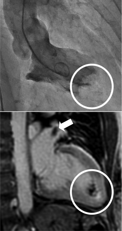
To the Editor: A 61-year-old postmenopausal woman has presented 3 times within 4 years with acute tako-tsubo cardiomyopathy (TTC). Each admission was precipitated by a heated argument and characterized by severe central chest pain, anterior ST-segment elevation, and elevated serum troponin levels; however, angiography revealed normal coronary arteries. Inotropic and noninvasive ventilatory support was required on all 3 occasions and intubation on 1 occasion. Left ventricular (LV) apical thrombus complicated the first episode but resolved with anticoagulation. Rapid normalization of LV systolic function on echocardiography, as well as resolution of the ST-segment elevation on electrocardiography, was observed after each presentation. During the patient's latest admission, coronary angiography (on day 10) revealed a large and mobile LV apical thrombus (Figure 1, top). This thrombus had not been observed on transthoracic echocardiography performed 6 days before angiography, and it had developed despite administration of aspirin and subcutaneous heparin and in the absence of atrial fibrillation on continuous monitoring.
FIGURE 1.
Top, Left ventricular apical thrombus (circle) as seen by ventriculography (day 10 of most recent admission). Bottom, Left ventricular apical thrombus (circle) and left atrial appendage thrombus (arrow) as seen on cardiac magnetic resonance imaging (day 12) with use of a gradient echo inversion recovery sequence 2 minutes after gadolinium-diethylenetriaminepentaacetic acid injection (0.2 mmol/kg; inversion recovery time, 350 ms).
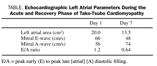
Cardiac magnetic resonance imaging was performed to exclude alternative myocardial pathology. A scan obtained on day 12 showed normalization of LV systolic function with no late gadolinium enhancement. However, in addition to the apical thrombus in the LV, the scan revealed a large thrombus in the left atrial appendage (LAA) (Figure 1, bottom, supplemental video). Echocardiographic assessment of left atrial (LA) function at presentation was suggestive of transient LA enlargement and dysfunction (Table). Screening for thrombophilia was negative.
TABLE.
Echocardiographic Left Atrial Parameters During the Acute and Recovery Phase of Tako-Tsubo Cardiomyopathy
Left ventricular apical thrombus formation is a known complication of TTC and has been reported in up to 8% of case series,1 with potential for embolic sequelae. To our knowledge, this is the first published report of LAA thrombus complicating TTC independent of atrial fibrillation. We hypothesize that the following mechanisms are important contributors to thrombus formation in the setting of acute TTC:
Catecholamine excess–induced platelet activation and aggregation. Plasma catecholamine levels have been found to be higher at presentation in patients with TTC than in those with ST-segment elevation myocardial infarction.2 Both norepinephrine and epinephrine have been shown to induce platelet activation.3
Catecholamine-mediated endothelial and cardiomyocyte injury. Surges of catecholamines may induce cardiac myocyte injury via cyclic adenosine monophosphate–mediated calcium overload4 or indirectly due to catecholamine-induced endothelial dysfunction and associated damage to underlying myocytes.5 Myocytes in the LV apex are thought to be particularly sensitive to excessive levels of catecholamines, partly as a result of the higher endothelial to myocardial ratio.6 Our finding of an LAA thrombus suggests that myocytes in the left atrium may also be sensitive to the pathological milieu of TTC. Atrial dysfunction may be compounded by increased atrial filling pressures secondary to increases in LV end-diastolic pressure. Consistent with this and the current case, patients with TTC have been shown to have worse LA function compared with those with ST-segment elevation myocardial infarction.7
Although the occurrence of the LV apical thrombus in the episode of TTC we describe was most likely caused by acute ventricular wall akinesis, we suggest that increased platelet activation and aggregability due to catecholamine release in conjunction with LA stretch and subsequent endothelial injury may also act as contributory factors that favor thrombus development. The global nature of these factors may explain thrombosis in other cardiac chambers, such as the LAA as described in our case. Given the prothrombotic state that characterizes TTC episodes, a review of anticoagulation protocols for this condition may be warranted.
Supplementary Material
Acknowledgments
Supported by grants from the North Shore Heart Research Foundation and the Medical Foundation, University of Sydney, Australia. We acknowledge James House, MHlthMgmt, and Anne Bright, MAppSc(MedImaging), from North Shore Radiology for their assistance in the acquisition of the cardiac magnetic resonance images.
Footnotes
Supporting Online Material
www.mayoclinicproceedings.com/content/85/9/863/suppl/DC1 Supplemental video
References
- 1.Haghi D, Papavassiliu T, Heggemann F, Kaden JJ, Borggrefe M, Suselbeck T. Incidence and clinical significance of left ventricular thrombus in tako-tsubo cardiomyopathy assessed with echocardiography. Q J Med. 2008;101(5):381-386 [DOI] [PubMed] [Google Scholar]
- 2.Wittstein IS, Thiemann DR, Lima JAC, et al. Neurohumoral Features of myocardial stunning due to sudden emotional stress. N Engl J Med. 2005;352(6):539-548 [DOI] [PubMed] [Google Scholar]
- 3.Ardlie NG, Glew G, Schwartz CJ. Influence of catecholamines on nucleotide-induced platelet aggregation. Nature. 1966;212(5060):415-417 [DOI] [PubMed] [Google Scholar]
- 4.Mann DL, Kent RL, Parsons B, Cooper G., IV Adrenergic effects on the biology of the adult mammalian cardiocyte. Circulation. 1992;85:790-804 [DOI] [PubMed] [Google Scholar]
- 5.Brutsaert DL, Meulemans AL, Sipido KR, Sys SU. Effects of damaging the endocardial surface on the mechanical performance of isolated cardiac muscle. Circ Res. 1988;62:358-366 [DOI] [PubMed] [Google Scholar]
- 6.Brutsaert DL. Tako-tsubo cardiomyopathy [letter]. Eur J Heart Fail. 2007;9(8):854 [DOI] [PubMed] [Google Scholar]
- 7.Browning JA, Newell MC, Sharkey S, et al. Stress cardiomyopathy depresses left atrial function compared to acute anterior myocardial infarction: left atrial size and function by cardiac MRI [poster]. J Cardiovasc Magn Reson. 2010;12(suppl 1):196 [Google Scholar]
Associated Data
This section collects any data citations, data availability statements, or supplementary materials included in this article.


