Figure 6.

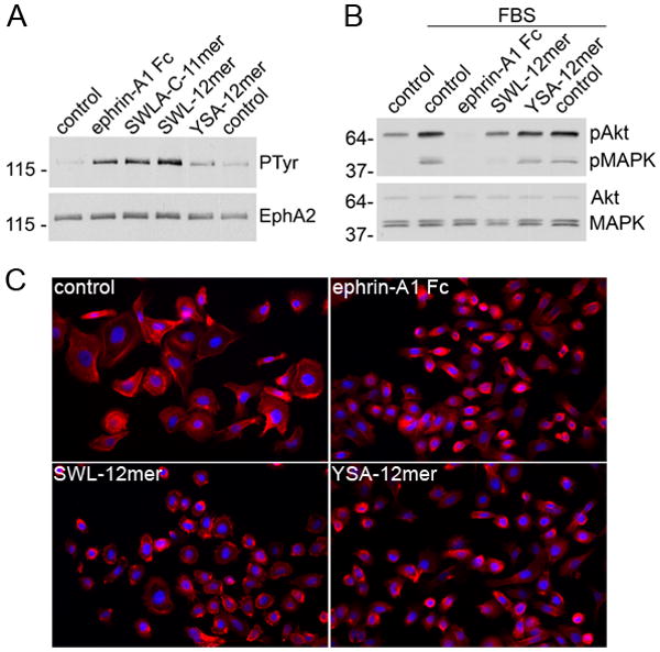
Biological effects of EphA2-targeting peptides. (A) PC3 cells were treated for 20 min with 0.1 μg/ml ephrin-A1 Fc, 50 μM of the indicated peptides, or left untreated as a control (in duplicate samples). EphA2 immunoprecipitates were probed with anti-phosphotyrosine (PTyr) antibodies and reprobed with anti-EphA2 antibodies. (B) Serum-starved PC3 cells were either left untreated (control), treated with 10% FBS (in duplicate samples), or treated with FBS together with 1μg/ml ephrin-A1 Fc or 50 μM of the indicated peptides. Cell lysates were probed with antibodies specific for Akt phosphorylated at T308 or phosphoErk1/Erk2 MAP kinases (pMAPK) and reprobed for total Akt and MAP kinases. (C) Cells were treated for 20 min with 1 μg/ml ephrin-A1 Fc, for 30 min with 100 μM SWL-12mer peptide or 100 μM YSA-12mer peptide, or left untreated as a control. The cells were then fixed in formaldehyde and labeled with phalloidin to stain actin filaments (red) and with DAPI to label nuclei (blue).
