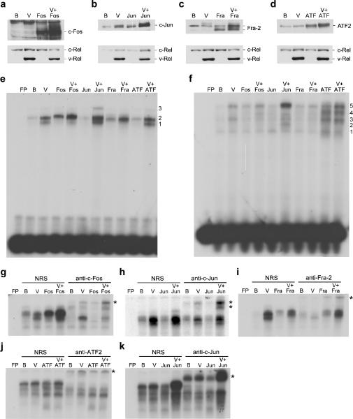
Figure 1.
Expression and DNA binding activity of AP-1 family members expressed from bicistronic retroviruses. DT40 cells were infected with control BIS retroviruses (B), BIS viruses expressing v-Rel (V), c-Fos, c-Jun, Fra-2, and ATF2 alone, or viruses that co-expressed v-Rel with each of the AP-1 family members. Whole cell lysates (25 μg) from infected cells were analyzed by Western blot to evaluate the expression of c-Fos, c-Jun, Fra-2, ATF2, and Rel proteins (panels a–d). Proteins detected by the antisera are indicated on the right of each panel. Nuclear extracts from infected cells were used in EMSAs with [32P]-labeled probes containing AP-1 DNA binding sites specific for Fos:Jun (panel e) or ATF2:Jun heterodimers (panel f). Free probe (FP) and the viruses expressed in the cells that extracts were derived from are indicated above each panel. Numbers to the right of each panel indicate the location of complexes described in the text. Supershift analyses were performed with the extracts described above employing normal rabbit serum (NRS) or antisera specific to c-Fos, c-Jun, Fra-2, and ATF2. Probes containing AP-1 DNA binding sites specific for Fos:Jun (panel g–i) or ATF2:Jun heterodimers (panel j–k) were employed for these studies. The asterisk to the right of each panel indicates the location of the supershifted band(s).

