Abstract
Previously, cell type-specific expression of AtHKT1;1, a sodium transporter, improved sodium (Na+) exclusion and salinity tolerance in Arabidopsis. In the current work, AtHKT1;1, was expressed specifically in the root cortical and epidermal cells of an Arabidopsis GAL4-GFP enhancer trap line. These transgenic plants were found to have significantly improved Na+ exclusion under conditions of salinity stress. The feasibility of a similar biotechnological approach in crop plants was explored using a GAL4-GFP enhancer trap rice line to drive expression of AtHKT1;1 specifically in the root cortex. Compared with the background GAL4-GFP line, the rice plants expressing AtHKT1;1 had a higher fresh weight under salinity stress, which was related to a lower concentration of Na+ in the shoots. The root-to-shoot transport of 22Na+ was also decreased and was correlated with an upregulation of OsHKT1;5, the native transporter responsible for Na+ retrieval from the transpiration stream. Interestingly, in the transgenic Arabidopsis plants overexpressing AtHKT1;1 in the cortex and epidermis, the native AtHKT1;1 gene responsible for Na+ retrieval from the transpiration stream, was also upregulated. Extra Na+ retrieved from the xylem was stored in the outer root cells and was correlated with a significant increase in expression of the vacuolar pyrophosphatases (in Arabidopsis and rice) the activity of which would be necessary to move the additional stored Na+ into the vacuoles of these cells. This work presents an important step in the development of abiotic stress tolerance in crop plants via targeted changes in mineral transport.
Introduction
Salinity stress affects crop production worldwide, particularly by limiting plant growth and reducing potential yield. Plants have three mechanisms for tolerating soil salinity, including tolerance of the osmotic effects of salt; a tolerance in leaf tissues of the negative effects of sodium ions (Na+) on cellular function through compartmentalisation of Na+ in specific tissues, cells, or cellular organelles; and exclusion of Na+ from the sensitive shoot tissue [1]. Of these mechanisms, Na+ exclusion from the shoot is best understood and is, therefore, the current best candidate for a targeted genetic modification approach to improving salinity tolerance.
The HKT gene family encodes one of the most widely studied groups of Na+ transporters in plants. Current understanding indicates that AtHKT1;1 from Arabidopsis is expressed primarily in the stele and has a role in the retrieval of Na+ from the transpiration stream [2], [3]. Reduced activity of this transporter leads to increased transfer of Na+ from the root to the shoot, leading to a higher shoot Na+ concentration and a decrease in salinity tolerance [4]. More recently, it was demonstrated that OsHKT1;5 in rice [5] and the HKT1;5 gene from wheat [6] are crucial in maintaining low shoot Na+ concentrations via a similar mechanism of Na+ retrieval from the transpiration stream into cells within the stele. A second HKT gene that is involved in sodium exclusion in cereal species is HKT1;4. The encoded transporter appears to function in combination with HKT1;5, by retrieving Na+ from the shoot transpiration stream and sequestering Na+ in the sheath tissue [7]. The endogenous function of the family of HKT transporters and their role in Na+ transport and provision of salinity tolerance to plants has been reviewed recently [8].
An obvious strategy for improving the salinity tolerance of plants is to increase the expression of HKT genes. Constitutive overexpression of plasma membrane Na+ transporters, however, has been unsuccessful in producing salinity tolerant plants [9]. It was hypothesized that such overexpression increases Na+ fluxes into all cells of the plant, this being, on balance, counter-productive to the reduction of Na+ transport to the shoot [9], [10]. In contrast, it was shown that overexpression of AtHKT1;1 specifically within the root xylem parenchyma cells of Arabidopsis leads to improved Na+ exclusion and salinity tolerance [9]. Interestingly, the excess Na+ in the root resulting from the improved Na+ exclusion from the shoot was stored within the cortical cells of the root [9].
The aim of experiments described in the current work was to determine if overexpression of AtHKT1;1 specifically within the outer cells (epidermis and cortex) of the root would result in opposite effects to overexpression in the stele, or whether a similar improvement of Na+ exclusion and salinity tolerance of Arabidopsis and rice would be observed. A secondary goal was to determine if expression of AtHKT1;1 in the epidermal and cortical cell layers of the root is the reason for the deleterious effects observed when AtHKT1;1 is constitutively overexpressed, as described previously [9].
In both species, we found that transgenic plants overexpressing AtHKT1;1 in mature root cortex had greater shoot Na+ exclusion and thus increased salinity tolerance.
Results
Expression of AtHKT1;1 in the root epidermis and cortex of Arabidopsis decreases Na+ accumulation in the shoot
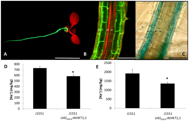
A homozygous single-insert GAL4-GFP enhancer trap line (J1551) with cell type-specific mGFP –ER expression in the epidermal and cortical cell layers of the root (Figures 1A and 1B) was isolated from a library described previously [9], [11]. Epidermal GFP fluorescence appeared slightly less intense than that found in the cortex. To confirm that a gene-of-interest could be transactivated specifically in the same cell-types as GAL4-GFP, the uidA gene was fused to a second UASGAL4 element and transformed into J1551. β-glucuronidase activity was detected in the same cell-types as the original GFP fluorescence pattern (Figure 1C) and both GFP and GUS expression was unaffected by NaCl treatment. Therefore, AtHKT1;1 was transactivated in the J1551 background (J1551 UASGAL4:AtHKT1;1) to express AtHKT1;1 specifically in the mature root epidermal and cortical cells.
Figure 1. Arabidopsis plants expressing AtHKT1;1 within the root epidermal and cortical cells.
(A) Representative fluorescence stereomicroscope and (B) confocal laser microscope images of J1551 showing GFP fluorescence specifically within the root epidermal and cortical cells. Tissue was stained with propidium iodide. GFP and propidium iodide images were captured separately and overlaid to create the composite image. Tissues labelled include the epidermis (ep), cortex (c), endodermis (en) and xylem parenchyma (xp). (C) Transactivation of uidA in J1551 produces GUS staining specifically within the root epidermal and cortical cells [Scale bars = 10 mm in (A), 75 µm in (B) and 75 µm in (C)]. (D) Concentration of Na+ within the leaves of T1 J1551 and J1551 UASGAL4:AtHKT1;1 plants grown on soil and watered with nutrient solution containing 2 mM NaCl (n = 12 for background and 45 independent events for AtHKT1;1, error bars represent SEM). (E) Accumulation of Na+ within the leaves of T2 J1551 and J1551 UASGAL4:AtHKT1;1 plants grown on soil and watered with nutrient solution containing 5 mM NaCl (n = 3 for background and 20 for AtHKT1;1 lines for each of 4 independent events, presented as a grand average, error bars represent SEM). Statistical significance from the J1551 line was determined using Student's t-test, *P<0.05.
Primary Arabidopsis J1551 UASGAL4:AtHKT1;1 transformants grown in soil and treated with a low concentration of NaCl (2 mM) showed 20% lower leaf Na (Figure 1D) and significantly higher K, P and Zn (averaged over all independent T1 lines produced) than J1551 (Table S1). Four independent lines were selected to test heritability in the T2 generation. The four lines were grown in soil and were sprayed with Basta to remove null segregants. Each of the four independent lines retained 16 to 37% lower shoot Na content (treated with 5 mM NaCl) than J1551 in the T2 generation (Figure 1E, Table S2) and maintained higher K (Table S2).
Expression of AtHKT1;1 in the root cortex of rice increases salinity tolerance and decreases shoot Na+ accumulation
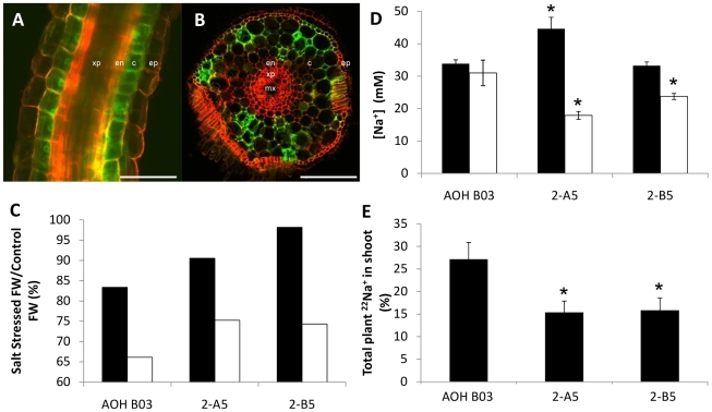
Two lines were selected from the GAL4-GFP enhancer trap library described previously [12], one with GFP fluorescence specifically within the xylem parenchyma of the root (ASG F03) (Figures S1A and S1B), and one with GFP fluorescence specifically within the cortical cell layer of the root (AOH B03) (Figures 2A and 2B). Compared with cortical cells, the GFP fluorescence in epidermal cells was extremely weak (Figure 2A).
Figure 2. Rice plants expressing AtHKT1;1 within the root epidermal and cortical cells.
(A + B) Representative confocal laser microscope images of line AOH B03 showing GFP fluorescence specifically within the root epidermis and cortical cells [Scale bars = 100 µm in (A) and 100 µm in (B)]. Tissue was stained with propidium iodide. GFP and propidium iodide images were captured separately and overlaid to create the composite image. Tissues labelled include the epidermis (ep), cortex (c), endodermis (en), xylem parenchyma (xp) and metaxylem (mx). (C) Salinity stress tolerance in AOH B03 and two independently transformed T2 AOH B03 UASGAL4:AtHKT1;1 rice lines (2-A5 and 2-B5). Plants were stressed for 5 d at 80 mM. Bars represent: ((stress FW/control FW) ×100) in the roots (black bars) and shoots (open bars) (n = 8). (D) Concentration of Na+ within the roots (black bars) and shoots (open bars) of AOH B03 and AOH B03 UASGAL4:AtHKT1;1 lines (2-A5 and 2-B5) stressed for 5 d at 80 mM (n = 8, error bars represent SEM). Statistical significance from the AOH BO3 line for each tissue was determined using Student's t-test, *P<0.05. (E) Total amount of 22Na+ in the rice plant that is contained within the shoot tissue of the plant (expressed as a percentage) after exposing the roots of intact plants to a solution containing 30 mM Na+ for 1 h (n = 8, error bars represent SEM). Statistical significance from the AOH BO3 line was determined using Student's t-test, *P<0.05.
The two GAL4 lines were transformed [13], [14] with a UASGAL4:AtHKT1;1 construct. Two independent T0 lines in each background were selected that accumulated levels of leaf Na+ which were average for their respective group of independent T0 lines. T2 lines derived from this material expressing AtHKT1;1 in the cortical cells of the root (AOH B03 UASGAL4:AtHKT1;1) maintained significantly higher fresh weight and lower shoot Na+ concentration (and higher K+ concentration) when challenged with a Na+ stress than AOH B03 (Figure S2). However, the lines expressing AtHKT1;1 in the xylem parenchyma cells of the root (ASG F03 UASGAL4:AtHKT1;1) had similar fresh weights and root Na+ concentrations and had higher shoot Na+ concentrations (and lower K+ concentrations) than ASG F03 (Figure S2). Therefore, the ASG F03 UASGAL4:AtHKT1;1 lines were eliminated from further analyses.
The two independent T2 AOH B03 UASGAL4:AtHKT1;1 lines (2-A5 and 2-B5) were better able to maintain growth in saline conditions compared to the parental line, AOH B03 (Figures 2C, S3A and S3B) and had lower shoot Na+ and higher shoot K+ than the parent, with a higher root Na+ concentration in one line (2-A5) (Figures 2D and S3C).
In order to determine which component of Na+ transport had been altered by the expression of AtHKT1;1 in the cortical root cells, resulting in the lowering of shoot Na+ concentration in those lines, radiotracer 22Na+ flux analysis was used to measure the rate of transfer of Na+ between various tissues in the plants. This analysis indicated that the root-to-shoot transfer of Na+ had been significantly decreased in the two AOH B03 UASGAL4:AtHKT1;1 lines (15 and 16% versus 27% in control) (Figure 2E). A significant proportion of Na+ influx into rice roots is reported to occur through the apoplastic pathway [15], thus excised roots were also analysed for unidirectional influx in an attempt to determine if transport protein-mediated Na+ influx had been affected by the expression of AtHKT1;1. No difference was observed between the AOH B03 and AOH B03 UASGAL4:AtHKT1;1 lines (Figure S4D). Furthermore, calculated unidirectional efflux and transfer of Na+ from the leaf sheath to the leaf blade, were not altered in response to the AtHKT1;1 expression (Figures S4A, S4B and S4C).
Wild-type rice (cv. Nipponbare) was also transformed with a construct containing the CaMV35S promoter fused to the AtHKT1;1 gene to produce rice plants expressing AtHKT1;1 constitutively. Many of these plants died in tissue culture, and those that did survive were stunted and generally sterile when grown on soil. Consequently, the plants constitutively overexpressing AtHKT1;1 were abandoned at this point, as no further analysis was possible.
Cell-specific overexpression of AtHKT1;1 increases expression of native AtHKT1;1 in Arabidopsis and OsHKT1;5 in rice which leads to decreased shoot Na+ accumulation
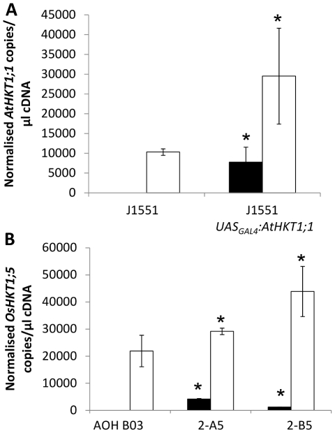
Pleiotropic regulation of other genes following misexpression of a gene-of-interest is commonly observed [e.g. 16] and it is possible that a component of the improved salinity tolerance in the transgenic lines may also relate to such an event. Stelar expression of AtHKT1;1 in Arabidopsis and OsHKT1;5 in rice regulates the amount of Na+ transferred to the shoot [3], [5], [6], [9]. Therefore, expression of the native HKT genes in the roots transgenic lines was examined as there was a decrease in root-to-shoot transfer of Na+ in these lines. To distinguish transgene copies of AtHKT1;1 from native AtHKT1;1 in transgenic Arabidopsis, unique sets of primers were designed (primer sequences are listed in Table S3). The reverse primer for the transgene AtHKT1;1 was located on a unique restriction site sequence between AtHKT1;1 and the nos terminator sequence, the reverse primer for the native AtHKT1;1 was located in the 3′ untranslated region of the native AtHKT1;1 gene. Use of these primer sets revealed a significant increase in transcript abundance of the native AtHKT1;1 in response to the expression of the transgenic AtHKT1;1 in the epidermal and cortical root cells (Figure 3A). Analysis of the rice lines for expression of the AtHKT1;1 transgene indicated a difference in root AtHKT1;1 expression between the two AOH B03 UASGAL4:AtHKT1;1 lines (Figure 3B). Similar analysis of the rice AOH B03 UASGAL4:AtHKT1;1 lines revealed that OsHKT1;5 was significantly more abundant compared with AOH B03 under conditions of salinity stress (Figure 3B). No other consistent changes in expression between both independent AOH B03 UASGAL4:AtHKT1;1 lines and the AOH B03 background line of any other member of the OsHKT gene family were observed (Figure S5). It should be noted that OsHKT1;2 is reported to be a pseudogene in japonica rice [17] and OsHKT2;2 is not present in japonica rice [17], thus expression of these genes was not examined.
Figure 3. Root expression of HKT genes measured using quantitative reverse transcriptase PCR.
(A) Normalized measurements of the expression of the AtHKT1;1 transgene (black bars) and the endogenous AtHKT1;1 (open bars) in the roots of J1551 and T2 J1551 UASGAL4:AtHKT1;1 plants (n = 12, error bars represent SEM). Statistical significance from the J1551 line was determined for each gene using Student's t-test, *P<0.05. (B) Normalized measurements of the expression of the AtHKT1;1 transgene (black bars) and the OsHKT1;5 gene (open bars) within the roots of AOH B03 and two independently transformed T2 AOH B03 UASGAL4:AtHKT1;1 lines (2-A5 and 2-B5) (n = 4, error bars represent SEM). Statistical significance from the AOH BO3 line for each gene was determined using Student's t-test, *P<0.05.
Outer root cells of transgenic rice overexpressing AtHKT1;1 accumulate more Na+
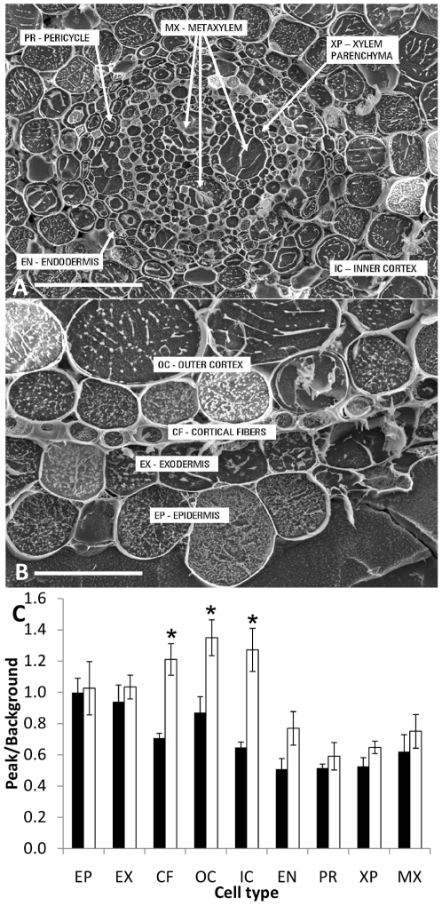
Rice root [Na+] was higher in the AOH B03 UASGAL4:AtHKT1;1 line (2-B5) compared to AOH B03, therefore to examine where the extra Na+ was stored cryo-scanning electron microscopy and x-ray microanalysis was performed on cell-specific vacuolar contents [9]. Given that extra Na+ was likely to be retrieved from the transpiration stream by the native stelar-localized HKT transporters, the aim was to determine where in the root tissue this Na+ was being stored. Since the expression of GFP (hence, AtHKT1;1) was observed in the cortical cells immediately behind the root apex, this tissue was analysed (Figures 4A and 4B). Interestingly, the analysis revealed that the cortical fibres, outer cortex and inner cortex but not the epidermal cells of the AOH B03 UASGAL4:AtHKT1;1 lines contained significantly higher Na+ contents than AOH B03 (Figures 4A–C). It should be noted the AOH B03 UASGAL4:AtHKT1;1 line (2-B5) chosen for this analysis had the lower whole root Na+ concentration of the two independent lines (Figure 2D). The AOH B03 UASGAL4:AtHKT1;1 lines contained less K+ in all cell-types than AOH B03 (Figure S6).
Figure 4. Scanning electron microscopy (SEM) images and measurements of amount of Na+ in specific root cell-types.
SEM images of the (A) stelar cell-types and (B) outer cell-types of the rice root [Scale bars = 100 µm in (A) and 20 µm in (B)]. (C) EDAX measurements of the amount of Na+ (software measurement of peak/background) present in 9 root cell-types of AOH B03 (black bars) and an AOH B03 UASGAL4:AtHKT1;1 line (2-B5) (open bars) including: EP - epidermis, EX - exodermis, CF – cortical fibres, OC – outer cortex, IC – inner cortex, EN - endodermis, PR - pericycle, XP – xylem parenchyma, MX - metaxylem. Each bar is an average of measurements made on three cells from each cell-type in the roots of three independent plants grown in a solution containing 50 mM Na+ for 5 d (error bars represent SEM). Statistical significance from the AOH BO3 line was determined using Student's t-test, *P<0.05.
Increased expression of AtAVP1 in Arabidopsis, and OsOVP4, OsOVP5, OsOVP6 in rice leads to increased vacuolar sequestration of Na+
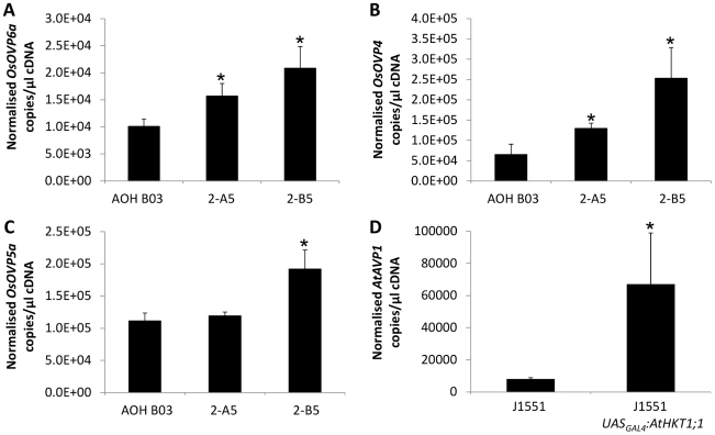
The expression of AtHKT1;1 in the cortical cells of the rice roots led to an increase in the vacuolar [Na+] of those cells. This may be expected as AtHKT1;1 is the protein that catalyses influx of Na+ across the plasma membrane and to avoid damage to cytoplasmic contents, plants must sequester Na+ into vacuoles. Two proteins are associated with vacuolar sequestration: the vacuolar Na+/H+ antiporter, NHX1, which transports Na+ into vacuoles; and the vacuolar H+-pyrophosphatase, which provides the electrochemical potential necessary to drive the NHX1-mediated movement of Na+ into vacuoles by moving protons into the vacuole. OsNHX1 (Os07g47100) expression showed no upregulation in Na+ treated rice roots of AOH B03 UASGAL4:AtHKT1;1 compared with AOH B03 (Figure S7). Interestingly, expression of OsOVP4 (Os02g55890), OsOVP5 (Os02g09150) and OsOVP6 (Os01g23580) in Na+ treated roots from the AOH B03 UASGAL4:AtHKT1;1 lines was significantly higher than in AOH B03 (Figures 5A, 5B and 5C) while expression of OsOVP1 (Os06g43660), OsOVP2 (Os06g08080) and OsOVP3 (Os05g06480) was decreased or unchanged in response to Na+ treatment between the AOH B03 UASGAL4:AtHKT1;1 and AOH B03 lines (Figure S8).
Figure 5. Root expression of vacuolar H+-pyrophosphatase genes measured using Quantitative Reverse Transcriptase PCR.
Normalized measurements of the expression of (A) OsOVP4, (B) OsOVP5a and (C) OsOVP6a within the roots of AOH B03 and two independently transformed T2 AOH B03 UASGAL4:AtHKT1;1 lines (2-A5 and 2-B5) (n = 4, error bars represent SEM). Statistical significance from the AOH BO3 line was determined using Student's t-test, *P<0.05. (D) Normalized measurements of the expression of the AtAVP1 gene in the roots of J1551 and T2 J1551 UASGAL4:AtHKT1;1 plants (n = 12, error bars represent SEM). Statistical significance from the J1551 line was determined using Student's t-test, *P<0.05.
In Arabidopsis, no difference in root expression of AtNHX1 (At5g27150) was observed between J1551 and J1551 UASGAL4:AtHKT1;1 plants, but expression of AtAVP1 (At1g15690) was significantly higher in the J1551 UASGAL4:AtHKT1;1 plants (Figure 5D).
Discussion
The expression of AtHKT1;1 specifically within the outer cells of the root led to reduced Na+ in the shoot in both Arabidopsis and rice. The data from rice experiments revealed that this reduction in shoot Na+ resulted from a decreased root-to-shoot flux of Na+, and led to increased fresh weight under salinity stress. The decrease in root-to-shoot flux of Na+ is related to increased gene expression of the HKT transporter responsible for retrieval of Na+ from the transpiration stream and the increased sequestration of Na+ in the root cortical cells. This is facilitated by AtHKT1;1 overexpression in these cells and the pleiotropic increase in transcript abundance of the vacuolar H+-translocating pyrophosphatases. However, the sequence of events underlying these observations is less clear.
One possible hypothesis is that the expression of AtHKT1;1 in outer root cells causes upregulation of AtHKT1;1 in Arabidopsis and OsHKT1;5 in rice. The mechanism for this is unknown. OsHKT1;5 transcript abundance was increased even in control conditions, when root Na+ is low, which points to a signalling mechanism that does not require high Na+. One possible mechanism could involve the movement from the cortex of small RNAs [18] upregulating AtHKT1;1/OsHKT1;5 transcription or preventing transcript degradation.
The consequence of increased stelar HKT abundance is an increased retrieval of Na+ from the transpiration stream, which must then be removed from the stelar cells to avoid Na+ toxicity in these relatively small cells which have only a small vacuole. Thus, the extra Na+ is transported back across the endodermis to the cortex of the root, and stored in the extra Na+ sink created by the cortical cells expressing AtHKT1;1. In order to protect the cytoplasm from Na+ toxicity, the accumulated Na+ is then further sequestered into the large vacuoles of the cortical cells. Movement into the vacuoles occurs through the Na+/H+ antiporter, energized by the increase in the electrochemical potential difference for H+ resulting from the increased H+-pyrophosphatase expression observed (and presumably function). Such an increased capacity for Na+ accumulation was described previously [9] in response to direct over-expression of AtHKT1;1 in the stele and appears to be an important component of limiting Na+ transport to the shoot in addition to Na+ retrieval from the transpiration stream.
Constitutive overexpression of the AtHKT1;1 gene was detrimental to plant growth and expression of the gene in stelar tissue did not improve salinity tolerance of rice, thus this study demonstrates the value of cell type-specific expression of Na+ transporters for engineering salinity tolerance in important crop plants. A further question arising from our work is why the expression of AtHKT1;1 in the stelar cells of Arabidopsis improved Na+ exclusion and salinity tolerance of Arabidopsis [9], but had no effect or even a detrimental effect on the transgenic rice in this study. Many differences could be cited for this discrepancy including the different stelar cell types expressing AtHKT1;1 in the previous study [9] and this one. As well, the morphology of Arabidopsis and rice roots are very different, thus creating a different radial root Na+ transport scenario for the two species. Finally, there are clear differences in the makeup of the HKT family between the two species (and many other important gene families) which could have an important impact on the results of seemingly analogous experiments. This highlights the importance of isolating species specific genetic elements which will enable the driving of cell type-specific expression of transgenes.
In conclusion, expression of AtHKT1;1 in the outer cells of the root in both Arabidopsis and rice led to lower shoot Na+ concentrations and improved salinity tolerance. In rice, this result appears to be due to an upregulation of OsHKT1;5 expression, as well as an increase in the Na+ sequestration capacity of the cortical cells due to increased expression of rice vacuolar H+-translocating pyrophosphatases. Several important questions follow from this study and will form the basis of future research. The mechanism leading to the upregulation of the native HKT genes when AtHKT1;1 is expressed in the outer root layers requires further analysis. Deep sequencing of the small RNA population of these roots may reveal an important regulatory molecule. Further work will also be required to determine whether the transgenic lines produced in this study have increased yield in the field when challenged by salinity stress. Regardless, expressing genes in a cell type-specific manner has improved the salinity tolerance of rice and may be useful in improving tolerance to a wide variety of crop stresses including ion toxicities, mineral deficiencies and potentially even biotic stresses of the roots or leaves.
Materials and Methods
Plant Materials and Growth Conditions
Arabidopsis thaliana GAL4-GFP enhancer trap line J1551 was produced by Dr. J. Haseloff (Univeristy of Cambridge) in the C24 ecotype background. The rice GAL4-GFP enhancer trap lines AOH B03 and ASG F03 were obtained from a library described previously [12]. Details on growth conditions can be found in Methods S1.
Microscopy
Fluorescence and confocal microscopy of Arabidopsis and rice was performed as previously described [9], [12], however 30 µm root sections were produced using a Leica VT 1200 S Vibrating Microtome (Leica, Germany). Cryo-scanning electron microscopy and x-ray microanalysis was performed as described previously [9] with modifications described in Methods S1.
DNA Constructs and Transformation
Binary vectors used to transform Arabidopsis have been described previously [8]. Details on binary vectors used to transform rice can be found in SI Materials and Methods. Rice transformation is as described previously [13], [14] with minor modifications noted in Methods S1.
GUS Staining
Staining of tissues for visualization of uidA expression was performed as described previously [9], [12].
Quantitative RT-PCR (Q-PCR)
Arabidopsis tissue samples were collected from the transformed plants when the plants were 5 weeks old. Approximately 15 mg of root and shoot tissue was collected from plants previously selected by spraying with phosphinotricin. Samples were freeze-dried and RNA was extracted from tissue powder using the Pure-Link 96 RNA Kit (Invitrogen) following the manufacturer's instructions. The RNA was treated using the DNA-free kit (Ambion) and cDNA was synthesized using SuperScript III First-Strand Synthesis System (Invitrogen) according the manufacturer's instructions.
Rice root tissue was ground under liquid nitrogen and total RNA was extracted from approximately 100 mg of ground tissue using the TRIzol reagent (Invitrogen) according to the manufacturer's instructions.
Q-PCR was performed as described previously [19] with modifications noted in Methods S1 and using primers in Table S3.
Tissue Elemental Analysis
Arabidopsis tissue was analyzed for elemental content via ICP-AES as described previously [9]. Rice leaf blades, sheaths and roots from individual plants were harvested into 50 mL Falcon tubes for Na+ and K+ accumulation analysis. Fresh weights of samples were taken, the tissue was dried in an oven at 70°C for 12 h and dry weights were recorded. Samples were digested in 10 mL of 1% nitric acid for 4 h at 80°C. Digested samples were then analysed using a flame photometer (Sherwood Scientific Ltd., Cambridge, U.K.) for Na+ and K+ content. Values from the flame photometer were converted into Na+ and K+ concentration values on a tissue water basis (millimolar).
22Na+ Fluxes
Experiments were conducted on rice plants that had been pretreated with 30 mM NaCl in the hydroponic solution prior to analysis in an influx solution containing 30 mM NaCl and labelled with 0.05 µCi/ml 22Na+. Experiments were conducted essentially as previously described [20] with modifications described in Methods S1.
Supporting Information
ICP-MS measurements of the concentration (in mg/kg) of several common elements in the leaf tissue of J1551 compared with independent T1 J1551 UASGAL4:AtHKT1;1 lines. Plants were grown on 2 mM NaCl.
(0.03 MB DOC)
ICP-AES measurements of the concentration (in mg/kg) of several common elements in the leaf tissue of J1551 (‘Background’) compared with independent T2 J1551 UASGAL4:AtHKT1;1 families. Plants were grown on 5 mM NaCl.
(0.04 MB DOC)
List of primers used for vector construction and Q-PCR analysis in Arabidopsis and rice. Forward and reverse primers are listed and the gene identifier for each gene analysed by Q-PCR is provided.
(0.05 MB DOC)
Images of the rice GAL4-GFP enhancer trap line ASG FO3. (A + B) Confocal laser microscope images of line ASG F03 showing GFP fluorescence specifically within the root xylem parenchyma [Scale bars = 100 µm in (A) and 100 µm in (B)]. Tissue was stained with propidium iodide. GFP and propidium iodide images were captured separately and overlaid to create the composite image. Tissues labelled include the epidermis (ep), cortex (c), endodermis (en), xylem parenchyma (xp) and metaxylem (mx).
(2.65 MB TIF)
Fresh weight (FW) measurements and tissue Na+ and K+ concentration of T1 rice lines. Lines express AtHKT1;1 within the root cortical cells (AOH B03 UASGAL4:AtHKT1;1) or the root xylem parenchyma cells (ASG F03 UASGAL4:AtHKT1;1) and are compared with their respective background lines (AOH B03 and ASG F03). Measurements of FW of (A) shoots and (B) roots of control (black bars) and salinity stressed (open bars) rice plants grown for 5 d on 80 mM NaCl (n = 8, error bars represent SEM). Statistical significance between treatments for each line was determined via the Student's t-test, *P<0.05. Tissue concentration of (C) Na+ and (D) K+ in roots (black bars) and shoots (open bars) of rice plants grown for 5 d on 80 mM NaCl (n = 8, error bars represent SEM). Statistical significance from the respective control line for each tissue was determined via the Student's t-test, *P<0.05.
(0.21 MB TIF)
Fresh weight measurements of T2 rice plants. The FW was measured in (A) shoots and (B) roots of rice plants grown in control conditions (black bars) or in salinity stress conditions (open bars). (C) Concentration of K+ in the roots (black bars) and shoots (open bars) under salinity stress conditions. AOH B03 and two independent T2 AOH B03 UASGAL4:AtHKT1;1 lines (2-A5 and 2-B5) were grown for 5 d on nutrient solution containing 80 mM NaCl (n = 8, error bars represent SEM). Statistical significance from the AOH BO3 line was determined via the Student's t-test, *P<0.05.
(0.12 MB TIF)
Flux measurements of Na+ inferred using the radiotracer 22Na+. Measurements were taken in AOH B03 and two independent T2 AOH B03 UASGAL4:AtHKT1;1 lines (2-A5 and 2-B5) grown for 3 d prior to experiment on nutrient solution containing 30 mM Na+ (n = 8, error bars represent SEM). (A) Measurement of the unidirectional influx of 22Na+ into non-excised rice roots exposed to an influx solution containing 30 mM Na+ for 2 min. (B) Root efflux of 22Na+ calculated by subtracting the net influx of 22Na+ into the plant (after 1 h exposure of the roots to a solution containing 30 mM Na+) from the unidirectional influx of 22Na+ (after a 2 min exposure of the roots to a solution containing 30 mM Na+). (C) Measurement of the sheath-to-blade transfer of 22Na+ expressed as a percentage of the total amount of 22Na+ in the shoot tissue that is contained within the leaf blades after exposing the roots of intact plants to a solution containing 30 mM Na+ for 1 h. (D) Measurement of the unidirectional influx of 22Na+ into rice roots excised immediately prior to experiment and exposed to an influx solution containing 30 mM Na+ for 2 min.
(0.19 MB TIF)
Expression of OsHKT family members measured using quantitative reverse transcriptase PCR. Included are AOH B03 and AOH B03 UASGAL4:AtHKT1;1 lines 2-A5 and 2-B5. Normalized expression of (A) OsHKT1;1, (B) OsHKT1;3, (C) OsHKT1;4, (D) OsHKT2;1, (E) OsHKT2;3 and (F) OsHKT2;4 in the roots of rice plants grown in 80 mM NaCl for 5 d. Each bar represents an average of four replicates (error bars represent SEM). Statistical significance from the AOH BO3 line was determined using Student's t-test, *P<0.05.
(0.43 MB TIF)
EDAX measurements of the amount of K+ (software measurement of peak/background) present in 9 root cell-types of rice. Lines include AOH B03 (black bars) and a AOH B03 UASGAL4:AtHKT1;1 line (2-B5) (open bars) and cell types are: EP - epidermis, EX - exodermis, CF - cortical fibres, OC - outer cortex, IC - inner cortex, EN - endodermis, PR - pericycle, XP - xylem parenchyma, MX - metaxylem. Each bar is an average of measurements made on three cells from each cell-type in the roots of three independent plants grown in a solution containing 50 mM Na+ for 5 d (error bars represent SEM). Statistical significance from the AOH BO3 line was determined via the Student's t-test, *P<0.05.
(0.10 MB TIF)
Expression of the Na+/H+ antiporter gene OsNHX1 measured using quantitative reverse transcriptase PCR. Included are AOH B03 and AOH B03 UASGAL4:AtHKT1;1 lines. Normalized expression of the vacuolar H+-pyrophosphatases (A) OsOVP1, (B) OsOVP2 and (C) OsOVP4 in the roots of rice plants grown in 80 mM NaCl for 5 d. Each bar represents an average of four replicates (error bars represent SEM). Statistical significance from the AOH BO3 line was determined using Student's t-test, *P<0.05.
(0.05 MB TIF)
Expression of several rice vacuolar H+-pyrophosphatase genes measured via quantitative reverse transcriptase PCR. Included are AOH B03 and AOH B03 UASGAL4:AtHKT1;1 lines. Normalized expression of the vacuolar H+-pyrophosphatases (A) OsOVP1, (B) OsOVP2 and (C) OsOVP4 in the roots of rice plants grown in 80 mM solution for 5 d. Each bar represents an average of 4 replicates (error bars represent SEM). Statistical significance from the AOH BO3 line was determined via the Student's t-test, *P<0.05.
(0.19 MB TIF)
Acknowledgments
We thank Lynette Waterhouse and Gwen Mayo for assistance with microscopy, Emmanuel Guiderdoni and Jim Haseloff for plant material and Adelaide Microscopy for access to equipment. We thank Romola Davenport for technical support and advice.
Footnotes
Competing Interests: The authors have declared that no competing interests exist.
Funding: This work was supported by a Biotechnology and Biological Research Sciences Council (http://www.bbsrc.ac.uk) grant to MT and Australian Research Council (http://www.arc.gov.au) grants FF0348367 and DP0557920 to MT. PhD students were supported by an International Postgraduate Research Scholarship (DP), a BP Egypt-Cambridge scholarship (GS) and the Danish Research Council (ISM). The funders had no role in study design, data collection and analysis, decision to publish, or preparation of the manuscript.
References
- 1.Munns R, Tester M. Mechanisms of salinity tolerance. Ann Rev of Plant Biol. 2008;59:651–681. doi: 10.1146/annurev.arplant.59.032607.092911. [DOI] [PubMed] [Google Scholar]
- 2.Sunarpi, Horie T, Motoda J, Kubo M, Yang H, et al. Enhanced salt tolerance mediated by AtHKT1 transporter induced Na+ unloading from xylem vessels to xylem parenchyma cells. Plant J. 2005;44:928–938. doi: 10.1111/j.1365-313X.2005.02595.x. [DOI] [PubMed] [Google Scholar]
- 3.Davenport RJ, Muñoz-Mayor A, Jha D, Essay PA, Rus A, et al. The Na+ transporter AtHKT1;1 controls retrieval of Na+ from the xylem in Arabidopsis. Plant, Cell and Environ. 2007;30:497–507. doi: 10.1111/j.1365-3040.2007.01637.x. [DOI] [PubMed] [Google Scholar]
- 4.Rus A, Lee B-h, Muñoz-Mayor A, Sharkhuu A, Miura K, et al. AtHKT1 Facilitates Na+ homeostasis and K+ nutrition in planta. Plant Phys. 2004;136:2500–2511. doi: 10.1104/pp.104.042234. [DOI] [PMC free article] [PubMed] [Google Scholar]
- 5.Ren Z-H, Gao J-P, Li L-G, Cai X-L, Huang W, et al. A rice quantitative trait locus for salt tolerance encodes a sodium transporter. Nature Biotech. 2005;37:1141–1146. doi: 10.1038/ng1643. [DOI] [PubMed] [Google Scholar]
- 6.Byrt CS, Platten JD, Spielmeyer W, James RA, Lagudah ES, et al. HKT1;5-Like cation transporters linked to Na+ exclusion loci in wheat, Nax2 and Kna1. Plant Phys. 2007;143:1918–1928. doi: 10.1104/pp.106.093476. [DOI] [PMC free article] [PubMed] [Google Scholar]
- 7.Huang S, Spielmeyer W, Laguday ES, James RA, Platten JD, et al. A sodium transporter (HKT7) is a candidate for Nax1, a gene for salt tolerance in durum wheat. Plant Phys. 2006;142:1718–1727. doi: 10.1104/pp.106.088864. [DOI] [PMC free article] [PubMed] [Google Scholar]
- 8.Hauser F, Horie T. A conserved primary salt tolerance mechanism mediated by HKT transporters: a mechanism for sodium exclusion and maintenance of high K+/Na+ ratio in leaves during salinity stress. Plant Cell Environ. 2010;33:552–565. doi: 10.1111/j.1365-3040.2009.02056.x. [DOI] [PubMed] [Google Scholar]
- 9.Møller IS, Gilliham M, Jha D, Mayo G, Roy S, et al. Shoot Na+ exclusion and increased salinity tolerance engineered by cell type-specific alteration of Na+ transport in Arabidopsis. Plant Cell. 2009;21:2163–2178.10. doi: 10.1105/tpc.108.064568. [DOI] [PMC free article] [PubMed] [Google Scholar]
- 10.Tester M, Davenport R. Na+ tolerance and Na+ transport in higher plants. Ann Bot. 2003;91:503–527. doi: 10.1093/aob/mcg058. [DOI] [PMC free article] [PubMed] [Google Scholar]
- 11.Haseloff J. GFP variants for multispectral imaging of living cells. Met Cell Biol. 1999;58:139–151. doi: 10.1016/s0091-679x(08)61953-6. [DOI] [PubMed] [Google Scholar]
- 12.Johnson AAT, Hibberd JM, Gal CI, Essah PA, Haseloff J, et al. Spatial control of transgene expression in rice (Oryza sativa L.) using the GAL4 enhancer trapping system. Plant J. 2005;41:779–789. doi: 10.1111/j.1365-313X.2005.02339.x. [DOI] [PubMed] [Google Scholar]
- 13.Sallaud C, Meynard D, Boxtel, Jv, Gay C, Bés M, et al. Highly efficient production and characterization of T-DNA plants for rice (Oryza sativa L.) functional genomics. TAG. 2003;106:1396–1408. doi: 10.1007/s00122-002-1184-x. [DOI] [PubMed] [Google Scholar]
- 14.Sallaud C, Gay CI, Larmande P, Bés M, Piffanelli P, et al. High throughput T-DNA insertion mutagenesis in rice: a first step towards in silico reverse genetics. Plant J. 2004;39:450–464. doi: 10.1111/j.1365-313X.2004.02145.x. [DOI] [PubMed] [Google Scholar]
- 15.Yeo AR, Yeo ME, Flowers TJ. The contribution of an apoplastic pathway to sodium uptake by rice roots in saline conditions. J Exp Bot. 1987;38:1141–1153. [Google Scholar]
- 16.Sottosanto JB, Saranga Y, Blumwald E. Impact of AtNHX1, a vacuolar Na+/H+ antiporter, upon gene expression during short- and long-term salt stress in Arabidopsis thaliana. BMC Plant Biol. 2007;7:18. doi: 10.1186/1471-2229-7-18. [DOI] [PMC free article] [PubMed] [Google Scholar]
- 17.Garciadeblás B, Senn ME, Bañuelos MA, Rodríguez-Navarro A. Sodium transport and HKT transporters: the rice model. Plant J. 2003;34:788–801.18. doi: 10.1046/j.1365-313x.2003.01764.x. [DOI] [PubMed] [Google Scholar]
- 18.Wu X, Weigel D, Wigge PA. Signaling in plants by intercellular RNA and protein movement. Genes Dev. 2002;16:151–158. doi: 10.1101/gad.952002. [DOI] [PubMed] [Google Scholar]
- 19.Burton RA, Shirley NJ, King BJ, Harvey AJ, Fincher GB. The CesA gene family of barley. Quantitative analysis of transcripts reveals two groups of co-expressed genes. Plant Phys. 2004;134:224–236. doi: 10.1104/pp.103.032904. [DOI] [PMC free article] [PubMed] [Google Scholar]
- 20.Davenport R, James RA, Zakrisson-Plogander A, Tester M, Munns R. Control of sodium transport in durum wheat. Plant Phys. 2005;137:807–818. doi: 10.1104/pp.104.057307. [DOI] [PMC free article] [PubMed] [Google Scholar]
Associated Data
This section collects any data citations, data availability statements, or supplementary materials included in this article.
Supplementary Materials
ICP-MS measurements of the concentration (in mg/kg) of several common elements in the leaf tissue of J1551 compared with independent T1 J1551 UASGAL4:AtHKT1;1 lines. Plants were grown on 2 mM NaCl.
(0.03 MB DOC)
ICP-AES measurements of the concentration (in mg/kg) of several common elements in the leaf tissue of J1551 (‘Background’) compared with independent T2 J1551 UASGAL4:AtHKT1;1 families. Plants were grown on 5 mM NaCl.
(0.04 MB DOC)
List of primers used for vector construction and Q-PCR analysis in Arabidopsis and rice. Forward and reverse primers are listed and the gene identifier for each gene analysed by Q-PCR is provided.
(0.05 MB DOC)
Images of the rice GAL4-GFP enhancer trap line ASG FO3. (A + B) Confocal laser microscope images of line ASG F03 showing GFP fluorescence specifically within the root xylem parenchyma [Scale bars = 100 µm in (A) and 100 µm in (B)]. Tissue was stained with propidium iodide. GFP and propidium iodide images were captured separately and overlaid to create the composite image. Tissues labelled include the epidermis (ep), cortex (c), endodermis (en), xylem parenchyma (xp) and metaxylem (mx).
(2.65 MB TIF)
Fresh weight (FW) measurements and tissue Na+ and K+ concentration of T1 rice lines. Lines express AtHKT1;1 within the root cortical cells (AOH B03 UASGAL4:AtHKT1;1) or the root xylem parenchyma cells (ASG F03 UASGAL4:AtHKT1;1) and are compared with their respective background lines (AOH B03 and ASG F03). Measurements of FW of (A) shoots and (B) roots of control (black bars) and salinity stressed (open bars) rice plants grown for 5 d on 80 mM NaCl (n = 8, error bars represent SEM). Statistical significance between treatments for each line was determined via the Student's t-test, *P<0.05. Tissue concentration of (C) Na+ and (D) K+ in roots (black bars) and shoots (open bars) of rice plants grown for 5 d on 80 mM NaCl (n = 8, error bars represent SEM). Statistical significance from the respective control line for each tissue was determined via the Student's t-test, *P<0.05.
(0.21 MB TIF)
Fresh weight measurements of T2 rice plants. The FW was measured in (A) shoots and (B) roots of rice plants grown in control conditions (black bars) or in salinity stress conditions (open bars). (C) Concentration of K+ in the roots (black bars) and shoots (open bars) under salinity stress conditions. AOH B03 and two independent T2 AOH B03 UASGAL4:AtHKT1;1 lines (2-A5 and 2-B5) were grown for 5 d on nutrient solution containing 80 mM NaCl (n = 8, error bars represent SEM). Statistical significance from the AOH BO3 line was determined via the Student's t-test, *P<0.05.
(0.12 MB TIF)
Flux measurements of Na+ inferred using the radiotracer 22Na+. Measurements were taken in AOH B03 and two independent T2 AOH B03 UASGAL4:AtHKT1;1 lines (2-A5 and 2-B5) grown for 3 d prior to experiment on nutrient solution containing 30 mM Na+ (n = 8, error bars represent SEM). (A) Measurement of the unidirectional influx of 22Na+ into non-excised rice roots exposed to an influx solution containing 30 mM Na+ for 2 min. (B) Root efflux of 22Na+ calculated by subtracting the net influx of 22Na+ into the plant (after 1 h exposure of the roots to a solution containing 30 mM Na+) from the unidirectional influx of 22Na+ (after a 2 min exposure of the roots to a solution containing 30 mM Na+). (C) Measurement of the sheath-to-blade transfer of 22Na+ expressed as a percentage of the total amount of 22Na+ in the shoot tissue that is contained within the leaf blades after exposing the roots of intact plants to a solution containing 30 mM Na+ for 1 h. (D) Measurement of the unidirectional influx of 22Na+ into rice roots excised immediately prior to experiment and exposed to an influx solution containing 30 mM Na+ for 2 min.
(0.19 MB TIF)
Expression of OsHKT family members measured using quantitative reverse transcriptase PCR. Included are AOH B03 and AOH B03 UASGAL4:AtHKT1;1 lines 2-A5 and 2-B5. Normalized expression of (A) OsHKT1;1, (B) OsHKT1;3, (C) OsHKT1;4, (D) OsHKT2;1, (E) OsHKT2;3 and (F) OsHKT2;4 in the roots of rice plants grown in 80 mM NaCl for 5 d. Each bar represents an average of four replicates (error bars represent SEM). Statistical significance from the AOH BO3 line was determined using Student's t-test, *P<0.05.
(0.43 MB TIF)
EDAX measurements of the amount of K+ (software measurement of peak/background) present in 9 root cell-types of rice. Lines include AOH B03 (black bars) and a AOH B03 UASGAL4:AtHKT1;1 line (2-B5) (open bars) and cell types are: EP - epidermis, EX - exodermis, CF - cortical fibres, OC - outer cortex, IC - inner cortex, EN - endodermis, PR - pericycle, XP - xylem parenchyma, MX - metaxylem. Each bar is an average of measurements made on three cells from each cell-type in the roots of three independent plants grown in a solution containing 50 mM Na+ for 5 d (error bars represent SEM). Statistical significance from the AOH BO3 line was determined via the Student's t-test, *P<0.05.
(0.10 MB TIF)
Expression of the Na+/H+ antiporter gene OsNHX1 measured using quantitative reverse transcriptase PCR. Included are AOH B03 and AOH B03 UASGAL4:AtHKT1;1 lines. Normalized expression of the vacuolar H+-pyrophosphatases (A) OsOVP1, (B) OsOVP2 and (C) OsOVP4 in the roots of rice plants grown in 80 mM NaCl for 5 d. Each bar represents an average of four replicates (error bars represent SEM). Statistical significance from the AOH BO3 line was determined using Student's t-test, *P<0.05.
(0.05 MB TIF)
Expression of several rice vacuolar H+-pyrophosphatase genes measured via quantitative reverse transcriptase PCR. Included are AOH B03 and AOH B03 UASGAL4:AtHKT1;1 lines. Normalized expression of the vacuolar H+-pyrophosphatases (A) OsOVP1, (B) OsOVP2 and (C) OsOVP4 in the roots of rice plants grown in 80 mM solution for 5 d. Each bar represents an average of 4 replicates (error bars represent SEM). Statistical significance from the AOH BO3 line was determined via the Student's t-test, *P<0.05.
(0.19 MB TIF)


