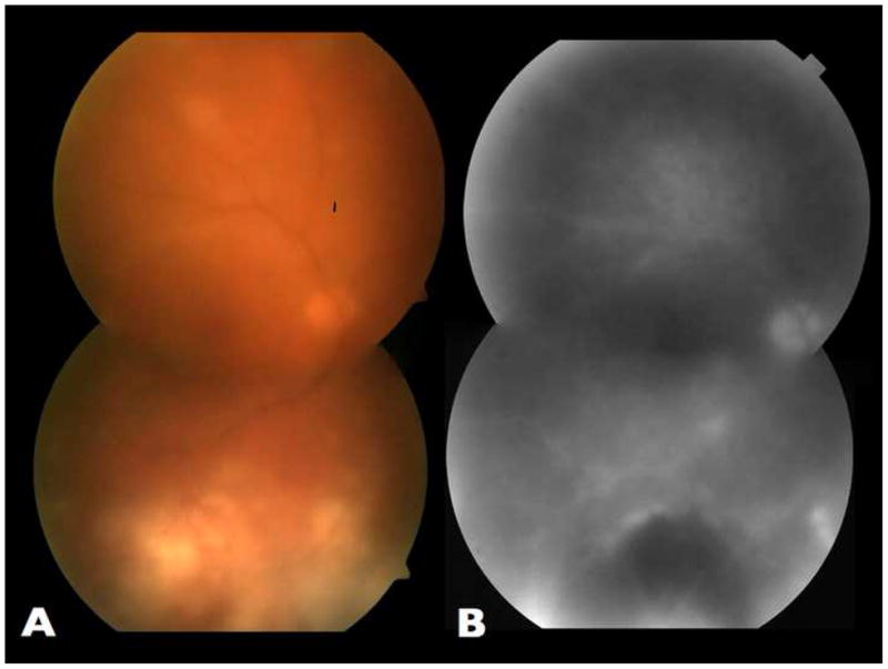
Figure 1.

Fundus aspect of the right eye at presentation. A. Montage color fundus photograph showing significant vitreous haze and multiple grey-yellowish chorioretinal lesions. Note that inferiorly these lesions are distributed around an area of chorioretinal scarring. B. Montage of late phase fluorescein angiogram frame, showing disc leakage, diffuse venular staining and hypofluorescence of the large lesion inferiorly.
