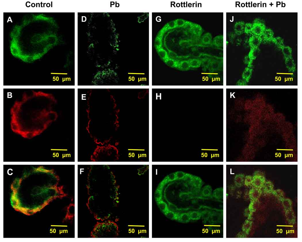
Fig. 3.
Inhibition of Pb-induced migration of LRP1 by rottlerin. Rat CP tissues were isolated and pre-treated with rottlerin (2 µM, 20 min) alone or followed by Pb (10 µM, 1 h) exposure in vitro. (A–C) Confocal image of LRP1 and PKC-δ in the cytosol of a representative CP tissue from a control rat. (D–F) Confocal image of LRP1 and PKC-δ in a representative CP tissue from a Pb-exposed tissue in absence of rottlerin pre-treatment. (G–I) Confocal image of a representative CP tissue pre-treated with rottlerin in the absence of Pb. (J–L) Representative image of a CP tissue pre-treated with rottlerin followed by Pb exposure. LRP1 is stained in green (A, D, G, J), and PKC-δ in red (B, E, H, K). The data are representative of experiments performed in triplicate.

