Crystals of 5-aminolaevulinic acid dehydratase from B. subtilis are tetragonal, belonging to space group P42212, and diffract to 2.7 Å resolution.
Keywords: 5-aminolaevulinic acid dehydratase, Bacillus subtilis
Abstract
5-Aminolaevulinic acid dehydratase (ALAD), a crucial enzyme in the biosynthesis of tetrapyrrole, catalyses the condensation of two 5-aminolaevulinic acid (ALA) molecules to form porphobilinogen (PBG). The gene encoding ALAD was amplified from genomic DNA of Bacillus subtilis and the protein was overexpressed in Escherichia coli strain BL21 (DE3). The protein was purified and crystallized with an additional MGSSHHHHHHSSGLVPRGSH– tag at the N-terminus of the target protein. Diffraction-quality single crystals were obtained by the hanging-drop vapour-diffusion method. An X-ray diffraction data set was collected at a resolution of 2.7 Å.
1. Introduction
5-Aminolaevulinic acid dehydratase (ALAD) catalyses the condensation of two 5-aminolaevulinic acid (ALA) molecules to form the pyrrole porphobilinogen (PBG). Four PBG molecules are linked together by porphobilinogen deaminase and then cyclized by uroporphyrinogen synthase to form uroporphyrinogen III, which can be further modified to produce different metallo-prosthetic groups such as haem, chlorophyll and vitamin B12 (Jordan, 1991 ▶, 1994 ▶; Warren & Scott, 1990 ▶; Jaffe, 1995 ▶). ALADs from different species share a high degree of sequence identity and contain about 350 amino acids per subunit. Most wild-type ALADs are octameric, but they can also be converted to other quaternary-structure isoforms such as hexamers (Jaffe, 2000 ▶; Breinig et al., 2003 ▶; Bollivar et al., 2004 ▶). Substrates and other compounds can induce this conversion, but little is known about the mechanism of formation of the other quaternary-structure isoforms and the structural basis of the kinetic differences between the different isoforms (Tang et al., 2005 ▶; Lawrence et al., 2009 ▶).
ALAD catalyzes the formation of an asymmetric pyrrole from two identical substrates. Single-turnover experiments have proven that the first substrate molecule entering the active site finally forms the propionate side of the product PBG and the second molecule forms the acetate side. According to this, the two substrates are distinguished as the ‘P-site’ and ‘A-site’ substrates (Jordan & Gibbs, 1985 ▶). Each active site of ALAD contains two invariant lysine residues that can form Schiff bases with the substrates (195 and 248 in ALAD from Bacillus subtilis). The P-site substrate forms a direct Schiff base with one of the Schiff-base lysine residues (Gibbs & Jordan, 1986 ▶), whilst there is some controversy about how the A-site substrate forms the second Schiff base to achieve its position and activation in the active site (Neier, 1996 ▶; Erskine, Newbold et al., 2001 ▶; Jaffe, 2004 ▶).
Most ALADs are dependent on divalent cations for activity and these metal ions participate in catalysis or allosteric regulation. The conserved metal ion-binding sites in the primary sequence of B. subtilis ALAD indicate that it requires both a zinc ion in the active site and a magnesium ion in the allosteric site (Jaffe, 2003 ▶). In this paper, we report the crystallization and preliminary crystallographic analysis of B. subtilis ALAD in order to obtain a better understanding of the reaction mechanism and properties of ALAD.
2. Materials and methods
2.1. Protein expression and purification
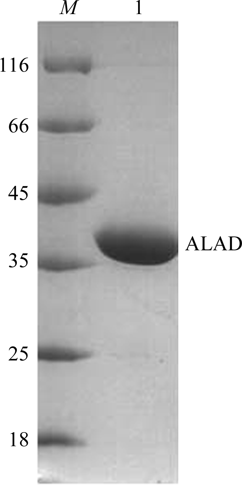
The gene (Entrez Gene ID 936972) encoding the full-length ALAD protein (residues 1–324) was amplified by polymerase chain reaction (PCR) from B. subtilis genomic DNA with primer A (5′-GGAATTCCATATGAGTCAATCATTTAATAGACACCGCC-3′; forward) and primer B (5′-CGGGATCCTTACTCCGCAAGCCATTTCGCTG-3′; reverse). The PCR product was digested by the restriction endonucleases NdeI and BamHI and then inserted into the pET15b (Novagen) vector predigested with the same two restriction enzymes. The pET15b vector added an additional MGSSHHHHHHSSGLVPRGSH– tag to the N-terminus of the target protein for purification. Successful cloning was confirmed by DNA sequencing. The recombinant plasmid was transformed into Escherichia coli BL21 (DE3) cells (Stratagene). The cells were first cultured in Luria–Bertani medium containing 100 µg ml−1 ampicillin at 310 K until the OD600 reached 0.8. To overexpress the protein, a final concentration of 0.5 mM isopropyl β-d-1-thiogalactopyranoside (IPTG) was added to the culture and the cells were cultured for a further 4 h at the same temperature. The cells were harvested by centrifugation, resuspended in buffer A (50 mM Na2HPO4 pH 8.0, 500 mM NaCl) and lysed by sonication. The supernatant containing the target protein was separated from the cell lysate by centrifugation at 15 000g for 30 min and was loaded onto a nickel-chelating column (GE Healthcare) pre-equilibrated with buffer A. Ten column volumes of buffer A were used to wash the column and 20 column volumes of buffer A supplemented with 50 mM imidazole were then used to remove the remaining impurities. Buffer A containing 400 mM imidazole was used to elute the target protein. The eluted fraction containing the target protein was concentrated by ultrafiltration using an Amicon Ultra-4 Centrifugal Filter Unit with an Ultracel-3 membrane (Millipore, 3 kDa cutoff) and a swinging-bucket rotor (Beckman); the condensed protein solution was then loaded onto a Superdex 200 column (GE Healthcare) pre-equilibrated with buffer A. The fraction consistent with an octamer of 38 kDa monomers of the target protein was concentrated and desalted by ultrafiltration using the same method. Finally, buffer B (20 mM Na2HPO4 pH 8.0, 50 mM NaCl) was used to store the purified target protein; the final concentration was about 20 mg ml−1 as measured using the BCA Protein Assay Kit (Pierce). The purity of the target protein was estimated by SDS–PAGE to be greater than 95% (Fig. 1 ▶).
Figure 1.

SDS–PAGE of ALAD. Lane 1, purified ALAD; lane M, molecular-weight markers (kDa).
2.2. Crystallization
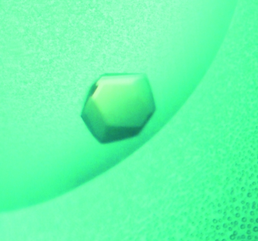
Initial crystallization trials were performed using the hanging-drop vapour-diffusion method with the Crystal Screen and Crystal Screen 2 reagent kits (Hampton Research) at 287 K. 4,6-Dioxoheptanoic acid was added to the protein solution to a final concentration of 1 mM (in buffer B) and the mixture was kept at room temperature for 1 h. Hanging drops made up of 1 µl protein solution and 1 µl reservoir solution were equilibrated against 150 µl reservoir solution. Small crystals were observed after 3 d in condition No. 11 of Crystal Screen 2, which consists of 10 mM CoCl2, 0.1 M sodium acetate trihydrate pH 4.6, 1.0 M 1,6-hexanediol (Hampton Research). After several rounds of optimization of the buffer pH, precipitant concentration, ionic strength, protein concentration, temperature and protein:reservoir volume ratio, single crystals (Fig. 2 ▶) with good diffraction ability were obtained within two weeks at 295 K using a reservoir solution consisting of 15 mM CoCl2, 0.1 M sodium acetate trihydrate pH 4.6, 0.75 M 1,6-hexanediol. The hanging drops were made up of 1 µl reservoir solution and 2 µl protein solution with an optimized protein concentration of 8 mg ml−1; the final concentration of 4,6-dioxoheptanoic acid was adjusted to 1 mM in the final optimized crystallization mixture.
Figure 2.
Photomicrograph of an ALAD crystal. The dimensions of this single crystal are about 0.3 × 0.2 × 0.1 mm.
2.3. Data collection
X-ray diffraction experiments were performed on beamline BL17U at SSRF (Shanghai Synchrotron Radiation Facility) and diffraction images were collected using an MX-225 CCD imaging-plate detector (MAR Research). Before data collection, each crystal was transferred into a solution consisting of 15 mM CoCl2, 0.1 M sodium acetate trihydrate pH 4.6, 3 M 1,6-hexanediol and exposed to air at room temperature for half an hour for dehydration. The dehydrated crystals were then quickly passed through cryoprotectant buffer comprising 20%(v/v) glycerol, 15 mM CoCl2, 0.1 M sodium acetate trihydrate pH 4.6, 0.75 M 1,6-hexanediol and flash-cooled to 100 K in a stream of cold nitrogen gas provided by an Oxford Cryosystems cooling device (Oxford Cryosystems Ltd). The discovery of the sensitivity of the crystals to dehydration was a fortunate accident and could be used to improve the diffraction ability of the crystals. A complete diffraction data set consisting of 180 images was collected from a single crystal with an oscillation angle of 1° per image at 100 K. Diffraction data were processed using MOSFLM (Leslie, 1992 ▶) and scaled with SCALA from the CCP4 suite (Collaborative Computational Project, Number 4, 1994 ▶). Data-collection and processing statistics are listed in Table 1 ▶.
Table 1. X-ray data-collection and processing statistics.
Values in parentheses are for the last shell.
| Wavelength (Å) | 1.0 |
| Space group | P42212 |
| Unit-cell parameters (Å) | a = b = 129.4, c = 111.4 |
| Resolution range (Å) | 40–2.70 (2.85–2.70) |
| No. of reflections | 388976 |
| No. of unique reflections | 50848 |
| Rmerge (%) | 11.5 (32.1) |
| 〈I/σ(I)〉 | 12.9 (5.5) |
| Multiplicity | 7.7 (7.7) |
| Completeness (%) | 99.8 (100.0) |
| Molecules per asymmetric unit | 3 |
| VM (Å3 Da−1) | 2.16 |
| Solvent content (%) | 43.1 |
3. Results and discussion
Analysis of a self-rotation function indicated that there were two molecules per asymmetric unit in the crystal of B. subtilis ALAD. While the Matthews analysis suggested that a more reasonable number might be three molecules, with a Matthews coefficient of 2.16 Å3 Da−1 (solvent content 41.3%), it did not rule out the presence of only two (Matthews coefficient of 3.24 Å3 Da−1; solvent content 62.1%). We could not find a reasonable answer to this conflict at this point and it seems that this ambiguity will only be solved by solution of the B. subtilis ALAD structure. For the purposes of this presentation, we will assume the former possibility for now.
Search models for molecular replacement are available from several species, such as E. coli, Chlorobium vibrioforme and yeast (Coates et al., 2004 ▶; Erskine et al., 1999 ▶; Erskine, Coates et al., 2001 ▶), and share an average of 50% amino-acid sequence identity with B. subtilis ALAD. The differences between B. subtilis ALAD and these deposited structures should help to illuminate the structural basis of the kinetic differences between ALADs from different species (Jaffe, 2004 ▶) and to obtain a deeper insight into the reaction mechanism of ALAD from the structure of B. subtilis ALAD with its inhibitor 4,6-dioxoheptanoic acid.
Acknowledgments
Financial support for this project was provided by research grants from the Chinese National Natural Science Foundation (grant Nos. 30025012 and 10979039), the Chinese Ministry of Science and Technology (grant Nos. 2006CB806500, 2006CB910200 and 2006AA02A318) and the Chinese Academy of Sciences (grant No. KSCX2-YW-R-60).
References
- Bollivar, D. W., Clauson, C., Lighthall, R., Forbes, S., Kokona, B., Fairman, R., Kundrat, L. & Jaffe, E. K. (2004). BMC Biochem.5, 17. [DOI] [PMC free article] [PubMed]
- Breinig, S., Kervinen, J., Stith, L., Wasson, A. S., Fairman, R., Wlodawer, A., Zdanov, A. & Jaffe, E. K. (2003). Nature Struct. Biol.10, 757–763. [DOI] [PubMed]
- Coates, L., Beaven, G., Erskine, P. T., Beale, S. I., Avissar, Y. J., Gill, R., Mohammed, F., Wood, S. P., Shoolingin-Jordan, P. & Cooper, J. B. (2004). J. Mol. Biol.342, 563–570. [DOI] [PubMed]
- Collaborative Computational Project, Number 4 (1994). Acta Cryst. D50, 760–763.
- Erskine, P. T., Coates, L., Newbold, R., Brindley, A. A., Stauffer, F., Wood, S. P., Warren, M. J., Cooper, J. B., Shoolingin-Jordan, P. M. & Neier, R. (2001). FEBS Lett.503, 196–200. [DOI] [PubMed]
- Erskine, P. T., Newbold, R., Brindley, A. A., Wood, S. P., Shoolingin-Jordan, P. M., Warren, M. J. & Cooper, J. B. (2001). J. Mol. Biol.312, 133–141. [DOI] [PubMed]
- Erskine, P. T., Norton, E., Cooper, J. B., Lambert, R., Coker, A., Lewis, G., Spencer, P., Sarwar, M., Wood, S. P., Warren, M. J. & Shoolingin-Jordan, P. M. (1999). Biochemistry, 38, 4266–4276. [DOI] [PubMed]
- Gibbs, P. N. B. & Jordan, P. M. (1986). Biochem. J.236, 447–451. [DOI] [PMC free article] [PubMed]
- Jaffe, E. K. (1995). J. Bioenerg. Biomembr.27, 169–179. [DOI] [PubMed]
- Jaffe, E. K. (2000). Acta Cryst. D56, 115–128. [DOI] [PubMed]
- Jaffe, E. K. (2003). Chem. Biol.10, 25–34. [DOI] [PubMed]
- Jaffe, E. K. (2004). Bioorg. Chem.32, 316–325. [DOI] [PubMed]
- Jordan, P. M. (1991). Editor. Biosynthesis of Tetrapyrroles, pp. 1–65. Amsterdam: Elsevier.
- Jordan, P. M. (1994). Curr. Opin. Struct. Biol.4, 902–911. [DOI] [PubMed]
- Jordan, P. M. & Gibbs, P. N. B. (1985). Biochem. J.227, 1015–1020. [DOI] [PMC free article] [PubMed]
- Lawrence, S. H., Ramirez, U. D., Selwood, T., Stith, L. & Jaffe, E. K. (2009). J. Biol. Chem.284, 35807–35817. [DOI] [PMC free article] [PubMed]
- Leslie, A. G. W. (1992). Jnt CCP4/ESF–EACBM Newsl. Protein Crystallogr.26
- Neier, R. (1996). Advances in Nitrogen Heterocycles, edited by C. M. Moody, Vol. 2, pp. 35–146. Stamford, USA: JAI Press.
- Tang, L., Stith, L. & Jaffe, E. K. (2005). J. Biol. Chem.280, 15786–15793. [DOI] [PubMed]
- Warren, M. J. & Scott, A. I. (1990). Trends Biochem. Sci.15, 486–491. [DOI] [PubMed]


