

Abstract
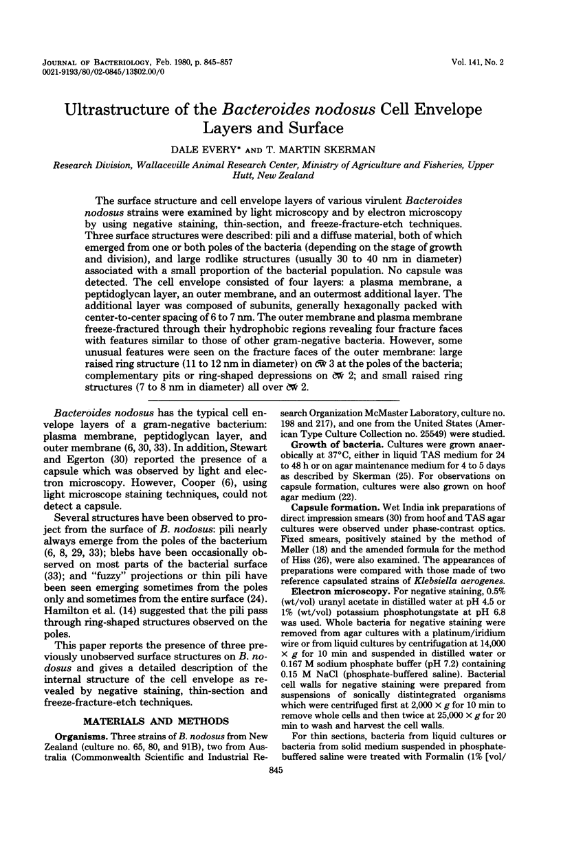
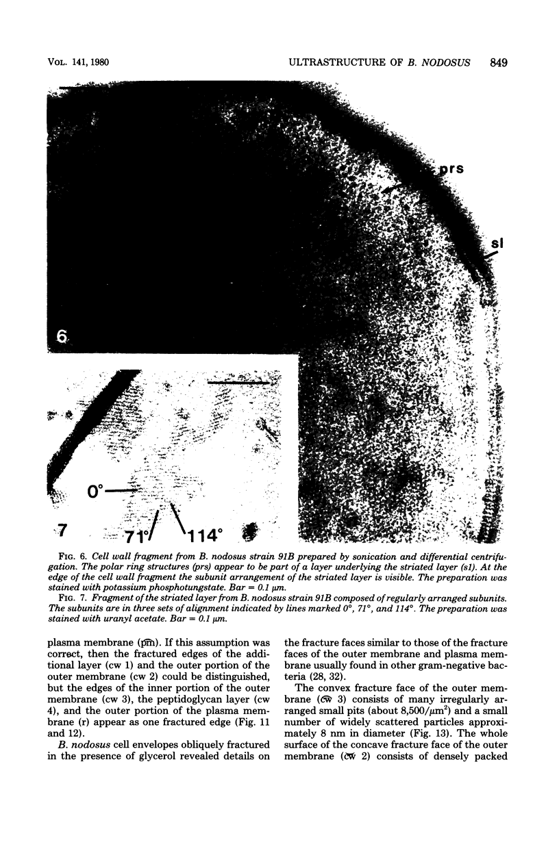
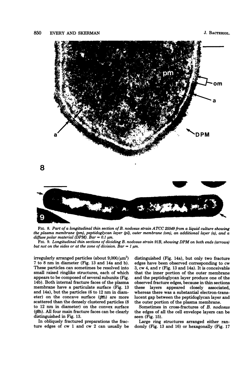
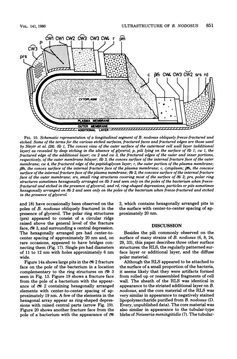
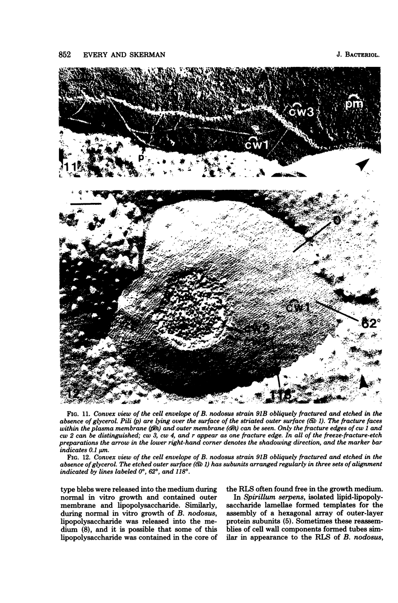
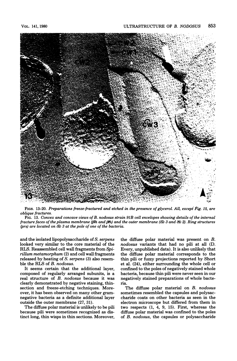
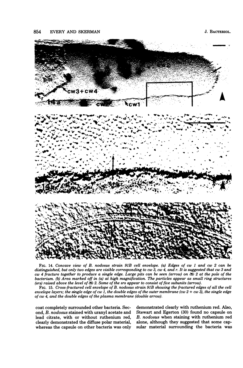
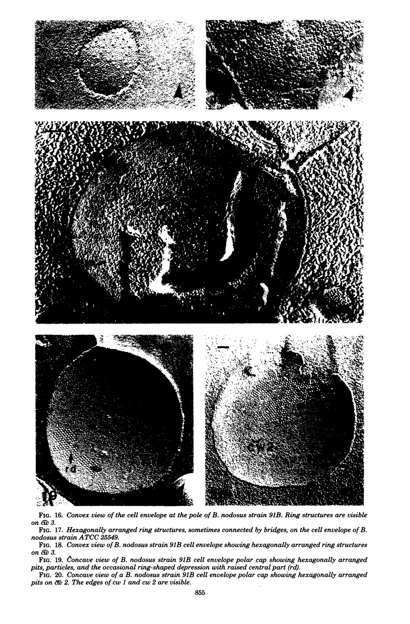
The surface structure and cell envelope layers of various virulent Bacteroides nodosus strains were examined by light microscopy and by electron microscopy by using negative staining, thin-section, and freeze-fracture-etch techniques. Three surface structures were described: pili and a diffuse material, both of which emerged from one or both poles of the bacteria (depending on the stage of growth and division), and large rodlike structures (usually 30 to 40 nm in diameter) associated with a small proportion of the bacterial population. No capsule was detected. The cell envelope consisted of four layers: a plasma membrane, a peptidoglycan layer, an outer membrane, and an outermost additional layer. The additional layer was composed of subunits, generally hexagonally packed with center-to-center spacing of 6 to 7 nm. The outer membrane and plasma membrane freeze-fractured through their hydrophobic regions revealing four fracture faces with features similar to those of other gram-negative bacteria. However, some unusual features were seen on the fracture faces of the outer membrane: large raised ring structure (11 to 12 nm in diameter) on cw 3 at the poles of the bacteria; complementary pits or ring-shaped depressions on cw 2; and small raised ring structures (7 to 8 nm in diameter) all over cw 2.
Full text
PDF
Images in this article
Selected References
These references are in PubMed. This may not be the complete list of references from this article.
- Bayer M. E., Thurow H. Polysaccharide capsule of Escherichia coli: microscope study of its size, structure, and sites of synthesis. J Bacteriol. 1977 May;130(2):911–936. doi: 10.1128/jb.130.2.911-936.1977. [DOI] [PMC free article] [PubMed] [Google Scholar]
- Beveridge T. J., Murray R. G. Surface arrays on the cell wall of Spirillum metamorphum. J Bacteriol. 1975 Dec;124(3):1529–1544. doi: 10.1128/jb.124.3.1529-1544.1975. [DOI] [PMC free article] [PubMed] [Google Scholar]
- Buckmire F. L., Murray R. G. Studies on the cell wall of Spirillum serpens. 1. Isolation and partial purification of the outermost cell wall layer. Can J Microbiol. 1970 Oct;16(10):1011–1022. doi: 10.1139/m70-171. [DOI] [PubMed] [Google Scholar]
- Cagle G. D., Pfister R. M., Vela G. R. Improved staining of extracellular polymer for electron microscopy: examination of Azotobacter, Zoogloea, Leuconostoc, and Bacillus. Appl Microbiol. 1972 Sep;24(3):477–487. doi: 10.1128/am.24.3.477-487.1972. [DOI] [PMC free article] [PubMed] [Google Scholar]
- Chester I. R., Murray R. G. Protein-lipid-lipopolysaccharide association in the superficial layer of Spirillum serpens cell walls. J Bacteriol. 1978 Feb;133(2):932–941. doi: 10.1128/jb.133.2.932-941.1978. [DOI] [PMC free article] [PubMed] [Google Scholar]
- Cooper B. S. Differences in morphology of Bacteroides nodosus attributable to culture media. N Z Vet J. 1977 Jan-Feb;25(1-2):16–25. doi: 10.1080/00480169.1977.34341. [DOI] [PubMed] [Google Scholar]
- Every D. Purification of pili from Bacteroides nodosus and an examination of their chemical, physical and serological properties. J Gen Microbiol. 1979 Dec;115(2):309–316. doi: 10.1099/00221287-115-2-309. [DOI] [PubMed] [Google Scholar]
- Gilleland H. E., Jr, Stinnett J. D., Roth I. L., Eagon R. G. Freeze-etch study of Pseudomonas aeruginosa: localization within the cell wall of an ethylenediaminetetraacetate-extractable. J Bacteriol. 1973 Jan;113(1):417–432. doi: 10.1128/jb.113.1.417-432.1973. [DOI] [PMC free article] [PubMed] [Google Scholar]
- Gross H., Bas E., Moor H. Freeze-fracturing in ultrahigh vacuum at -196 degrees C. J Cell Biol. 1978 Mar;76(3):712–728. doi: 10.1083/jcb.76.3.712. [DOI] [PMC free article] [PubMed] [Google Scholar]
- Hamilton R. C., Bover F. G., Mason T. J. An association between fimbriae and pores in the wall of Fusiformis nodosus. J Gen Microbiol. 1975 Dec;91(2):421–424. doi: 10.1099/00221287-91-2-421. [DOI] [PubMed] [Google Scholar]
- Kasper D. L. The polysaccharide capsule of Bacteroides fragilis subspecies fragilis: immunochemical and morphologic definition. J Infect Dis. 1976 Jan;133(1):79–87. doi: 10.1093/infdis/133.1.79. [DOI] [PubMed] [Google Scholar]
- Kübler O., Gross H., Moor H. Complementary structures of membrane fracture faces obtained by ultrahigh vacuum freeze-fracturing at -196 degrees C and digital image processing. Ultramicroscopy. 1978;3(2):161–168. doi: 10.1016/s0304-3991(78)80022-9. [DOI] [PubMed] [Google Scholar]
- MOLLER O. A new method for staining bacterial capsules. Acta Pathol Microbiol Scand. 1951;28(2):127–130. [PubMed] [Google Scholar]
- MacRae T. H., Dobson W. J., McCurdy H. D. Fimbriation in gliding bacteria. Can J Microbiol. 1977 Aug;23(8):1096–1108. doi: 10.1139/m77-165. [DOI] [PubMed] [Google Scholar]
- Novotny P., Short J. A., Hughes M., Miler J. J., Syrett C., Turner W. H., Harris J. R., MacLennan I. P. Studies on the mechanism of pathogenicity of Neisseria gonorrhoeae. J Med Microbiol. 1977 Aug;10(3):347–365. doi: 10.1099/00222615-10-3-347. [DOI] [PubMed] [Google Scholar]
- Novotny P., Short J. A., Walker P. D. An electron-microscope study of naturally occurring and cultured cells of Neisseria Gonorrhoeae. J Med Microbiol. 1975 Aug;8(3):413–427. doi: 10.1099/00222615-8-3-413. [DOI] [PubMed] [Google Scholar]
- Parsonson I. M., Egerton J. R., Roberts D. S. Ovine interdigital dermatitis. J Comp Pathol. 1967 Jul;77(3):309–313. doi: 10.1016/0021-9975(67)90040-0. [DOI] [PubMed] [Google Scholar]
- Remsen C. C., Watson S. W. Freeze-etching of bacteria. Int Rev Cytol. 1972;33:253–296. doi: 10.1016/s0074-7696(08)61452-7. [DOI] [PubMed] [Google Scholar]
- Short J. A., Thorley C. M., Walker P. D. An electron microscope study of Bacteroides nodusus: ultrastructure of organisms from primary isolates and different colony types. J Appl Bacteriol. 1976 Jun;40(3):311–315. doi: 10.1111/j.1365-2672.1976.tb04179.x. [DOI] [PubMed] [Google Scholar]
- Skerman T. M. Determination of some in vitro growth requirements of Bacteroides nodosus. J Gen Microbiol. 1975 Mar;87(1):107–119. doi: 10.1099/00221287-87-1-107. [DOI] [PubMed] [Google Scholar]
- Sleytr U. B. Regular arrays of macromolecules on bacterial cell walls: structure, chemistry, assembly, and function. Int Rev Cytol. 1978;53:1–62. doi: 10.1016/s0074-7696(08)62240-8. [DOI] [PubMed] [Google Scholar]
- Sleytr U. B., Thornley M. J., Glauert A. M. Location of the fracture faces within the cell envelope of Acinetobacter species strain MJT-F5-5. J Bacteriol. 1974 May;118(2):693–707. doi: 10.1128/jb.118.2.693-707.1974. [DOI] [PMC free article] [PubMed] [Google Scholar]
- Steward D. J., Egerton J. R. Studies on the ultrastructural morphology of Bacteroides nodosus. Res Vet Sci. 1979 Mar;26(2):227–235. [PubMed] [Google Scholar]
- Stewart D. J. An electron microscopic study of Fusiformis nodosus. Res Vet Sci. 1973 Jan;14(1):132–134. [PubMed] [Google Scholar]
- Thorne K. J. Regularly arranged protein on the surfaces of Gram-negative bacteria. Biol Rev Camb Philos Soc. 1977 May;52(2):219–234. doi: 10.1111/j.1469-185x.1977.tb01351.x. [DOI] [PubMed] [Google Scholar]
- Verkleij A. J., Ververgaert P. H. Freeze-fracture morphology of biological membranes. Biochim Biophys Acta. 1978 Sep 29;515(3):303–327. doi: 10.1016/0304-4157(78)90017-5. [DOI] [PubMed] [Google Scholar]
- Walker P. D., Short J., Thomson R. O., Roberts D. S. The fine structure of Fusiformis nodosus with special reference to the location of antigens associated with immunogenicity. J Gen Microbiol. 1973 Aug;77(2):351–361. doi: 10.1099/00221287-77-2-351. [DOI] [PubMed] [Google Scholar]