Abstract
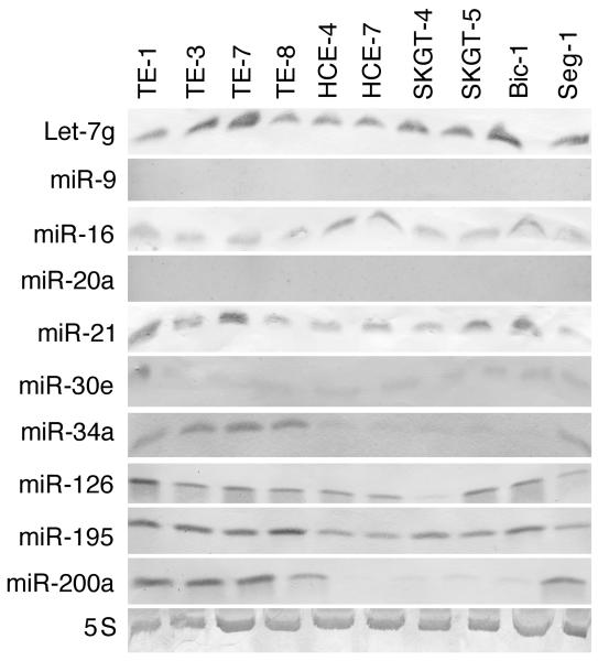
Altered microRNA (miRNA) expression has been found to promote carcinogenesis, but little is known about the role of miRNAs in esophageal cancer. In this study, we selected 10 miRNAs and analyzed their expression in 10 esophageal cancer cell lines and 158 tissue specimens using Northern blotting and in situ hybridization, respectively. We found that Let-7g, miR-21, and miR-195p were expressed in all 10 cell lines, miR-9 and miR-20a were not expressed in any of the cell lines, and miR-16-2, miR-30e, miR-34a, miR-126, and miR-200a were expressed in some of the cell lines but not others. In addition, transient transfection of miR-34a inhibited c-Met and cyclin D1 expression and esophageal cancer cell proliferation, whereas miR-16-2 suppressed RAR-β2 expression and increased tumor cell proliferation. Furthermore, we found that miR-126 expression was associated with tumor cell de-differentiation and lymph node metastasis, miR-16-2 was associated with lymph node metastasis, and miR-195p was associated with higher pathologic disease stages in patients with esophageal adenocarcinoma. Kaplan-Meier analysis showed that miR-16-2 expression and miR-30e expression were associated with shorter overall and disease-free survival in all esophageal cancer patients. In addition, miR-16-2, miR-30e, and miR-200a expression were associated with shorter overall and disease-free survival in esophageal adenocarcinoma patients; however, miR-16-2, miR-30e, and miR-200a expression was not associated with overall or disease-free survival in squamous cell carcinoma patients. Our data indicate that further evaluation of miR-30e and miR-16-2 as prognostic biomarkers is warranted in patients with esophageal adenocarcinoma. In addition, the role of miR-34a in esophageal cancer also warrants further study.
Keywords: miRNA, cell viability, gene regulation, prognosis, esophageal cancer
Esophageal cancer is one of the deadliest cancers and the incidence of esophageal adenocarcinoma has increased rapidly in the United States and other developed countries in recent years.1-3 Esophageal adenocarcinoma now accounts for approximately 7% of all esophageal cancer cases in the United States. The factors underlying this changing pattern are unknown, but Barrett esophagus may be involved.1-5 Epidemiologic studies demonstrated that tobacco and alcohol use and low fruit and vegetable consumption are significant risk factors for both esophageal squamous cell carcinoma and esophageal adenocarcinoma, and frequent gastroesophageal reflux, overweight, and obesity are linked to the increasing incidence of esophageal adenocarcinoma.1-9 These risk factors may contribute to esophageal cancer development through multiple genetic alterations, such as oncogene activation and tumor-suppressor gene dysfunction.3-10
Our previous studies have found that some common risk factors are associated with altered gene expression in esophageal cancer. For example, we found that benzo[a]pyrene diol epoxide (BPDE, a carcinogen present in tobacco and environmental pollution) and bile acid (a tumor promoter in gastrointestinal cancer) suppressed expression of retinoic acid receptor-β;2 and upregulated the expression of epidermal growth factor receptor, activating factor-1, phosphorylated extracellular signal-regulated protein kinases 1/2, and cyclooxygenase-2 in esophageal cancer cells.11-13 Although these disparate observations have improved our understanding of esophageal cancers, more needs to be learned.
Recently, a class of naturally occurring small noncoding RNAs, 18 to 22 nucleotides in length, called microRNAs (miRNAs), has been discovered that can postranscriptionally silence protein expression by binding to complementary target messenger RNAs, thereby degrading these messenger RNAs or inhibiting them from translating into proteins.14-19 A number of studies have found that various miRNAs play an important role in cell growth, differentiation, apoptosis, and carcinogenesis.14-19 For example, miR-15a and miR-16-1 are downregulated in most human chronic lymphocytic leukemias.,20 and miR-143 and miR-145 are downregulated in human colon cancer.21 Let-7 is downregulated in human lung cancer and associated with shortened survival in these patients; however, restoration of Let-7 expression induces growth inhibition of lung cancer cells.22 miR-155 is strongly upregulated in some cases of Burkitt lymphoma and several other types of lymphoma.23,24 miR-21 expression is increased in glioblastoma cells and tissues, where it acts as an antiapoptotic factor.25 miR-103/107 expression is associated with poor survival rates for esophageal squamous cell carcinoma patients.26 Thus, the detection of miRNAs will not only provide us with potential biomarkers for the early detection and prognostic assessment of human cancer but will also provide future and functional analyses of miRNAs for increasing our understanding of their role in esophageal carcinogenesis.
To date, there are few published data on miRNAs in esophageal cancer. Thus, we conducted the current study by choosing 10 miRNAs previously found to be differentially expressed in esophageal cancer27 and determined their expression levels in esophageal adenocarcinoma and squamous cell carcinoma cell lines and human tissue specimens. We then correlated the expression of the 10 miRNAs with the patients’ clinicopathologic data. Our goal was to determine whether miRNAs might be useful prognostic biomarkers in patients with esophageal cancer.
Materials and Methods
Cell culture and Drug treatment
Esophageal squamous cell carcinoma cell lines (TE-1, TE-3, TE-7, TE-8, HCE-4, and HCE-7) and adenocarcinoma cell lines (SKGT-4, SKGT-5, Seg-1, and Bic-1) were grown in Dulbecco's modified Eagle's medium with 10% fetal bovine serum at 37°C in a humidified atmosphere of 95% air and 5% CO2. For BPDE treatment, the cells were plated for 24 h in regular medium. The medium was then replaced with either control medium (containing 1 μL of tetrahydrofuran) or medium containing 1 μM of BPDE (Midwest Research Institute, Kansas City, MO) dissolved in tetrahydrofuran (Sigma) and then incubated for 12 h. The cells were then subjected to northern blotting analysis of miR-16-2 expression.
miRNA probes
We chose 10 miRNAs (Let-7g, miR-9, miR-16-2, miR-20, miR-21, miR-30e, miR-34a, miR-126, miR-195p, and miR-200a) on the basis of a miRNA microarray study27 showing that these miRNAs had different expression levels in esophageal cancer tissues and cell lines compared to normal tissues. We used a 5S probe as the positive control. A miRNA target prediction program showed that the 10 miRNAs chosen may target the genes we have recently been studying. DNA sequences were downloaded from the miRNA database (http://www.microrna.org), and the probes were customarily synthesized by Integrated DNA Technologies (San Diego, CA) with locked nucleic acid modifications and 3′-end labeling with digoxigenin (Table 1).
TABLE 1.
SEQUENCES OF MIRNA PROBES, LNA-MODIFICATION SITES, AND PREDICTED GENE TARGETS
| miRNA | Sequence | Predicted target(s) |
|---|---|---|
| Let-7g | 5′-ct+Gt+A+C+Aa+A+Ct+Actacctca-3′ | RAR-β, EGFR |
| miR-9 | 5′- tcatacag+C+T+Agataaccaaaga-3′ | RXR-α, MAPK |
| miR-16-2 | 5′- cgcc+Aat+Atttac+Gtgctgcta-3′ | RAR-β, SH3GL2 |
| miR-20 | 5′- gatggacgtgatat+T+Cgtgaaat-3′ | MAPK, cyclin D1 |
| miR-21 | 5′- tcaacat+Cag+T+Ctgataagcta-3′ | MAPK, TP53INP1, NFIB |
| miR-30e | 5′- tccagtCAAGgatgtttaca-3′ | RAR-β, Jun |
| miR-34a | 5′- aacaaccagctaagacactgcc-3′ | Cyclin D1, c-Met |
| miR-126 | 5′- gcattattac+T+Cacggtacga-3′ | VEGF |
| miR-195p | 5′- gccaa+Tattt+Ctgtgctgcta-3′ | Cyclin D1, MAPK |
| miR-200a | 5′- acatcgtta+Ccagacagtgtta-3′ | MAPK, RAR-β |
| 5S | 5′- ttagcttccgagatcagacg-3′ | N/A |
LNA, locked nucleic acid; N/A, not available.
Note: The plus sign (+) followed by a capital letter indicates LNA-modified nucleic acid. Tm (DNA melting temperature) of these miRNAs was justified to approximately 58°C after LNA-modification. The predicted gene targets of miRNAs were obtained from the miRNA database (http://www.microrna.org/). N/A indicates that no targets are available to date.
RNA isolation and Northern blotting
Total RNA was isolated from the esophageal cancer cell lines using Tri-reagent (Molecular Research Center, Cincinnati, OH), as described previously,28,29 and subjected to Northern blotting. Briefly, 20 μg of total RNA from the esophageal cancer cell lines was separated onto 12.5% denatured polyacrylamide gels. The blots were incubated in hybridization buffer for 1 h at 42°C and then overnight in hybridization buffer containing 2 nM probe at 42°C. After washing (2 times for 30 min each at 42°C in 2X standard saline citrate (SSC)/0.1° sodium dodecyl sulfate), the blots were rinsed in washing buffer (0.1 M maleic acid and 0.15 M NaCl [pH 7.5]) and blocked in washing buffer containing 5° milk powder for 30 min at room temperature. Subsequently, the blots were incubated with anti-digoxigenin-alkaline phosphatase (AP) antibody (Roche, Indianapolis, IN) in blocking buffer for 1 h and washed 3 times for 10 min each in washing buffer and, briefly, with AP buffer (0.1 M Tris-HCl [pH 9.5], 50 mM MgCl2, and 0.1 M NaCl). Positive signals were detected using a chromogen solution (45 μL of nitroblue tetrazolium and 35 μL of an X-phosphate solution in 10 mL of AP buffer) for 6 h, with occasional observation for color development.
miRNA expression vector construction and gene transfection
The miR-34 and miR-16-2 gene sequences were obtained from www.microrng.org, and the sense and antisense nucleic acids were then synthesized by Sigma-Genosys (The Woodlands, TX), heated to 95°C, cooled down at room temperature, and purified by 10% polyacrylamide gel electrophoresis. The annealized oligonucleotides were then ligated to pRNA-u6.3 expression vector (GenScript, Piscataway, NJ) and amplified. After sequence verification, the miRNA expression vectors were amplified using a maxi DNA preparation kit (QIAGEN, Valencia, CA). For gene transfection, esophageal cancer cell cells were seeded overnight in 10-cm cell culture dishes and transfected with 2 μg pRNA-u6.3/miR-34a, pRNA-u6.3/miR-16-2, or vector only with FuGENE6 and screened with 400 μg/mL of G418 for 3-4 days (FuGENE6 and G418 both supplied by Roche). The transfected cells were then subjected to Western blotting, RT-PCR, and cell viability assays.
Protein extraction and Western blotting
Total cellular protein was extracted from cells as described previously.11-13 Samples containing 50 μg of protein from each cell line were then separated by 10% to 14% sodium dodecyl sulfate-polyacrylamide gel electrophoresis and transferred electrophoretically to a Hybond-C nitrocellulose membrane (GE Healthcare, Arlington Heights, IL) at 500 mA for 2 h at 4°C. To confirm that the proteins were loaded equally and to verify transfer efficiency, the membrane was stained with 0.5% Ponceau S containing 1% acetic acid. The membranes were then subjected to Western blotting by overnight incubation in a blocking solution containing 5% bovine skim milk and 0.1% Tween 20 in phosphate-buffered saline at 4°C. The next day, the membranes were incubated first with primary antibodies and then with horse anti-mouse or goat anti-rabbit secondary antibodies (GE Healthcare) for enhanced chemiluminescence detection of antibody signals. The antibodies used were anti-cyclin D1 (diluted at 1:50) and anti-c-Met (diluted at 1:50; all from Cell Signaling Technology, Danvers, MA) and anti-β-actin (Sigma-Aldrich, St. Louis, MO).
Immunohistochemical staining of Ki-67 after transient miRNA transfection
Esophageal cancer cells were grown in monolayer overnight and transiently transfected with either pCMS/EGFP (Clontech, Mountain View, CA) plus pRNA-u6.3/miR-34a vector or pCMS/EGFP plus pRNA-u6.3 vector (GenScript) using Lipofectamine 2000 (Invitrogen, Carlsbad, CA) for 36 h. The cells were then treated with 400 μg/mL G418 for an additional 24 h, fixed with 4% paraformaldehyde for 10 min at room temperature to preserve the green fluorescent protein (GFP), and permeabilized in 0.5% Triton X-100 for 10 min at room temperature. The cells were then subjected to Ki-67 immunostaining as described previously.30 Ki-67 antibody was purchased from Vector Laboratories (Burlingame, CA) and diluted at 1:50 in phosphate-buffered saline. Approximately 200 cells in 10 fields were then counted at 20x magnification for positive GFP staining (green) and positive or negative Ki-67 staining (red). The percentage of cell proliferation control was calculated using the following equation: % control = N34a/NVector × 100, where N34a and NVector represent the numbers of Ki-67-positive cells in GFP-positive cells of miRNA-transfected and control cultures, respectively.
Reverse transcription-polymerase chain reaction (RT-PCR)
After the cells was transfected with miR-16-2 expression vector and treated with G418 for 3 days, RNA from the cells was isolated and subjected to RT-PCR analysis of RAR-β2 mRNA expression as described previously.28 The primers for RAR-β2 expression were 5′-CAAACCGAATGGCAGCATCGG-3′ and 5′-GCGGAAAAAGCCCTTACATCCC-3′, which amplified a 195-bp band. GAPDH primers were 5′-CCCTTCATTGACCTCAACTACATGG-3′ and 5′-CATGGTGGTGAAGACGCCAG-3′, which generated 192-bp band.
Methyl thiazolyl tetrazolium (MTT) assay
The cells were transiently transfected with miR-16-2 expression vector for 8 h and then treated with G418 for 4 days. After that, 20 μL of MTT (5 mg/ml, Sigma, St Louis, MO) was added to each well of the 96-well plates and incubated for an additional 2 h. After the growth medium was removed, 100 μL of DMSO was added to the wells to dissolve the MTT crystal, and the optical densities were measured with an automated spectrophotometric plate reader at a single wavelength of 540 nm. The percentage of cell proliferation was calculated using the formula: % control = ODt/ODc × 100, where ODt and ODc are the optical densities for treated and control cells, respectively.
Tissue specimens
Our institutional review board approved our protocol for the use of patient samples in this study. We identified 172 consecutive patients who had undergone esophagectomy without preoperative chemotherapy or radiotherapy between 1986 and 1998 at The University of Texas M. D. Anderson Cancer Center. To create a tissue microarray, each patient's tumor specimen was first identified on hematoxylin and eosin–stained slides, and the corresponding formalin-fixed, paraffin-embedded tissue blocks were obtained. One-millimeter tissue cores in triplicate were obtained from each tumor and arrayed onto a recipient paraffin block using a tissue arrayer (Beecher Instruments, Sun Prairie, WI), as previously described.31 After cutting and staining of these microarrays, there were 158 cases with available tissue spots for evaluation.
In situ hybridization
To detect miRNA expression in the tissue samples, nonradioactive in situ hybridization was performed as described previously, with some modifications.29,32 Briefly, the tissue arrays were subjected to treatment with 0.2 N HCl and proteinase K after deparaffinization and rehydration, respectively. The sections were then postfixed with 4% paraformaldehyde and acetylated in freshly prepared 0.25% acetic anhydride in a 0.1-M triethanolamine buffer. The sections were prehybridized at 37°C with a hybridization solution containing 50% formamide, SSC, 2X Denhardt's solution, 10% dextran sulfate, 400 μg/mL yeast tRNA, and 20 μM dithiothreitol in diethylpyrocarbonate-treated water. Next, the sections were incubated for 4 h at 37°C in 150 μL of hybridization solution/section containing 100 nM of a freshly denatured digoxigenin-labeled miRNA probe. The slides were washed for 2 h in 2X SSC containing 2% normal sheep serum and 0.05% Triton X-100. For color reaction, the sections were incubated for 30 min at 23°C in 0.1 M maleic acid and 0.15 M NaCl containing 2% normal sheep serum and 0.3% Triton X-100 and then incubated overnight at 4°C with a sheep anti-digoxigenin antibody. After sections were washed in buffer twice, the color was developed in a chromogen solution (45 μL of nitroblue tetrazolium and 35 μL of an X-phosphate solution in 10 mL of AP buffer, which consists of 0.1 M Tris, 0.1 M NaCl, and 0.05 M MgCl2 [pH 9.5]) for 16 h, with occasional observation for color development. The sections were then mounted with cover glass in Aqua mounting medium. For negative controls, the paraffin sections were pretreated with 4 μg/100 μL RNase A per slide before being subjected to in situ hybridization. We also performed in situ hybridization in esophageal cancer cell lines with positive and negative-expressed miRNA as additional control of the specificity of the miRNA probes (supplemental Figure 1).
Review and scoring of sections
The stained sections were reviewed under an Olympus microscope (Melville, NY) and scored based on the staining intensity and the percentage of cells stained. Staining intensity was scored as negative (−), weakly positive (+/−), positive (+), strongly positive (++), or very strongly positive (+++). The percentage of cells stained was scored as negative (−) if none of the three tissue spots were stained and positive (+) if one or more tissue spots were stained positively. High miRNA expression was then defined for each case as positive staining intensity (+, ++, or +++) and positive percentage of tumor cells stained; low expression was defined as negative (−) or weakly positive (+/−) staining intensity with negative percentage of tumor cells stained.
Statistical analysis
Disease-free and overall survival curves were plotted separately for patients with low versus high expression of each miRNA using the Kaplan-Meier method. The log-rank test was used to compare the survival times of the patients with high and low expression of specific miRNAs. Disease-free and overall survival times were calculated from the date of surgery to the date of disease recurrence or death. To determine the prognostic value of each miRNA in terms of disease-free and overall survival among esophageal cancer patients, we used Cox proportional hazards regression to calculate the hazard ratios (HRs) and 95% confidence intervals (CIs) for each model. For the multivariate analyses, we adjusted for other known prognostic factors, such as age, sex, clinical and pathologic disease stage, tumor histology, differentiation, metastasis, tumor size, and tumor-free margins. All statistical analyses were performed using SPSS statistical package, version 11 (SPSS Inc., Chicago, IL). Two-sided t tests were considered statistically significant if p values were < 0.05.
Results
miRNA expression in esophageal cancer cell lines
Using digoxigenin-labeled and locked nucleic acid-modified miRNA probes, we performed nonradioactive Northern blotting to detect the expression of the 10 miRNAs in the esophageal cancer cell lines. We found that the 10 miRNAs were differentially expressed in these esophageal cancer cell lines (Figure 1). In particular, Let-7g, miR-21, and miR-195p were expressed in all of the cell lines; miR-9 and miR-20a were not expressed in any of the cell lines; and miR-16-2, miR-30e, miR-34a, miR-126, and miR-200a were expressed in some of the cell lines but not others (Figure 1).
Figure 1.
Nonradioactive Northern blotting analysis of miRNA expression. Esophageal squamous cell carcinoma cell lines (TE-1, TE-3, TE-7, TE-8, HCE-4, and HCE-7) and adenocarcinoma cell lines (SKGT-4, SKGT-5, Seg-1 and Bic-1) were grown in Dulbecco's modified Eagle medium, and total RNA was extracted for Northern blot analysis with digoxigenin-labeled and locked nucleic acid–modified miRNA probes. The blots were then scanned and plotted.
Impact of miR-34a and miR-16-2 on gene expression and cancer cell proliferation
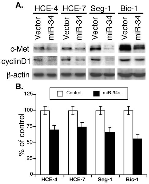
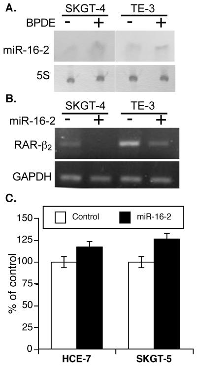
Previous studies have shown that miR-34a expression is controlled by the p53 protein,33 and p53 is frequently mutated in esophageal cancer.3-10 Moreover, transactivation of miR-34a by p53 broadly influenced gene expression and promoted apoptosis.34 Thus, we chose to study miR-34a further by constructing an expression vector carrying miR-34a that was transiently transfected into four esophageal cancer cell lines (HCE-4, HCE-7, Seg-1, and Bic-1). Western blotting showed that miR-34a inhibited c-Met and cyclin D1 protein expression (Figure 2A). In addition, cell proliferation assays showed that transient transfection of miR-34a reduced the viability of these four cell lines (Figure 2B). Furthermore, miR-16-2 was predicted to target RAR-β2 expression by an internet tool (http://www.microrna.org/) and BPDE was shown to suppress RAR-β2 expression.11,13 We found that BPDE was able to induce miR-16-2 expression and that transfection of miR-16-2 inhibited RAR-β2 expression, but induced proliferation of esophageal cancer cells (Figure 3).
Figure 2.
miR-34a suppression of gene expression and proliferation of esophageal cancer cells. A, Western blotting. Esophageal squamous cell carcinoma lines HCE-4 and HCE-7 and adenocarcinoma cell lines Seg-1 and Bic-1 were grown in monolayer and transiently transfected with pRNA-u6.3/miR-34a vector or vector-only control and treated with G418 for 3 days. The total cellular protein was then extracted and subjected to Western blotting analysis of c-Met and cyclin D1 expression. B, Cell proliferation assay. Cells were grown in monolayer and transiently transfected with either pCMS/EGFP plus pRNA-u6.3/miR-34a vector or pCMS/EGFP plus pRNA-u6.3 vector. Cells were then fixed with 4% paraformaldehyde and permeabilized in Triton X-100. Afterward, cells were immunostained for Ki-67 expression, and approximately 200 cells were counted for positive GFP staining f (green) and positive or negative Ki-67 staining (red). The percentage of control of cell proliferation was then calculated.
Figure 3.
The role of miR-16-2 in esophageal cancer cells. A, Northern blotting. Esophageal adenocarcinoma SKGT-4 cells and squamous cell carcinoma TE-3 cells were grown and treated with 1 μl BPDE for 12 h and RNA was isolated and subjected to northern blot analysis. B, SKGT-4 and TE-3 cells were grown in monolayer and transiently transfected with pRNA-u6.3/miR-16-2 vector or vector-only control and treated with G418 for 3 days. RNA from the cells was isolated and subjected to RT-PCR analysis of RAR-β2 expression. C, Esophageal adenocarcinoma SKGT-5 cells and squamous cell carcinoma HEC-7 cells were grown and transiently transfected with pRNA-u6.3/miR-16-2 vector or vector-only control and treated with G418 for 4 days. The cells were then subjected to cell viability assay.
miRNA expression in esophageal cancer tissue specimens and relationship between miRNA expression and clinicopathologic features
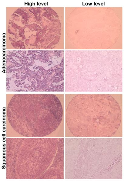
On in situ hybridization, we found that the 10 miRNAs were expressed in the esophageal cancer tissue microarray containing 172 esophageal cancer specimens but only 158 cases are available for review (Figure 4). The clinicopathologic data from these patients are shown in supplemental Table 1. The positive staining rates for each miRNA were as follows: Let-7g, 56.7%; miR-9, 78.5%; miR-16-2, 51.9%; miR-20, 73.0%; miR-21, 81.6%; miR-30e, 63.3%; miR-34a, 39.5%; miR-126, 44.3%; miR-195p, 62.4%; and miR-200a, 43.9%. We then associated the individual miRNA expression levels with the patients' clinicopathologic data. We found that miR-126 expression was associated with tumor cell de-differentiation (p = 0.0079) and lymph node metastasis (p = 0.01), miR-16-2 expression was associated with lymph node metastasis (p = 0.03), and miR-195p expression was associated with higher pathologic disease stages (p = 0.0008) in esophageal adenocarcinoma patients. However, the other clinicopathologic parameters (e.g., age, sex, or tumor size) were not significantly associated with the expression of any of the other miRNAs (data not shown).
Figure 4.
Detection of miRNA expression in esophageal tissue microarrays using in situ hybridization. A tissue microarray contained 158 available cases of esophageal cancer, and three tissue spots for each case were on the same sections. A total of three sections were hybridized with each digoxigenin-labeled miRNA probe and then scored for expression of each miRNA.
Association of miRNA expression with overall and disease-free survival
We then performed Kaplan-Meier analyses to determine the relationship between miRNA expression and prognosis. The patients' median overall survival was 16.25 months (range, 0.37 to 256.43 months). miR-16-2 expression and miR-30e expression were correlated with shorter overall and disease-free survival in esophageal cancer patients overall (Figure 5). Cox proportional hazards analyses showed similar results: miR-30e expression was associated with shorter overall survival (HR 1.80 [95% CI: 1.26-2.57]; p = 0.001) and disease-free survival (HR 1.67 [95% CI: 1.17-2.38]; p = 0.005), and miR-16-2 expression was also associated with shorter overall survival (HR 1.40 [95% CI: 1.00-1.95]; p = 0.048) and disease-free survival (HR 1.46 [95% CI: 1.00-2.04]; p = 0.024) in esophageal cancer patients overall.
Figure 5.
Relationship between miRNA expression and survival in all esophageal cancer patients. The tissue microarrays were hybridized in situ with digoxigenin-labeled miRNA probes and then scored for high and low expression of each miRNA. The data on the high and low miRNA expression were then statistically analyzed against the overall and disease-free survival data of the 158 patients with esophageal cancer using the Kaplan-Meier method.
Because of the relatively different etiologies and gene expression profiles of esophageal squamous cell carcinoma and esophageal adenocarcinoma, we separated the patients with squamous cell carcinoma from those with adenocarcinoma for the Kaplan-Meier plot, Cox proportional hazards, and univariate and multivariate hazard ratio analyses. There were 99 available cases of adenocarcinoma and 59 available cases of squamous cell carcinoma.
Adenocarcinoma
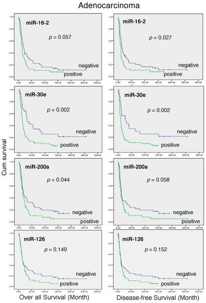
Figure 6 shows that in patients with adenocarcinoma, miR-30e expression and miR-200a expression were significantly associated with poor overall survival, while miR-16-2 expression and miR-126 expression were borderline significant for worse overall survival. miR-16-2 and miR-30e expression were also significantly associated with poor disease-free survival, while miR-200a and miR-126 expression was borderline significant for disease-free survival in the adenocarcinoma patients. On univariate analysis, miR-30e expression was associated with shorter overall survival (HR = 2.21 [95% CI, 1.31-3.73]; p < 0.003; Table 2). On multivariate analysis, miR-30e expression retained borderline significance for shorter overall survival (HR 2.0 [95% CI” 0.93-4.31]; p < 0.078). Similarly, miR-30e expression was significantly associated with poor disease-free survival on univariate and multivariate analysis (Table 3). After surgery, the risk of recurrence for adenocarcinoma patients was 2.5-fold greater for patients with miR-30e expression than for patients without miR-30e expression.
Figure 6.
Relationship between miRNA expression and survival in the patients with esophageal adenocarcinoma. The tissue microarrays were hybridized in situ with digoxigenin-labeled miRNA probes and then scored for high and low expression of each miRNA. The data on the high and low miRNA expression were then statistically analyzed against the overall and disease-free survival data of the 99 patients with esophageal adenocarcinoma using the Kaplan-Meier method.
TABLE 2.
UNIVARIATE AND MULTIVARIATE ANALYSES OF THE RELATIONSHIP BETWEEN MIRNA EXPRESSION AND OVERALL SURVIVAL IN ESOPHAGEAL ADENOCARCINOMA PATIENTS
| Univariate analysis | Multivariate analysis | ||||||
|---|---|---|---|---|---|---|---|
| miRNA | Frequency (N) |
HR | 95% CI |
p value |
HR | 95% CI |
p value |
| Let-7g | − 37 | 1.00 | 1.00 | ||||
| + 62 | 0.73 | 0.47-1.12 | 0.15 | 0.55 | 0.29-1.05 | 0.069 | |
| miR-9 | − 11 | 1.00 | 1.00 | ||||
| + 88 | 0.92 | 0.49-1.73 | 0.80 | 0.62 | 0.26-1.48 | 0.28 | |
| miR-16-2 | − 52 | 1.00 | 1.00 | ||||
| + 47 | 1.50 | 0.99-2.29 | 0.06 | 0.83 | 0.45-1.55 | 0.56 | |
| miR-20 | − 16 | 1.00 | 1.00 | ||||
| + 83 | 1.17 | 0.65-2.12 | 0.61 | 0.69 | 0.26-4.31 | 0.47 | |
| miR-21 | − 11 | 1.00 | 1.00 | ||||
| + 88 | 1.73 | 0.86-3.47 | 0.13 | 0.87 | 0.34-2.23 | 0.77 | |
| miR-30e | − 25 | 1.00 | 1.00 | ||||
| + 74 | 2.21 | 1.31-3.73 | 0.003 | 2.00 | 0.93-4.31 | 0.078 | |
| miR-34a | − 58 | 1.00 | 1.00 | ||||
| + 41 | 0.81 | 0.53-1.24 | 0.33 | 0.71 | 0.41-1.24 | 0.23 | |
| miR-126 | − 46 | 1.00 | 1.00 | ||||
| + 53 | 1.37 | 0.89-2.11 | 0.15 | 0.90 | 0.52-1.57 | 0.71 | |
| miR-195p | − 37 | 1.00 | 1.00 | ||||
| + 62 | 1.52 | 0.97-2.39 | 0.07 | 2.21 | 1.15-4.24 | 0.02 | |
| miR-200a | − 46 | 1.00 | 1.00 | ||||
| + 53 | 1.55 | 1.01-2.37 | 0.046 | 1.29 | 0.45-1.55 | 0.41 | |
TABLE 3.
UNIVARIATE AND MULTIVARIATE ANALYSES OF THE RELATIONSHIP BETWEEN MIRNA EXPRESSION AND DISEASE-FREE SURVIVAL IN ESOPHAGEAL ADENOCARCINOMA PATIENTS
| Univariate analysis | Multivariate analysis | ||||||
|---|---|---|---|---|---|---|---|
| miRNA | Frequency (N) |
HR | 95% CI |
p value |
HR | 95% CI |
p value |
| Let-7g | − 37 | 1.00 | 1.00 | ||||
| + 62 | 0.77 | 0.50-1.19 | 0.24 | 0.64 | 0.33-1.23 | 0.18 | |
| miR-9 | − 11 | 1.00 | 1.00 | ||||
| + 88 | 0.75 | 0.40-1.41 | 0.37 | 0.45 | 0.18-1.12 | 0.086 | |
| miR-16-2 | − 52 | 1.00 | 1.00 | ||||
| + 47 | 1.60 | 1.05-2.44 | 0.028 | 0.88 | 0.47-1.64 | 0.69 | |
| miR-20 | − 16 | 1.00 | 1.00 | ||||
| + 83 | 1.09 | 0.60-1.98 | 0.77 | 0.66 | 0.24-1.81 | 0.42 | |
| miR-21 | − 11 | 1.00 | 1.00 | ||||
| + 88 | 1.62 | 0.81-3.26 | 0.18 | 0.96 | 0.37-2.48 | 0.93 | |
| miR-30e | − 25 | 1.00 | 1.00 | ||||
| + 74 | 2.21 | 1.31-3.76 | 0.003 | 2.53 | 1.13-5.67 | 0.024 | |
| miR-34a | − 58 | 1.00 | 1.00 | ||||
| + 41 | 0.74 | 0.48-1.14 | 0.17 | 0.72 | 0.43-1.22 | 0.22 | |
| miR-126 | − 46 | 1.00 | 1.00 | ||||
| + 53 | 1.37 | 0.89-2.11 | 0.15 | 0.83 | 0.48-1.44 | 0.51 | |
| miR-195p | − 37 | 1.00 | 1.00 | ||||
| + 62 | 1.52 | 0.97-2.39 | 0.067 | 2.05 | 1.08-3.90 | 0.029 | |
| miR-200a | − 46 | 1.00 | 1.00 | ||||
| + 53 | 1.51 | 0.98-2.31 | 0.06 | 1.30 | 0.69-2.42 | 0.42 | |
Squamous cell carcinoma
For patients with squamous cell carcinoma, Kaplan-Meier analysis didn't show any association between survival and miRNA expression. However, multivariate analysis showed an association between overall or disease-free survival and miR-9 (p = 0.03, p = 0.03), miR-16-2 (p = 0.04, p = 0.02), miR-20 (p = 0.03, p = 0.06), and miR-200a (p = 0.047, p = 0.056) expression.
In addition, our data demonstrated that positive section margins (p = 0.036), advanced clinical disease stage (p=0.025), pathologic disease stage (p=0.0001), poor tumor differentiation (p=0.009), positive lymph nodes (p=0.008), and tumor metastasis (p=0.001) were associated with shorter overall survival in adenocarcinoma patients, and positive section margins (p = 0.029), pathologic disease stage (p= 0.007), and positive lymph nodes (p=0.047) were associated with shorter overall survival in squamous cell carcinoma patients. These same factors were statistically significant for shorter disease-free survival in both adenocarcinoma and squamous cell carcinoma patients.
Discussion
In the current study, we analyzed the expression of 10 miRNAs in esophageal cancer cell lines and tissue specimens and determined the role of miR-34a and miR-16-2 in regulating proliferation and gene expression in esophageal cancer cell lines. We found that most of the 10 miRNAs were expressed in the esophageal cancer cell lines and tissues. The expression of some of the miRNAs was associated with tumor cell de-differentiation (miR-126), lymph node metastasis (miR-126 and miR-16-2), or higher pathologic tumor stage (miR-195p) in esophageal adenocarcinoma patients. We also found that miR-34a was able to suppress c-Met and cyclin D1 expression and proliferation of esophageal cancer cell lines, whereas miR-16-2 was upregulated by BPDE treatment and miR-16-2 suppressed RAR-β2 expression but induced esophageal cancer proliferation. Moreover, miR-16-2 and miR-30e expression was significantly associated with poor overall and disease-free survival in esophageal cancer patients, especially in the adenocarcinoma patients. The data from this study indicate that detection of miR-30e expression may serve as an adjunct marker in predicting the survival of esophageal adenocarcinoma patients, although prospective validation is warranted. In addition, the role of miR-34a in esophageal cancer warrants further investigation.
To date, there are few published data on miRNAs in esophageal cancer.26,35,36 In a recent study,26 Guo et al analyzed the expression profiles of 509 miRNAs in 31 esophageal squamous cell carcinoma and adjacent normal tissue samples by using miRNA microarrays. They found that the expression levels of some miRNAs were different between esophageal squamous cell carcinoma and normal tissues, and miR-103/107 expression was associated with poor survival.26 Another recent study showed that elevated miR-21 expression in noncancerous tissues of SCC patients was associated with worse prognosis of the patients.36 However, our current study didn't find such an association. Like most previous studies on miRNA expression, the studies by Guo et al26 and Mathe et al36 were performed on microarrays using RNA extracted from human cancer tissue samples27,37-39 and containing a mixture of tumor and normal cells. Our current study instead utilized the in situ hybridization technique. The advantage of this technique is that it can precisely identify positive signals at the cellular level.29 Indeed, our most recent data have demonstrated that some miRNAs had very high expression levels in stromal cells but not in tumor cells, which is similar to what we observed in our current study (Figure 3). Using in situ hybridization to detect the expression of miRNAs may help us develop biomarkers for use in the early detection or risk assessment of esophageal cancer.
Our current study demonstrated that miR-30e expression was associated with poor overall survival and disease-free survival in adenocarcinoma patients. However, there was not a significant association between miRNA expression and overall survival and disease-free survival in squamous cell carcinoma patients. In this study, we confirmed that multiple factors can predict the survival of esophageal cancer patients, such as clinical and pathologic TNM stages, tumor differentiation and size, tumor-free section margins, and gene expression. It remains unknown which gene(s) miR-30e regulates or how miR-30e is regulated in esophageal cancer. To date, several other studies have successfully discovered a role for and target genes of some miRNAs; for example, Let-7 was found to regulate RAS gene expression, p53 was found to modulate miR-34a expression.33,34 A recent study demonstrated that an ectopic expression of mir-34a in IMR90 cells led to substantial inhibition of growth (i.e., the fraction of S-phase cells decreased, but G1 and G2 cells increased). Molecularly, miR-34a suppressed expression of c-MET and cell cycle-related genes.33 Our current study confirmed these data in esophageal cancer cells and showed that transfection of miR-34a into various esophageal cancer cell lines suppressed tumor cell growth and expression of c-MET and cyclinD1 although we didn't know whether inhibition of both genes by miR-34a is direct or indirect. However, the data from He et al33 demonstrated that miR-34 can directly bind to c-MET and then suppress c-MET expression, but inhibition of cyclinD1 (cell growth arrest) is indirect effect of miR-34a. Our current study also found that the altered expression of miR-34a in esophageal cancer tissue specimens did not show a significant association with the patients' clinicopathologic parameters and survival. Further investigations will focus on miRNA-targeted gene expression.
Our current data also demonstrated an association between miR-16-2 or miR-200a expression and worse overall and disease-free survival in adenocarcinoma patients. Nevertheless, the role of miR-16-2 and miR-200a in tumor progression is unclear. We noted that a similar miRNA, miR-16-1, was downregulated in chronic lymphocytic lymphoma, pituitary adenoma, and prostate cancer. Induction of miR-16-1 expression inhibited cell proliferation, promoted apoptosis of tumor cells, and suppressed tumorigenicity in vitro and in vivo. miR-16-1 functions by targeting multiple oncogenes, such as BCL2, MCL1, CCND1, and WNT3A.40 The miR-16-1 gene is localized in chromosome 13, while the miR-16-2 gene is localized at chromosome 3. Thus, miR-16-1 and miR-16-2 are different genes and have different gene targets (see details in www.microrna.org). In addition, a previous study showed that miR-200a was overexpressed in ovarian cancer tissues compared to normal tissues.41 Another study reported that miR-200a expression was significantly associated with fewer cancer recurrences and better overall survival in patients with ovarian cancer.42 That study showed that overexpression of miR-200a led to a reduction of 39% in cancer cell mobility,42 which is in contrast to our current data that demonstrated that expression of miR-200a was associated with poor survival in esophageal adenocarcinoma patients.
The finding that miR-16-2 and miR-30e expression associated with worse survival rates of patients with esophageal adenocarcinoma in the current study will need to be verified in other population before these miRNA could be used as biomarkers for clinical usage. We will determine whether any of the 10 miRNAs studied here can regulate expression of EGFR, AP-1, RAR-β, and MAPK in esophageal cancer cells, as these genes are frequently altered in esophageal cancer and may play a role in esophageal cancer development or progression.10
Supplementary Material
Acknowledgments
Grant sponsor: National Cancer Institute Grant R03 CA123568.
Footnotes
Novelty: Use miRNAs (miR-16-2 and miR-30e) as biomarkers for prognosis of esophageal cancer patients.
Impact: After verified, these miRNAs would be used to predict survival of the patients. In addition, these data will guide our future study of miRNA function in esophageal cancer.
None of the authors have any conflicts of interest to disclose.
References
- 1.Blot W. Esophageal cancer trends and risk factors. Semin Oncol. 1994;21:403–10. [PubMed] [Google Scholar]
- 2.van Soest EM, Dieleman JP, Siersema PD, Sturkenboom MC, Kuipers EJ. Increasing incidence of Barrett's oesophagus in the general population. Gut. 2005;54:1062–6. doi: 10.1136/gut.2004.063685. [DOI] [PMC free article] [PubMed] [Google Scholar]
- 3.Chen X, Yang CS. Esophageal adenocarcinoma: a review and perspectives on the mechanism of carcinogenesis and chemoprevention. Carcinogenesis. 2001;22:1119–29. doi: 10.1093/carcin/22.8.1119. [DOI] [PubMed] [Google Scholar]
- 4.Reid BJ, Blount PL, Rabinovitch PS. Biomarkers in Barrett's esophagus. Gastrointest Endosc Clin N Am. 2003;13:369–97. doi: 10.1016/s1052-5157(03)00006-0. [DOI] [PubMed] [Google Scholar]
- 5.Spechler SJ. Barrett's esophagus: a molecular perspective. Curr Gastroenterol Rep. 2005;7:177–81. doi: 10.1007/s11894-005-0031-z. [DOI] [PubMed] [Google Scholar]
- 6.Mandard AM, Hainaut P, Hollstein M. Genetic steps in the development of squamous cell carcinoma of the esophagus. Mutat Res. 2000;462:335–42. doi: 10.1016/s1383-5742(00)00019-3. [DOI] [PubMed] [Google Scholar]
- 7.Montesano R, Hollstein M, Hainaut P. Genetic alterations in esophageal cancer and their relevance to etiology and pathogenesis: a review. Int J Cancer. 1996;69:225–35. doi: 10.1002/(SICI)1097-0215(19960621)69:3<225::AID-IJC13>3.0.CO;2-6. [DOI] [PubMed] [Google Scholar]
- 8.Smith KJ, O'Brien SM, Smithers BM, Gotley DC, Webb PM, Green AC, Whiteman DC. Interactions among smoking, obesity, and symptoms of acid reflux in Barrett's esophagus. Cancer Epidemiol Biomarkers Prev. 2005;14:2481–6. doi: 10.1158/1055-9965.EPI-05-0370. [DOI] [PMC free article] [PubMed] [Google Scholar]
- 9.McManus DT, Olaru A, Meltzer SJ. Biomarkers of esophageal adenocarcinoma and Barrett's esophagus. Cancer Res. 2004;64:1561–9. doi: 10.1158/0008-5472.can-03-2438. [DOI] [PubMed] [Google Scholar]
- 10.Xu X-C. Risk factors and altered gene expression in esophageal cancer. In: Verma M, editor. Cancer epidemiology. Humana Press; New York: 2009. pp. 335–60. [DOI] [PubMed] [Google Scholar]
- 11.Song S, Xu X-C. Effect of benzo[a]pyrene diol epoxide on expression of retinoic acid receptor-beta in immortalized esophageal epithelial cells and esophageal cancer cells. Biochem Biophys Res Comm. 2001;281:872–7. doi: 10.1006/bbrc.2001.4433. [DOI] [PubMed] [Google Scholar]
- 12.Li M, Song S, Lippman SM, Zhang XK, Liu X, Lotan R, Xu X-C. Induction of retinoic acid receptor-beta suppresses cyclooxygenase-2 expression in esophageal cancer cells. Oncogene. 2002;21:411–8. doi: 10.1038/sj.onc.1205106. [DOI] [PubMed] [Google Scholar]
- 13.Song S, Lippman SM, Zou Y, Ye X, Ajani JA, Xu X-C. Induction of cyclooxygenase-2 by benzo[a]pyrene diol epoxide through inhibition of retinoic acid receptor-β2 expression. Oncogene. 2005;24:8268–76. doi: 10.1038/sj.onc.1208992. [DOI] [PubMed] [Google Scholar]
- 14.Bartel DP. MicroRNAs: genomics, biogenesis, mechanism, and function. Cell. 2004;116:281–97. doi: 10.1016/s0092-8674(04)00045-5. [DOI] [PubMed] [Google Scholar]
- 15.Alvarez-Garcia I, Miska EA. MicroRNA functions in animal development and human disease. Development. 2005;132:4653–62. doi: 10.1242/dev.02073. [DOI] [PubMed] [Google Scholar]
- 16.Gregory RI, Shiekhattar R. MicroRNA biogenesis and cancer. Cancer Res. 2005;65:3509–12. doi: 10.1158/0008-5472.CAN-05-0298. [DOI] [PubMed] [Google Scholar]
- 17.McManus MT. MicroRNAs and cancer. Semin Cancer Biol. 2003;13:253–8. doi: 10.1016/s1044-579x(03)00038-5. [DOI] [PubMed] [Google Scholar]
- 18.Croce CM, Calin GA. miRNAs, cancer, and stem cell division. Cell. 2005;122:6–7. doi: 10.1016/j.cell.2005.06.036. [DOI] [PubMed] [Google Scholar]
- 19.Mendell JT. MicroRNAs: critical regulators of development, cellular physiology and malignancy. Cell Cycle. 2005;4:1179–84. doi: 10.4161/cc.4.9.2032. [DOI] [PubMed] [Google Scholar]
- 20.Calin GA, Dumitru CD, Shimizu M, Bichi R, Zupo S, Noch E, Aldler H, Rattan S, Keating M, Rai K, Rassenti L, Kipps T, et al. Frequent deletions and down-regulation of micro-RNA genes miR15 and miR16 at 13q14 in chronic lymphocytic leukemia. Proc Natl Acad Sci U S A. 2002;99:15524–9. doi: 10.1073/pnas.242606799. [DOI] [PMC free article] [PubMed] [Google Scholar]
- 21.Michael MZ, SM OC, van Holst Pellekaan NG, Young GP, James RJ. Reduced accumulation of specific microRNAs in colorectal neoplasia. Mol Cancer Res. 2003;1:882–91. [PubMed] [Google Scholar]
- 22.Takamizawa J, Konishi H, Yanagisawa K, Tomida S, Osada H, Endoh H, Harano T, Yatabe Y, Nagino M, Nimura Y, Mitsudomi T, Takahashi T. Reduced expression of the let-7 microRNAs in human lung cancers in association with shortened postoperative survival. Cancer Res. 2004;64:3753–6. doi: 10.1158/0008-5472.CAN-04-0637. [DOI] [PubMed] [Google Scholar]
- 23.Metzler M, Wilda M, Busch K, Viehmann S, Borkhardt A. High expression of precursor microRNA-155/BIC RNA in children with Burkitt lymphoma. Genes Chromosomes Cancer. 2004;39:167–9. doi: 10.1002/gcc.10316. [DOI] [PubMed] [Google Scholar]
- 24.Eis PS, Tam W, Sun L, Chadburn A, Li Z, Gomez MF, Lund E, Dahlberg JE. Accumulation of miR-155 and BIC RNA in human B cell lymphomas. Proc Natl Acad Sci U S A. 2005;102:3627–32. doi: 10.1073/pnas.0500613102. [DOI] [PMC free article] [PubMed] [Google Scholar]
- 25.Chan JA, Krichevsky AM, Kosik KS. MicroRNA-21 is an antiapoptotic factor in human glioblastoma cells. Cancer Res. 2005;65:6029–33. doi: 10.1158/0008-5472.CAN-05-0137. [DOI] [PubMed] [Google Scholar]
- 26.Guo Y, Chen Z, Zhang L, Zhou F, Shi S, Feng X, Li B, Meng X, Ma X, Luo M, Shao K, Li N. Distinctive microRNA profiles relating to patient survival in esophageal squamous cell carcinoma. Cancer Res. 2008;68:26–33. doi: 10.1158/0008-5472.CAN-06-4418. [DOI] [PubMed] [Google Scholar]
- 27.Lu J, Getz G, Miska EA, Alvarez-Saavedra E, Lamb J, Peck D, Sweet-Cordero A, Ebert BL, Mak RH, Ferrando AA, Downing JR, Jacks T, et al. MicroRNA expression profiles classify human cancers. Nature. 2005;435:834–8. doi: 10.1038/nature03702. [DOI] [PubMed] [Google Scholar]
- 28.Xu XC, Liu X, Tahara E, Lippman SM, Lotan R. Expression and up-regulation of retinoic acid receptor-beta is associated with retinoid sensitivity and colony formation in esophageal cancer cell lines. Cancer Res. 1999;59:2477–83. [PubMed] [Google Scholar]
- 29.Xu XC, Clifford JL, Hong WK, Lotan R. Detection of nuclear retinoic acid receptor mRNA in histological tissue sections using nonradioactive in situ hybridization histochemistry. Diagn Mol Pathol. 1994;3:122–31. doi: 10.1097/00019606-199406000-00009. [DOI] [PubMed] [Google Scholar]
- 30.Liang ZD, Lippman SM, Wu TT, Lotan R, Xu XC. RRIG1 mediates effects of retinoic acid receptor-β2 on tumor cell growth and gene expression through binding to and inhibiting RhoA. Cancer Res. 2006;66:7111–8. doi: 10.1158/0008-5472.CAN-06-0812. [DOI] [PubMed] [Google Scholar]
- 31.Wang KL, Wu TT, Resetkova E, Wang H, Correa AM, Hofstetter WL. Swisher SG, Ajani JA, Rashid A, Hamilton SR, Albarracin CT. Expression of annexin A1 in esophageal and esophagogastric junction adenocarcinomas: association with poor outcome. Clin Cancer Res. 2006;12:4598–604. doi: 10.1158/1078-0432.CCR-06-0483. [DOI] [PubMed] [Google Scholar]
- 32.Wienholds E, Kloosterman WP, Miska E, Alvarez-Saavedra E, Berezikov E, de Bruijn E, Horvitz HR, Kauppinen S, Plasterk RH. MicroRNA expression in zebrafish embryonic development. Science. 2005;309:310–1. doi: 10.1126/science.1114519. [DOI] [PubMed] [Google Scholar]
- 33.He L, He X, Lim LP, de Stanchina E, Xuan Z, Liang Y, Xue W, Zender L, Magnus J, Ridzon D, Jackson AL, Linsley PS, et al. A microRNA component of the p53 tumour suppressor network. Nature. 2007;447:1130–4. doi: 10.1038/nature05939. [DOI] [PMC free article] [PubMed] [Google Scholar]
- 34.Chang TC, Wentzel EA, Kent OA, Ramachandran K, Mullendore M, Lee KH, Feldmann G, Yamakuchi M, Ferlito M, Lowenstein CJ, Arking DE, Beer MA, et al. Transactivation of miR-34a by p53 broadly influences gene expression and promotes apoptosis. Molecular Cell. 2007;26:745–52. doi: 10.1016/j.molcel.2007.05.010. [DOI] [PMC free article] [PubMed] [Google Scholar]
- 35.Hiyoshi Y, Kamohara H, Karashima R, Sato N, Imamura Y, Nagai Y, Yoshida N, Toyama E, Hayashi N, Watanabe M, Baba H. MicroRNA-21 regulates the proliferation and invasion in esophageal squamous cell carcinoma. Clin Cancer Res. 2009;15:1915–22. doi: 10.1158/1078-0432.CCR-08-2545. [DOI] [PubMed] [Google Scholar]
- 36.Mathe EA, Nguyen GH, Bowman ED, Zhao Y, Budhu A, Schetter AJ, Braun R, Reimers M, Kumamoto K, Hughes D, Altorki NK, Casson AG, et al. MicroRNA expression in squamous cell carcinoma and adenocarcinoma of the esophagus: associations with survival. Clin Cancer Res. 2009;15:6192–200. doi: 10.1158/1078-0432.CCR-09-1467. [DOI] [PMC free article] [PubMed] [Google Scholar]
- 37.Bloomston M, Frankel WL, Petrocca F, Volinia S, Alder H, Hagan JP, Liu CG, Bhatt D, Taccioli C, Croce CM. MicroRNA expression patterns to differentiate pancreatic adenocarcinoma from normal pancreas and chronic pancreatitis. JAMA. 2007;297:1901–8. doi: 10.1001/jama.297.17.1901. [DOI] [PubMed] [Google Scholar]
- 38.Yanaihara N, Caplen N, Bowman E, Seike M, Kumamoto K, Yi M, Stephens RM, Okamoto A, Yokota J, Tanaka T, Cali GA, Liu CG, et al. Unique microRNA molecular profiles in lung cancer diagnosis and prognosis. Cancer Cell. 2006;9:189–98. doi: 10.1016/j.ccr.2006.01.025. [DOI] [PubMed] [Google Scholar]
- 39.Volinia S, Calin GA, Liu CG, Ambs S, Cimmino A, Petrocca F, Visone R, Iorio M, Roldo C, Ferracin M, Prueitt RL, Yanaihara N, et al. A microRNA expression signature of human solid tumors defines cancer gene targets. Proc Natl Acad Sci U S A. 2006;103:2257–61. doi: 10.1073/pnas.0510565103. [DOI] [PMC free article] [PubMed] [Google Scholar]
- 40.Aqeilan RI, Calin GA, Croce CM. miR-15a and miR-16-1 in cancer: discovery, function and future perspectives. Cell Death differentiation. doi: 10.1038/cdd.2009.69. in press. [DOI] [PubMed] [Google Scholar]
- 41.Iorio MV, Visone R, Di Leva G, Donati V, Petrocca F, Casalini P, Taccioli C, Volinia S, Liu CG, Alder H, et al. MicroRNA signatures in human ovarian cancer. Cancer Res. 2007;67:8699–707. doi: 10.1158/0008-5472.CAN-07-1936. [DOI] [PubMed] [Google Scholar]
- 42.Hu X, Macdonald DM, Huettner PC, Feng Z, El Naqa IM, Schwarz JK, Mutch DG, Grigsby PW, Powell SN, Wang X. A miR-200 microRNA cluster as prognostic marker in advanced ovarian cancer. Gynecol Oncol. 2009;114:457–64. doi: 10.1016/j.ygyno.2009.05.022. [DOI] [PubMed] [Google Scholar]
Associated Data
This section collects any data citations, data availability statements, or supplementary materials included in this article.


