Abstract
Maintenance of avian influenza virus in waterfowl populations requires that virions remain infectious while in the environment. Temperature has been shown to negatively correlate with persistence time, which is the duration for which virions are infectious. However, thermostability can vary between isolates regardless of subtype, and it is not known whether this variation occurs when host and geographic location of isolation are controlled. In this study, we analyzed the thermostabilities of 7 H2N3 viruses isolated from mallard ducks in Alberta, Canada. Virus samples were incubated at 37°C and 55°C, and infectivity titers were calculated at different time points. Based on the rate of infectivity inactivation at 37°C, isolates could be grouped into either a thermosensitive or thermostable fraction for both egg- and MDCK-grown virus populations. Titers decreased more rapidly for isolates incubated at 55°C, and this loss of infectivity occurred in a nonlinear, 2-step process, which is in contrast with the consensus on thermostability. This suggests that stock samples contain a mixture of subpopulations with different thermostabilities. The rate of decrease for the sensitive fraction was approximately 14 times higher than that for the stable fraction. The presence of subpopulations is further supported by selection experiments and plaque purification, both of which result in homogenous populations that exhibit linear decreases of infectivity titer. Therefore, variation of thermostability of influenza virus isolates begins at the level of the population. The presence of subpopulations with high thermostability suggests that avian viruses can persist in water longer than previously estimated, thus increasing the probability of transmission to susceptible hosts.
Influenza A viruses belong to the Orthomyxoviridae and are classified into subtypes according to their surface hemagglutinin (HA) and neuraminidase (NA) glycoproteins. All 16 subtypes of HA and 9 subtypes of NA have been isolated from aquatic birds, but not all subtype combinations have been detected in nature (9, 19). The virus primarily infects the intestine, yet replication in the respiratory tract has also been observed (37). Although aerosol transmission may be important for viruses that preferentially infect the lungs and respiratory tract, low pathogenic avian influenza (LPAI) viruses are shed in large quantities in the feces of waterfowl for several days to weeks (37). Susceptible individuals then acquire the infection when feeding or drinking in the aquatic system. Thus, transmission of LPAI is fecal-oral (36).
The fecal-oral route of transmission requires the virus to remain infectious while virions are in the environment. The probability of successful transmission depends on several factors such as the number of virions in the environment, the size of the population of susceptible individuals, and the duration of time for which the virus remains infective. When ducks roost on a pond, as many as 1010 50% egg infectious doses (EID50)·g−1·day−1 of infectious virions can enter the aquatic habitat for every infected duck (37). The length of the infectious period can vary from as little as 4 days (37) to more than 200 days (34). Several factors can affect the duration of infectivity. For example, deviation from the optimal pH of 7.4 to 8.2 decreases persistence time (3, 33). Virus incubated in low-pH environments loses infectivity because of a conformational change in HA (17, 28, 32). Persistence times are reasonably constant for salinities up to 20 ppt, but further increases in salinity significantly decrease the infectious period of influenza virus (3). The relationship among pH, salinity, and temperature is complex in terms of their effect on the infectivity of avian influenza viruses (AIV) in aquatic habitats. AIV persist longer at pH 6 than at pH 8 in a saline environment of 20 ppt, but the relationship is reversed under freshwater conditions of 0 ppt (33). Furthermore, differences in stability under several saline conditions are magnified at lower temperatures relative to higher temperatures (4).
In nature, virions are most likely to encounter temperatures of 4°C to 37°C. The duration of infectivity decreases with increasing temperature for all isolates tested (3, 4, 34, 37). The exact mechanism of thermal inactivation is unknown, but comparative studies on untreated and gamma-irradiated samples of A2/Aichi/2/68 suggest that RNA chain breakage is the primary cause for thermal inactivation (6). This is further supported in a study on the effectiveness of household cleaning agents, which demonstrated a decrease in genome copy number, as measured by quantitative reverse transcription-PCR (RT-PCR), following a 60-min incubation at 55°C in the absence of disinfectants (10). The rate of inactivation can be accelerated further at higher temperatures through the effect of temperature on enzyme kinetics (4), as evidenced by a decrease in NA activity of 2 A/Wuhan/395/95-like viruses (obtained by reverse genetics) after incubation at more than 48°C (39). Hemagglutination activity does not appear to be affected by temperatures below 60°C, which suggests that the thermostability trait is influenced by gene products other than HA (32).
The thermostabilities of influenza virus at 17°C and 28°C have been shown to differ among various subtypes (3, 34) and between isolates of the same subtype (4). Above 50°C, variation in thermostability between isolates persists, regardless of subtype (21, 32). Variation within a subtype is interesting, but interpretation of previous work could be confounded by two primary factors: host and geographic location. For example, Brown et al. (4) examined the stability of three H7N3 and two H5N1 viruses. Two of the H7N3 viruses were isolated from Delaware, one from a laughing gull (Leucophaeus atricilla) and the other from a ruddy turnstone (Arenaria interpres), and the third was isolated from a mallard duck (Anas platyrhynchos) in Minnesota. The 2 human H5N1 viruses were isolated from Mongolia and Anyang, China. The areas of isolation possibly have different environmental conditions, which in turn can affect transmission and epidemiology of the virus. Furthermore, some subtypes have been isolated only from ducks or only from gulls and shorebirds (18, 19). Individual isolates of AIV are likely adapted for efficient transmission in the host of isolation and at the habitat where the host resides. Thus, thermostability of isolates may be affected by the location and host individual, which would influence the observed differences in thermostability of influenza virus isolates (4).
Waterfowl in the same area test positive for influenza virus infection every year; therefore, the virus could maximize the probability of transmission to susceptible individuals by having a long persistence time in the aquatic habitat. The persistence of infectious virions in the nesting and staging areas could influence the increase in prevalence seen every year after the breeding season (27). Although AIV isolates have been reported to vary in thermostability, it remains unclear whether this variation persists when controlling for host species and location from where the sample was initially isolated. In this study, we determined the thermostability of 7 H2N3 viruses isolated from mallard ducks in Alberta, Canada. Infectivity titers of the isolates were determined following incubation at 37°C and 55°C, and subpopulations varying in thermostability were isolated to determine the pattern of thermal inactivation.
MATERIALS AND METHODS
Viruses.
Seven H2N3 field isolates were selected from the influenza virus repository at St. Jude Children's Research Hospital (see Table 1). The viruses were isolated from cloacal swabs of mallard ducks in Alberta, Canada, from 1977 to 2003. The viruses were grown in 10-day-old embryonated chicken eggs (ECE) according to standard procedures (35). All the isolates have been sequenced (26), and the sequences are publicly available (GenBank accession numbers CY003847 to CY003854, CY003879 to CY003886, CY003936 to CY003951, CY003968 to CY003983, and CY003992 to CY003999).
TABLE 1.
Slopes of the regression coefficients for H2N3 virus stocks incubated at 37°C and 55°C
| Virusa | Slope (±SE) for incubation at: |
||||
|---|---|---|---|---|---|
| 37°C (log10 PFU/h)b |
55°C (log10 PFU/min) |
||||
| Egg grown | MDCK grown | Linearc | Stabled | Sensitived | |
| A/Mallard/Alberta/77/1977 (P2E1) | −0.031 ± 0.003* | −0.031 ± 0.002* | −0.031 ± 0.004 | −0.005 ± 0.003 | −0.078 ± 0.005 |
| A/Mallard/Alberta/376/1985 (P1E1) | −0.038 ± 0.001† | −0.041 ± 0.001† | −0.053 ± 0.004 | −0.027 ± 0.002 | −0.093 ± 0.003 |
| A/Mallard/Alberta/353/1988 (P2E1) | −0.028 ± 0.002* | −0.029 ± 0.002* | −0.047 ± 0.003 | −0.029 ± 0.005 | −0.072 ± 0.009 |
| A/Mallard/Alberta/201/1996 (P2E1) | −0.026 ± 0.002* | −0.025 ± 0.002* | −0.042 ± 0.002 | −0.032 ± 0.002 | −0.075 ± 0.006 |
| A/Mallard/Alberta/205/1998 (P2E1) | −0.039 ± 0.002† | −0.031 ± 0.002* | −0.036 ± 0.006 | −0.010 ± 0.004 | −0.120 ± 0.007 |
| A/Mallard/Alberta/226/1998 (E4) | −0.039 ± 0.002† | −0.028 ± 0.001* | −0.047 ± 0.006 | −0.016 ± 0.005 | −0.115 ± 0.010 |
| A/Mallard/Alberta/79/2003 (E2) | −0.026 ± 0.002* | −0.036 ± 0.002† | −0.032 ± 0.006 | −0.003 ± 0.004 | −0.125 ± 0.008 |
The passage history of the stock viruses is included in parentheses (P, unknown passage; E, embryonated chicken egg).
Regression analysis suggested two groups for the various slope coefficients; values within a column with the same symbol (* or †) are not significantly different.
Linear fit of the data was not appropriate, but the slopes are included for comparison to the 37°C data.
Segmented regression produced a fit for 2 lines; the shallow slope is the stable fraction of the population, and the steep slope is the sensitive fraction.
Assessment of thermostability.
Initial titers (PFU/ml) for stock viruses in the allantoic fluid were determined by plaque assay (39). Overlay medium for the plaque assays consisted of either 0.9% Bacto agar (BD, Franklin Lakes, NJ) or 0.9% Avicel RC/CL (FMC BioPolymer, Newark, DE). The choice of overlay medium did not affect titers; however, plaque sizes were larger and plaques were easier to count when Avicel was used as an overlay (23). Prior to the incubations, starting titers were standardized by diluting virus samples in MDCK cell infection medium prepared with 1× minimum essential medium (MEM; Invitrogen, Carlsbad, CA) and supplemented with 0.3% bovine serum albumin fraction V (Sigma, St. Louis, MO), 0.225% sodium bicarbonate, 1× MEM vitamin solution (Sigma, St. Louis, MO), 1× antibiotic-antimycotic solution (Sigma, St. Louis, MO), 200 mM l-glutamine (Invitrogen, Carlsbad, CA), and 40 μg/ml gentamicin. Two temperatures were selected to assess thermostability: (i) sealed aliquots of virus were incubated at 37°C for up to 5 days in an incubator and (ii) aliquots of virus were incubated at 55°C for up to 2 h on a thermocycler. After incubation at 37°C and 55°C, aliquots were quickly cooled in ice water and either used immediately in a plaque assay or frozen on dry ice and stored at −80°C until titer determination. Titers were also determined as 50% egg infectious doses (EID50)/ml by following standard procedures (35).
Selection of subpopulations.
Two methods of selection were used to isolate subpopulations that varied in thermostability. In the first method, virus in phosphate-buffered saline (PBS) with 2,000 U/ml penicillin, 400 U/ml streptomycin, 200 U/ml polymyxin B, and 50 μg/ml gentamicin was incubated at 55°C for 120 min and 0.3 ml was injected into eggs. In theory, virions surviving the 2 h of incubation would replicate in ECE and produce a thermostable stock. In the second method, virus was diluted in MDCK cell infection medium and incubated at 55°C for 120 min. Instead of virus being injected into eggs, MDCK cells were infected with the virus and infectivity was measured by a plaque assay (39). Before removing the overlay media, individual plaques were picked with a glass pipette. Virus from plaques was propagated in ECE and MDCK cells. For both methods of selection, virus stocks were also passaged in ECE or MDCK cells to ensure that passage histories of tested samples were identical to those of stock samples.
Sequencing of populations.
Two populations of plaque-purified virus were sequenced. Viral RNA was extracted from egg allantoic fluid using a QIAmp viral RNA minikit (Qiagen Inc., Valencia, CA). Amplification of gene segments was accomplished with a OneStep RT-PCR kit (Qiagen Inc., Valencia, CA) and a universal primer set for influenza virus (14). PCR products were extracted and purified from a 1% agarose gel (QIAquick gel extraction kit; Qiagen Inc., Valencia, CA). Sequencing reactions were performed by the Hartwell Center for Biotechnology at St. Jude.
Imaging of virions.
Stock virus of isolate A/Mallard/Alberta/205/1998 and samples of two plaque-purified populations were imaged under transmission electron microscopy (TEM). All viruses were inactivated (heat inactivation at 70°C or 2.5% glutaraldehyde in 0.1 M sodium cacodylate buffer) prior to negative staining with 2% phosphotungstic acid. The TEM grids were initially surveyed to obtain a consensus on the general shape of the virions (spherical or filamentous), and then length and width measurements were obtained. Overall sizes were compared by analyzing length and width measurements. Differences in shape were investigated by assuming that virions were elliptical (filamentous particles were absent) and calculating the eccentricity for each virion. Because eccentricity is constrained between 0 and 1, the arcsine transformation was applied prior to statistical analysis.
Determination of plaque sizes.
Images of a few plaque assay plates were acquired using a flatbed scanner set to a maximum resolution of 600 dots per inch (dpi). Plaque size was measured in the GNU image manipulation program, version 2.2.15 (www.gimp.org). Moderate image quality of the plates prevented the counting of all pinhole-sized plaques, which caused a discrepancy between the numbers of plaques counted visually and those counted digitally. To account for this difference, pinhole plaques were assumed to be 1 pixel in diameter because the smallest visible plaques were 2 pixels in diameter.
Statistical analyses.
Plaque assays were performed in triplicate for each sample. Regression analysis was used to determine and compare thermostabilities of different isolates. Thermostability was calculated as the slope of the regression line; a large slope (more negative regression coefficient for the interaction term) indicated that an isolate is less stable at the experimental temperature. Samples from late time points, i.e., 90 or 120 min, that did not produce plaques were excluded from analysis if the best-fit line appeared to cross the x axis before those time points. All statistical analyses were performed in R, version 2.9.1 (www.R-project.org).
RESULTS
Thermostability of stock viruses.
Fecal-oral transmission of avian influenza virus requires that the virions remain infectious while in the environment. The probability of transmission to a susceptible host is influenced by the number of infectious virions in the environment, which is affected by the thermostability of the individual virions. For the H2N3 samples, virus incubated at 37°C exhibited a decrease in titer over time. Linear regression analysis showed that titers of various isolates decreased at different rates (P < 0.0005). The 7 isolates were divided into 2 groups (thermostable and thermosensitive) on the basis of the slope of the regression line (Table 1). The stabilities at 37°C of the 2 groups differed (P < 0.0001), but this difference was not significant among virus isolates within the same group (thermostable group, P = 0.49; thermosensitive group, P = 0.94).
To determine if the host cell contributes to thermostability of the virion, stocks were also grown in MDCK cells (Table 1). As seen for egg-grown stocks, the rate of decrease of titers varied for each isolate (P < 0.0001). Isolates were divided into 2 groups on the basis of their slopes (P < 0.013), and isolates within a group had approximately equal thermostabilities (thermostable group, P = 0.144; thermosensitive group, P = 0.087). Interestingly, group membership changed when MDCK- and egg-grown stocks were compared. Both isolates from 1998 were more thermostable when grown in MDCK cells (A/Mallard/Alberta/205/1998, P = 0.023; A/Mallard/Alberta/226/1998, P < 0.0001) than in ECE, but the MDCK-grown A/Mallard/Alberta/79/2003 sample was less stable (P = 0.017) than the egg-grown stock.
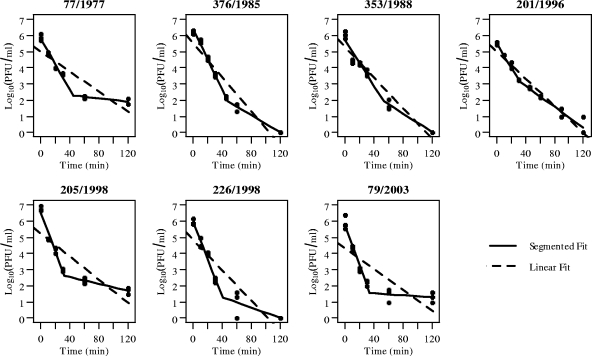
Persistence time decreases with increasing incubation temperature (3, 4, 34, 37). To confirm this relationship with the H2N3 isolates, a higher incubation temperature of 55°C was used. As expected, incubation at 55°C decreased the time required to reduce virus titers to zero (Table 1). Although r2 values of 0.62 to 0.95 were obtained from the regression analysis, there was a noticeable departure from linearity (lack of fit, P < 0.0005 for all isolates) (Fig. 1). The decrease in titer appeared curvilinear; therefore, a piecewise regression analysis was performed by using the segmented library in R (24). There was a rapid decrease in titer for all isolates during the first 30 to 45 min of incubation, after which the decrease became more gradual (Fig. 1), indicating that the samples comprise a mixed population of virions having subpopulations that differ in thermostability. On average, the rate of decrease in titer for the sensitive group was significantly higher (14-fold; Davies' test, P < 0.0001 for each isolate) than that for the stable group (Table 1). To confirm that the piecewise relationship was not an artifact of the plaque assay, isolates were incubated and titers in EID50/ml were calculated. Significant differences in slopes, calculated from a piecewise regression, were observed for isolates A/Mallard/Alberta/376/1985 (Davies' test, P < 0.0001), A/Mallard/Alberta/205/1998 (Davies' test, P < 0.02), and A/Mallard/Alberta/226/1998 (Davies' test, P < 0.0001).
FIG. 1.
Incubation at 55°C for up to 2 h revealed that loss of titer follows a 2-step process. Best-fit lines from a segmented (solid line) and linear regression (dashed line) are included to illustrate the lack of fit to a straight line.
Selection of subpopulations.
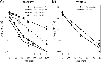
The curvilinear decrease of viral titers suggested that the stocks had a mixed population of the virus. Therefore, selection experiments were performed to purify thermostable and thermosensitive populations. Isolate A/Mallard/Alberta/205/1998 was incubated for 0 and 120 min at 55°C and injected into eggs. Two samples incubated at 55°C for 120 min had significantly different slopes (P < 0.0001) (Fig. 2). The samples incubated at 55°C for 0 min exhibited a curvilinear relationship, so a piecewise regression was performed. The slopes of the thermostable fractions (−0.023 ± 0.003 and −0.036 ± 0.001 log10 PFU/min for no selection no. 1 and 2, respectively) indicated similar or less stability than that of one of the samples incubated for 120 min (−0.023 ± 0.001 log10 PFU/min) and more stability than that of the other sample incubated at 120 min (−0.044 ± 0.002 log10 PFU/min). The same selection experiment was performed for isolate A/Mallard/Alberta/79/2003 (Fig. 2). Linear regression did not fit the data for the sample incubated for 0 min (P < 0.02). Thus, a piecewise regression was performed. After selection, the sample incubated for 120 min showed a decrease in titer (−0.047 ± 0.003), and this value was between the 2 values observed for the mixed population in the no-selection passage (−0.038 ± 0.004 and −0.066 ± 0.008 log10 PFU/min). Similar to results for isolate A/Mallard/Alberta/205/1998, the additional passage resulted in an increase in stability for the sensitive fraction relative to that for the stock population.
FIG. 2.
Selection by heating virus at 55°C for 2 h prior to propagation in eggs resulted in a population that exhibits a linear decrease in titers. Virus that was not incubated (no selection) was also grown in eggs, so passage histories are identical.
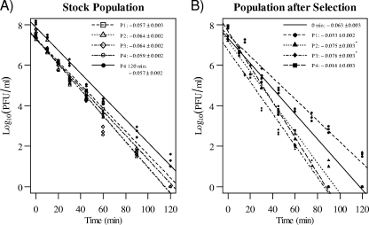
Selection of populations by incubating isolates at 55°C before injection into eggs yielded samples with thermostabilities between those of the thermostable and thermosensitive fractions of the stock virus. However, the allantoic fluid may still contain a mixed population of the virus. Therefore, isolate A/Mallard/Alberta/205/1998 was plaque purified after incubation at 55°C for 0 or 60 min. The 60-min time point was used because plaques at the 90- and 120-min time points were too small to visualize without fixing and staining the cell monolayer with crystal violet. Plaque purification of the stock yielded samples with regression slopes between −0.057 and −0.064 log10 PFU/min (Fig. 3 A), but the slopes were not significantly different (P > 0.13). While the thermostability of one of the plaque-purified samples for the 0-min group (plaque 4) was being assessed, a plaque was observed at the 120-min time point and purified. Although the titer was higher at every time point, the slope of the regression line for this additional passage was similar to that for the original sample from which the plaque was purified (P > 0.25) (Fig. 3A). The 4 plaques purified from the 60-min time point of the stock virus differ significantly in thermostability (P < 0.0001). Slopes of the regression lines were similar for 2 plaques (P > 0.48), but, when data for these 2 isolates were combined, the slopes of the regression lines remained different (P < 0.0001). Thus, from the 4 plaques purified, 3 populations of differing thermostabilities were obtained. Compared to the slope for 0-min plaque-purified samples (overall slope, −0.063 ± 0.003), the slope of the regression line for 3 plaque populations was steeper but 1 population exhibited a shallower slope.
FIG. 3.
Thermostability of single populations of isolate A/Mallard/Alberta/205/1998. Plaques were picked during a plaque assay of aliquots that were incubated at 55°C for 0 min to obtain representative virions from the stock population (A) and 60 min to obtain a broader selection of virions that differ in thermostability (B). The virus from the plaques was propagated in eggs, and linear regression was used to obtain best-fit lines. Slopes (±standard errors) are indicated, and those marked with asterisks are not significantly different (P > 0.05).
Two plaques (P1 and P4 from Fig. 3) and the corresponding stock virus of isolate A/Mallard/Alberta/205/1998 were imaged with TEM to investigate possible correlations between thermostability and size and shape of virus particles in the sample (Table 2). A single filamentous particle was observed for P4; the remaining virions observed in all 3 samples were either spherical or slightly oblong in shape. No difference in shape was observed when eccentricities (P < 0.26) and length-to-width ratios were compared (P < 0.24). Furthermore, overall sizes based on circumference (P < 0.13) and length (P < 0.21) were not different, but a difference between the average widths (P < 0.02) was detected. Specifically, virions from plaque P1 were wider than those from the stock (Tukey, P < 0.02), but no differences existed between the stock and P4 populations (Tukey, P < 0.60) or between the two plaque-purified populations (Tukey, P < 0.10).
TABLE 2.
Summary of sizes and shapes of randomly selected virions from the stock sample and 2 plaque-purified populations of isolate A/Mallard/Alberta/205/1998
| Parameter | Value (±SE) for: |
P | ||
|---|---|---|---|---|
| Stock virus | Plaque P1 | Plaque P4 | ||
| Stabilitya | Mixed | −0.053 ± 0.002 | −0.088 ± 0.003 | |
| Length (nm) | 114.4 ± 6.0 | 132.2 ± 5.7 | 138.6 ± 12.4 | <0.21 |
| Width (nm) | 87.4 ± 4.1 | 105.4 ± 4.3 | 93.5 ± 4.2 | <0.02c |
| Ratio | 1.34 ± 0.07 | 1.28 ± 0.05 | 1.52 ± 0.15 | <0.24 |
| Circumference (nm)b | 319.2 ± 14.1 | 375.1 ± 14.6 | 370.9 ± 25.0 | <0.13 |
| Eccentricityb | 0.56 ± 0.05 | 0.53 ± 0.04 | 0.61 ± 0.04 | <0.26 |
Calculated according to properties of an ellipse from the equations circumference = π(L/2 + W/2){1 + [3({L/2 − W/2}/{L/2 + W/2})2]/[10 + (4 − 3{[L/2 − W/2]/[L/2 + W/2]}2)1/2]} and eccentricity = {1 − [(W/2)/(L/2)]2}1/2, where L is length (nm) and W is width (nm).
Tukey's multiple comparison test revealed that the only comparison yielding a significant difference was that between P1 and the stock virus (P < 0.02).
Plaque size.
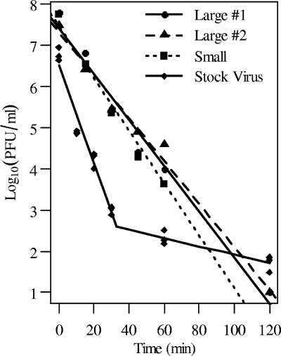
A shift in plaque size was observed during selection experiments. Virus populations grown in ECE after incubation at 55°C created pinhole-sized plaques. Because plaque size may be an indicator of thermostability, plaque sizes at different time points of incubation were compared for samples of virus. Stocks were passaged an additional time to allow comparison of the stock virus to samples that had undergone selection. Regardless of selective pressure, plaque sizes were consistent for each time point of incubation (P = 0.79). However, plaque sizes were significantly smaller for samples incubated for 2 h at 55°C and injected into eggs (P < 0.0001). One small plaque and two large plaques were purified from the A/Mallard/Alberta/205/1998 stock sample, and incubation experiments were performed (Fig. 4). Thermostabilities of the virus grown from each of the 3 plaques were not significantly different (P = 0.07).
FIG. 4.
Thermostabilities of 3 populations of isolate A/Mallard/Alberta/205/1998 were not different. Populations were selected by plaque purification of 2 large plaques and 1 small plaque. The thermostability profile of the stock virus is included for comparison.
Virus stocks are composed of a mixture of biologically active particles (22). It is possible that the pinhole-sized plaques were individual cells that had undergone apoptosis following infection by noninfectious cell-killing particles as described by Marcus and colleagues (22). To test this, plaque assays were performed using Avicel RC/CL as an overlay medium (23). The resulting plaques were larger than plaques observed when Bacto agar was used as an overlay, thus suggesting that the pinhole-sized plaques were indeed infected by plaque-forming particles.
DISCUSSION
Thermostability of AIV has been shown to vary between isolates of different subtypes (4, 32, 34). In this study, we found variations in thermostabilities at 37°C in 7 H2N3 isolates from the same host and location. Regression analysis suggested that the isolates could be divided into thermostable and thermosensitive groups on the basis of the rate of inactivation of infectivity. For example, given a starting titer of 106 PFU/ml, persistence time for thermostable isolates is 8.1 to 10.0 days whereas that for thermosensitive isolates is 6.1 to 6.9 days, regardless of whether the virus is grown in ECE or MDCK cells. These calculated persistence times are shorter than estimates of 11.4, 20.4, and 42.0 days reported for H11N6, H2N4, and H8N4 isolates, respectively (3). Although the persistence times of isolates from the thermostable and thermosensitive groups differ by approximately a day, this difference may be magnified at more biologically relevant temperatures (3).
A higher incubation temperature of 55°C was used to decrease the time required to observe significant reduction in titer. Interestingly, incubation at 55°C for up to 2 h revealed a nonlinear decrease in titer, and piecewise regression analysis suggested a 2-step pattern. A similar pattern has been observed for both untreated and gamma-irradiated A2/Aichi/2/68 after incubation at 56°C (6). Although not explicitly stated, nonlinear relationships can be inferred from studies on several other influenza A virus isolates (34) and for Newcastle disease virus (1). Although several factors can account for the 2-step process (6), presence of a heterogeneous virus population is the most likely reason.
In our study, the mixed population was composed primarily of thermosensitive fractions. A mixture of subpopulations was confirmed by selection experiments and plaque purification, which showed a linear decrease in titer for the stocks. Unsurprisingly, the slopes for the unselected populations (Fig. 3) were similar. Although more diversity of phenotypes should be present in the stock population, the ratio of thermosensitive to thermostable virions is strongly skewed to the thermosensitive fraction. Based on regression analysis (Fig. 1) and extrapolation to time zero, approximately 103 PFU/ml thermostable virions were present compared to almost 107 PFU/ml of the sensitive virions. Thus, 1 thermostable plaque is expected to be present in every 10,000 plaques observed in a plaque assay of the stock population. The stability of these 4 plaques was higher than that predicted based on the stability profile of the stock population (Fig. 1). An additional passage in eggs in the absence of selection still resulted in a piecewise loss of titer, which is in contrast to the linear results obtained from plaque purification. Interestingly, the sensitive fractions of these additional passages, like the plaque-purified stock viruses, are more stable than the equivalent fractions in the stock populations. Although only a single thermostable fraction was obtained during the selection experiments (Fig. 2), plaque purification of incubated samples resulted in 3 populations that varied in thermostability. The stabilities were similar to those calculated for the plaques picked from the stock population, but the postselection plaques were isolated from the 60-min time point. As such, the population of infectious virions at the 60-min time point likely contains a more even mixture of sensitive and stable virus particles. Thus, variation in thermostability occurs at the level of the population, which is then observed as the differences between isolates of the same subtype and different subtypes that have been previously reported (3, 4, 32, 34).
Thermostability of an influenza virus isolate can be affected by host and/or viral factors. We observed a change in thermostability grouping between stocks grown in ECE and those grown in MDCK cells, suggesting that host factors affect thermostability of resulting virions. On the other hand, the presence of subpopulations that differ in thermostability seems to indicate that stability is instead determined by genetic factors; a decrease in plaque size after selection may be indicative of such a relationship. Large- and small-plaque isolates of an H5N1 virus (A/Vietnam/1203/04) have been shown to differ at 5 amino acid positions: 2 in HA, 2 in PB1, and 1 in PA (15). In our study, virus propagated from both small and large plaques exhibited a linear decrease in titer but their slopes were not different, suggesting that some genetic factors affecting plaque size may be independent of those that determine the thermostability trait. Surprisingly, sequence analysis for 2 plaque-purified populations (P1 and P4 from Fig. 3) appears to refute the claim of a genetic underpinning for the observed differences in thermostability.
The complete genome for each of the viruses was sequenced (26). Compared to the published sequence for A/Mallard/Alberta/205/1998, both plaque-purified populations exhibited mutations in the HA1 (H3 numbering: K156E, G227R) and HA2 genes (H3 numbering: M84V, I133T). The generation of two mutations could reflect the high mutation rate of influenza virus (5) or amplification of a subpopulation from a mixture in the stock. Genetic differences between the 2 plaques were lacking, with the exception of a mutation in the PA gene (M579R; P4 to P1) and silent mutations in the PA (nucleotide [nt] 1095) and PB2 (nt 48) gene sequences. The single mutation in the PA gene could be related to replication rates (29), which would explain the observation that plaque sizes were smaller in virus that was amplified in ECE after being incubated at 55°C for 2 h. However, plaque sizes across the incubation times were not different, so plaque size is not an indicator of thermostability.
The ability to persist in the environment is essential for the maintenance of AIV in bird populations. A susceptible bird host is infected via the fecal-oral route of transmission (37), and the virion must remain infectious during the period of time it is outside the host. A longer persistence time increases the probability that the virus will successfully infect a new individual. Although this concept is intuitive, mathematical models that account for environmental transmission of AIV have only recently been developed (2, 30, 31), and the models suggest that low rates of environmental transmission are required to reproduce the periodicity and persistence of AIV observed in nature (2, 30). Furthermore, persistence in the environment results in a nonzero chance of an outbreak even when infectious individuals are not present and increases the duration of time for which prior outbreak areas may still contain infectious particles (31). The persistence time used in the mathematical models ranges from 4 days (31) to 207 days (30). These estimates were derived from several studies (3, 4, 33, 34), and all were calculated from a linear regression analysis. However, the upper limit of persistence time might have been underestimated because some fractions of a population can be extremely thermostable. This observation can influence the epidemiology of influenza in migratory birds, especially for flocks arriving at areas that may contain infectious virions. Specifically, stochastic transmission models show that virus concentration in a lake does not affect the probability of an outbreak, which is defined as infection of more than 10% of the population. However, it influences the cumulative fraction of the population that is infected, such that an increase in the initial virus concentration in the aquatic habitat results in a higher number of infected individuals when an outbreak occurs (31).
The influenza A virus has evolved to use waterfowl as a primary host (36). The prevalence of influenza A virus in ducks exhibits a cyclic pattern, peaking in the fall and declining until the birds return to the breeding areas in the spring (19, 25). How duck populations become infected in the spring is not known, but surveillance data suggest that this occurs because of year-round transmission within the migrating populations. Furthermore, the detection of influenza virus from lakes (11-13, 16, 20) and the high stability of virions in water suggest that persistence of the virus in the habitat during the winter may aid its perpetuation. In a study by Halvorson et al. (11), 9 of 10 influenza virus-positive water samples from Minnesota were obtained when water temperatures were less than 12°C. In accordance with the persistence times estimated by Brown et al. (3), stable fractions of the population should persist throughout the winter and remain infectious for the returning population of migratory birds.
Influenza virus virions are subject to the forces of selection. However, the observed fitness of influenza virus populations may not reflect the underlying fitness landscape (38). Specifically, these populations exist as quasispecies: the population is composed of closely related genetic variants instead of being a clonal population (8). The dynamics of quasispecies populations are such that traits with lower fitness could outcompete higher-fitness traits (8, 38). The high mutational rate of RNA viruses likely causes this phenomenon by pushing the population toward a minimum level of fitness, if it exists (8, 38). This “survival of the flattest” (in reference to a lower, flatter peak on the fitness landscape) could explain the variation in thermostability of influenza virus between different isolates of the same subtypes that is observed. The population of virions that once circulated in the waterfowl population could give rise to separate, individual populations when the surveillance swabs are tested by virus isolation. Selection would now act on these individual passages of virus populations instead of the larger influenza virus population and thus result in stocks with various degrees of thermostability. This requires that thermostability be determined at least partially by the genetic sequence of the virus. Although genetic differences between the 2 sequenced plaques were not observed, the method of gene amplification and sequencing identifies the sequence that is most prevalent in the population. Plaque purification of a presumably thermostable population may have resulted in selection of the less fit (less thermostable than expected) subpopulation in ECE. In essence, the high mutation rate of influenza virus could have produced less-thermostable variants that, as a group, can outcompete the now less common yet more fit thermostable variants, an idea that is consistent with quasispecies theory (8, 38). If the less-thermostable variants were present in both plaques, then both of the egg-grown plaque populations would probably consist of similar dominant subpopulations and therefore exhibit nearly identical genetic sequences (PA contained a single nonsynonymous mutation).
In nature, the thermostable trait is beneficial when persistence of influenza virus in the environment is considered. In theory, long persistence times should be under positive selection because this trait influences the probability of successful transmission. However, thermostability is only one factor that affects persistence time and hence transmission success. Several environmental factors influence the length of the infectious period (3, 33), while replication rates and the quantity of virions shed by infected individuals affect the probability that susceptible individuals contact and ingest infectious virions (2, 30, 31). Consequently, thermostability only partially determines fitness of the virus population, which is defined here as the ability to be transmitted to and replicate in the natural reservoir hosts. As such, the strength of selection for increased thermostability in nature could be much less than that for other traits related to transmission, which leads to populations that are less thermostable than what would be predicted.
Besides temperature, persistence of virus in the environment is also affected by pH and salinity. Low (33, 34)- and highly pathogenic isolates (4) have been seen to vary in stability in solutions of different pHs and salinities. As for thermostability studies, linear regression has been used to fit data and calculate persistence times under different environmental conditions. In a study by Brown et al. (3), virus titers were determined at 2 time points only, which is not enough to detect a nonlinear decrease. Also, lack-of-fit tests, which can reveal nonlinear trends of stability, were not conducted in previous studies; instead, these studies used the coefficient of determination as a measure of fit, and its value can be misleading (7). Thus, although the stability of isolates can vary with pH and salinity, it is unclear whether these variations also exist within populations as observed in our study.
The thermostability of AIV varies among isolates regardless of subtype. Our study demonstrates that this variation among isolates begins at the level of the population. Stocks grown from fecal swabs contained a mixture of subpopulations, each of which could differ in thermostability. A selective force of high temperatures on these populations or their plaque purification yielded a population that exhibited a linear decrease in titer, suggesting that the population had a homogenous thermostability. The presence of subpopulations with high thermostability suggests that these AIV can persist for a long time in water, thus increasing the probability of transmission to susceptible hosts. The mechanism that causes variation in thermostability remains unknown. Neither plaque size or shape nor sequence data provide convincing evidence that these factors significantly affect thermostability of influenza virus. However, a more thorough investigation on the size and shape of virions should be performed to conclusively determine if thermostability is correlated to virion morphology. Moreover, the thermostability trait may be in agreement with quasispecies theory in that less-stable fractions outcompete the more fit thermostable fractions when grown in ECE. Additional work, including sequencing of many more plaques to identify minor fractions of the total population (8), needs to be performed to confirm this hypothesis.
Acknowledgments
This work was supported by the National Institute of Allergy and Infectious Diseases (contract number HHSN266200700005C) and by ALSAC.
We thank Sharon Frase in the Cell and Tissue Imaging facility at St. Jude Children's Research Hospital for help in preparing and imaging the virus samples and Vani Shanker and Cherise Guess for editing the manuscript.
Footnotes
Published ahead of print on 7 July 2010.
REFERENCES
- 1.Boyd, R. J., and R. P. Hanson. 1958. Survival of Newcastle disease virus in nature. Avian Dis. 2:82-93. [Google Scholar]
- 2.Breban, R., J. M. Drake, D. E. Stallknecht, and P. Rohani. 2009. The role of environmental transmission in recurrent avian influenza epidemics. PLoS Comput. Biol. 5:e1000346. [DOI] [PMC free article] [PubMed] [Google Scholar]
- 3.Brown, J. D., G. Goekjian, R. Poulson, S. Valeika, and D. E. Stallknecht. 2009. Avian influenza virus in water: infectivity is dependent on pH, salinity and temperature. Vet. Microbiol. 136:20-26. [DOI] [PubMed] [Google Scholar]
- 4.Brown, J. D., D. E. Swayne, R. J. Cooper, R. E. Burns, and D. E. Stallknecht. 2007. Persistence of H5 and H7 avian influenza viruses in water. Avian Dis. 50:285-289. [DOI] [PubMed] [Google Scholar]
- 5.Comas, I., A. Moya, and F. Gonzalez-Candelas. 2005. Validating viral quasispecies with digital organisms: a re-examination of the critical mutation rate. BMC Evol. Biol. 5:5. [DOI] [PMC free article] [PubMed] [Google Scholar]
- 6.De Flora, S., and G. Badolati. 1973. Thermal inactivation of untreated and gamma-irradiated A2/Aichi/2/68 influenza virus. J. Gen. Virol. 20:261-265. [DOI] [PubMed] [Google Scholar]
- 7.Faraway, J. J. 2005. Linear models with R. Chapman & Hall/CRC, Boca Raton, FL.
- 8.Fishman, S. L., and A. D. Branch. 2009. The quasispecies nature and biological implications of the hepatitis C virus. Infect. Genet. Evol. 9:1158-1167. [DOI] [PMC free article] [PubMed] [Google Scholar]
- 9.Fouchier, R. A., and V. J. Munster. 2009. Epidemiology of low pathogenic avian influenza viruses in wild birds. Rev. Sci. Tech. 28:49-58. [DOI] [PubMed] [Google Scholar]
- 10.Greatorex, J. S., R. F. Page, M. D. Curran, P. Digard, J. E. Enstone, T. Wreghitt, P. P. Powell, D. W. Sexton, R. Vivancos, and J. S. Nguyen-Van-Tam. 2010. Effectiveness of common household cleaning agents in reducing the viability of human influenza A/H1N1. PLoS One 5:e8987. [DOI] [PMC free article] [PubMed] [Google Scholar]
- 11.Halvorson, D., D. Karunakaran, D. Senne, C. Kelleher, C. Bailey, A. Abraham, V. Hinshaw, and J. Newman. 1983. Epizootiology of avian influenza—simultaneous monitoring of sentinel ducks and turkeys in Minnesota. Avian Dis. 27:77-85. [PubMed] [Google Scholar]
- 12.Hinshaw, V. S., R. G. Webster, and B. Turner. 1980. The perpetuation of orthomyxoviruses and paramyxoviruses in Canadian waterfowl. Can. J. Microbiol. 26:622-629. [DOI] [PubMed] [Google Scholar]
- 13.Hinshaw, V. S., R. G. Webster, and B. Turner. 1979. Water-borne transmission of influenza A viruses? Intervirology 11:66-68. [DOI] [PubMed] [Google Scholar]
- 14.Hoffmann, E., J. Stech, Y. Guan, R. G. Webster, and D. R. Perez. 2001. Universal primer set for the full-length amplification of all influenza A viruses. Arch. Virol. 146:2275-2289. [DOI] [PubMed] [Google Scholar]
- 15.Hulse-Post, D. J., J. Franks, K. Boyd, R. Salomon, E. Hoffmann, H. L. Yen, R. J. Webby, D. Walker, T. D. Nguyen, and R. G. Webster. 2007. Molecular changes in the polymerase genes (PA and PB1) associated with high pathogenicity of H5N1 influenza virus in mallard ducks. J. Virol. 81:8515-8524. [DOI] [PMC free article] [PubMed] [Google Scholar]
- 16.Ito, T., K. Okazaki, Y. Kawaoka, A. Takada, R. G. Webster, and H. Kida. 1995. Perpetuation of influenza A viruses in Alaskan waterfowl reservoirs. Arch. Virol. 140:1163-1172. [DOI] [PubMed] [Google Scholar]
- 17.Junankar, P. R., and R. J. Cherry. 1986. Temperature and pH dependence of the haemolytic activity of influenza virus and of the rotational mobility of the spike glycoproteins. Biochim. Biophys. Acta 854:198-206. [DOI] [PubMed] [Google Scholar]
- 18.Kawaoka, Y., T. M. Chambers, W. L. Sladen, and R. G. Webster. 1988. Is the gene pool of influenza viruses in shorebirds and gulls different from that in wild ducks? Virology 163:247-250. [DOI] [PubMed] [Google Scholar]
- 19.Krauss, S., D. Walker, S. P. Pryor, L. Niles, L. Chenghong, V. S. Hinshaw, and R. G. Webster. 2004. Influenza A viruses of migrating wild aquatic birds in North America. Vector Borne Zoonotic Dis. 4:177-189. [DOI] [PubMed] [Google Scholar]
- 20.Lang, A. S., A. Kelly, and J. A. Runstadler. 2008. Prevalence and diversity of avian influenza viruses in environmental reservoirs. J. Gen. Virol. 89:509-519. [DOI] [PubMed] [Google Scholar]
- 21.Lu, H., A. E. Castro, K. Pennick, J. Liu, Q. Yang, P. Dunn, D. Weinstock, and D. Henzler. 2003. Survival of avian influenza virus H7N2 in SPF chickens and their environments. Avian Dis. 47:1015-1021. [DOI] [PubMed] [Google Scholar]
- 22.Marcus, P. I., J. M. Ngunjiri, and M. J. Sekellick. 2009. Dynamics of biologically active subpopulations of influenza virus: plaque-forming, noninfectious cell-killing, and defective interfering particles. J. Virol. 83:8122-8130. [DOI] [PMC free article] [PubMed] [Google Scholar]
- 23.Matrosovich, M., T. Matrosovich, W. Garten, and H. D. Klenk. 2006. New low-viscosity overlay medium for viral plaque assays. Virol. J. 3:63. [DOI] [PMC free article] [PubMed] [Google Scholar]
- 24.Muggeo, V. M. 2008. Modeling temperature effects on mortality: multiple segmented relationships with common break points. Biostatistics 9:613-620. [DOI] [PubMed] [Google Scholar]
- 25.Munster, V. J., C. Baas, P. Lexmond, J. Waldenstrom, A. Wallensten, T. Fransson, G. F. Rimmelzwaan, W. E. P. Beyer, M. Schutten, B. Olsen, A. Osterhaus, and R. A. M. Fouchier. 2007. Spatial, temporal, and species variation in prevalence of influenza A viruses in wild migratory birds. PLoS Pathog. 3:630-638. [DOI] [PMC free article] [PubMed] [Google Scholar]
- 26.Obenauer, J. C., J. Denson, P. K. Mehta, X. Su, S. Mukatira, D. B. Finkelstein, X. Xu, J. Wang, J. Ma, Y. Fan, K. M. Rakestraw, R. G. Webster, E. Hoffmann, S. Krauss, J. Zheng, Z. Zhang, and C. W. Naeve. 2006. Large-scale sequence analysis of avian influenza isolates. Science 311:1576-1580. [DOI] [PubMed] [Google Scholar]
- 27.Olsen, B., V. J. Munster, A. Wallensten, J. Waldenstrom, A. Osterhaus, and R. A. M. Fouchier. 2006. Global patterns of influenza A virus in wild birds. Science 312:384-388. [DOI] [PubMed] [Google Scholar]
- 28.Reed, M. L., H. L. Yen, R. M. DuBois, O. A. Bridges, R. Salomon, R. G. Webster, and C. J. Russell. 2009. Amino acid residues in the fusion peptide pocket regulate the pH of activation of the H5N1 influenza virus hemagglutinin protein. J. Virol. 83:3568-3580. [DOI] [PMC free article] [PubMed] [Google Scholar]
- 29.Regan, J. F., Y. Liang, and T. G. Parslow. 2006. Defective assembly of influenza A virus due to a mutation in the polymerase subunit PA. J. Virol. 80:252-261. [DOI] [PMC free article] [PubMed] [Google Scholar]
- 30.Roche, B., C. Lebarbenchon, M. Gauthier-Clerc, C. M. Chang, F. Thomas, F. Renaud, S. van der Werf, and J. F. Guegan. 2009. Water-borne transmission drives avian influenza dynamics in wild birds: the case of the 2005-2006 epidemics in the Camargue area. Infect. Genet. Evol. 9:800-805. [DOI] [PubMed] [Google Scholar]
- 31.Rohani, P., R. Breban, D. E. Stallknecht, and J. M. Drake. 2009. Environmental transmission of low pathogenicity avian influenza viruses and its implications for pathogen invasion. Proc. Natl. Acad. Sci. U. S. A. 106:10365-10369. [DOI] [PMC free article] [PubMed] [Google Scholar]
- 32.Scholtissek, C. 1985. Stability of infectious influenza A viruses at low pH and at elevated temperature. Vaccine 3:215-218. [DOI] [PubMed] [Google Scholar]
- 33.Stallknecht, D. E., M. T. Kearney, S. M. Shane, and P. J. Zwank. 1990. Effects of pH, temperature, and salinity on persistence of avian influenza viruses in water. Avian Dis. 34:412-418. [PubMed] [Google Scholar]
- 34.Stallknecht, D. E., S. M. Shane, M. T. Kearney, and P. J. Zwank. 1990. Persistence of avian influenza viruses in water. Avian Dis. 34:406-411. [PubMed] [Google Scholar]
- 35.Szretter, K. J., A. L. Balish, and J. M. Katz. 2006. Influenza: propagation, quantification, and storage, p. 15G.1.1-15G.1.22. In R. Coico, T. Kowalik, J. Quarles, B. Stevenson, and R. Taylor (ed.), Current protocols in microbiology. Wiley and Sons, Inc., Hoboken, NJ. [DOI] [PubMed]
- 36.Webster, R. G., W. J. Bean, O. T. Gorman, T. M. Chambers, and Y. Kawaoka. 1992. Evolution and ecology of influenza A viruses. Microbiol. Rev. 56:152-179. [DOI] [PMC free article] [PubMed] [Google Scholar]
- 37.Webster, R. G., M. Yakhno, V. S. Hinshaw, W. J. Bean, and K. G. Murti. 1978. Intestinal influenza: replication and characterization of influenza viruses in ducks. Virology 84:268-278. [DOI] [PMC free article] [PubMed] [Google Scholar]
- 38.Wilke, C. O. 2005. Quasispecies theory in the context of population genetics. BMC Evol. Biol. 5:44. [DOI] [PMC free article] [PubMed] [Google Scholar]
- 39.Yen, H. L., L. M. Herlocher, E. Hoffmann, M. N. Matrosovich, A. S. Monto, R. G. Webster, and E. A. Govorkova. 2005. Neuraminidase inhibitor-resistant influenza viruses may differ substantially in fitness and transmissibility. Antimicrob. Agents Chemother. 49:4075-4084. [DOI] [PMC free article] [PubMed] [Google Scholar]


