



Abstract
The existence of an immunological homunculus has been proposed, but evidence for location-specific response of the central nervous system to immunological stimulation is lacking. In this study, we show that inflammation induced by injection of casein into one of the hindlimbs causes c-fos expression in the paraventricular nucleus of the hypothalamus (PVN) in an asymmetrical manner: much stronger activation is always induced in the contralateral PVN. Unilateral sciatic nerve transection abolished the casein-induced PVN activation if casein was injected into the hindlimb with the nerve transection, but had no effect if casein was injected into the hindlimb with intact nerve innervation. Injection of casein into one the forelimbs also caused contralateral PNV activation. Further, stronger PVN activation was found in the anterior PVN after the forelimb injection, but in the posterior PVN after the hindlimb injection. Casein-induced PVN activation is absent in IL-1R1 KO, IL-6 KO, TNFα KO, and in C3H/HeJ (TLR4 mutant) animals. In comparison, injection of LPS, a systemic inflammagen, into one hindlimb induced bilateral PVN activation but injection of live E. coli into one hindlimb induced contralateral PVN activation. These results support the notion that local inflammation may activate the PVN by neural routes in a location-specific manner.
Keywords: c-fos, neural route, immune-to-brain
Introduction
Both humoral and neural routes have been found to relay peripheral immune signals to the central nervous system (CNS) (Quan and Banks, 2007). By humoral routes, inflammatory cytokines relay signals of peripheral inflammation via circulation. The circulating cytokines activate the CNS either indirectly through brain endothelium or directly on neurons in the brain parenchyma after they navigated the blood-brain barrier (BBB) or on neurons in the circumventricular organs where the BBB is leaky. By neural routes, inflammation activates vagal(Goehler et al., 1995) or primary sensory nerves to relay immune signals to the brain. Only the neural routes can transmit the information regarding the location of a given immune response to the brain because local inflammation may be hardwired to the CNS. This possibility has prompted Kevin Tracey to speculate that there exists an immunological homunculus in the brain (Oke and Tracey, 2008), although experimental support for this is lacking.
We showed in a previous study that local administration of casein induces localized inflammation that activates CNS without inducing blood and brain cytokines and without inducing brain cyclooxygenase-2 (COX-2) (Zhang et al., 2008). Therefore, casein-induced localized inflammation may activate CNS primarily by nerve pathways that project to the CNS from the site of inflammation. In this study, we investigated the neural pathway by which local inflammation might stimulate the CNS using the casein model, with a special emphasis on whether CNS activation patterns reflect the location of peripheral inflammation.
Materials and Methods
Animals
Male normal FVB mice, C3H/HeJ (a TLR4 mutant), IL-1R1 KO, TNFα KO, and IL-6 KO mice were purchased from the Jackson Laboratory. They were group housed (3/cage) and fed with food and water ad libitum in a light (0600 h to 1800 h) and temperature-controlled environment (20–22°C). The animals weighed 20–25 g when they were used in an experiment. All the procedures were approved by the Ohio State University Animal care and Use committee.
Reagents
Casein at 5% was prepared by dissolving 5 g casein powder (ICN Biomedicals, Aurora, OH) into 80 ml of 50 mM sodium bicarbonate and water was added to make a final volume of 100 ml. The mixture was stirred in a water bath at 65°C until the casein was completely dissolved. The solution was filtered through coarse filter paper (Fisher Scientific, Pittsburgh, PA) and stored at −20°C.
Ten milligrams of LPS (E. coli, serotype 055:B5, Sigma) was dissolved in 40 ml of pyrogen-free saline to achieve the final concentration of 0.25 mg/ml and stored at −20°C for later use. E. coli strain K12 (ATCC) was cultured in trypticase soy broth. The live stationary phase bacteria were collected and washed in PBS and re-suspended in PBS at 3 × 10⁹/ml before injection.
Experimental procedures
Animals were given intramuscular injections of 100 μl of casein, LPS, saline, or live E. coli. Home cage animals that did not receive any injection were used as controls. The intramuscular injections were made either in the right hindlimb (gastrocnemeius muscle) or in the right forelimb (triceps). Home cage control animals were used to establish basal c-fos expression patterns in the brain and spinal cord. Saline injected animals were used as controls for c-fos expression that maybe induced by the injection procedure. Casein and LPS injected animals were used to compare how local and systemic inflammation might induce different c-fos expression patterns in the brain. Live E coli injected animals were studied to determine whether casein-induced c-fos expression patterns are also present after localized bacterial infection. All the injections were made at 8 am to avoid the effects of circadian rhythm. Animals were sacrificed at 4, 6, or 10 h after the saline or casein injections; they were sacrificed 2 h or 6 h after the LPS or E. coli injections, respectively. The time course for the casein injection was designed to cover the time period during which casein injection is known to induce local inflammation and fever (Zhang et al., 2008). In most experiments animals were sacrificed 6 h post-injection because previous study showed that local inflammation peaks this time point (Zhang et al., 2008). To compare results obtained from injection of casein into the left side of the body, casein or saline was also injected into the left hindlimb and animals were sacrificed 4, 6, and 10 h post injection. Saline or casein was also injected into the right hindlimb of IL-1R1 KO, IL-6 KO, TNFα KO, and C3H/HeJ mice to test whether local inflammation-induced CNS c-fos expression required these inflammatory factors. To test the involvement of peripheral nerves, the left sciatic nerve was transected in 4 groups of FVB mice. Two weeks after the nerve transection, they were injected with saline or casein into either the left or the right hindlimb. Animals were sacrificed 6 h after the injection. All the injection conditions are tabulated in Table 1.
Table 1.
Experimental groups. All animals, except home cage controls in group 1, were given im injections at 8 am.
| Experimental Groups (n=6/group) | Animal | Sciatic Nerve Transection | Injectate | Injection Site | Time of Sacrifice |
|---|---|---|---|---|---|
| 1 | Wildtype | None | None | 2 pm | |
| 2 | Wildtype | None | Saline | Right Hindlimb | 12 pm |
| 3 | Wildtype | None | Saline | Right Hindlimb | 2 pm |
| 4 | Wildtype | None | Saline | Right Hindlimb | 6 pm |
| 5 | Wildtype | None | Casein | Right Hindlimb | 12 pm |
| 6 | Wildtype | None | Casein | Left Hindlimb | 12 pm |
| 7 | Wildtype | None | Casein | Right Hindlimb | 2 pm |
| 8 | Wildtype | None | Casein | Left Hindlimb | 2 pm |
| 9 | Wildtype | None | Casein | Right Hindlimb | 6 pm |
| 10 | Wildtype | None | Casein | Left Hindlimb | 6 pm |
| 11 | Wildtype | Left Sciatic | Saline | Right Hindlimb | 2 pm |
| 12 | Wildtype | Left Sciatic | Saline | Left Hindlimb | 2 pm |
| 13 | Wildtype | Left Sciatic | Casein | Right Hindlimb | 2 pm |
| 14 | Wildtype | Left Sciatic | Casein | Left Hindlimb | 2 pm |
| 15 | Wildtype | None | Casein | Right Forelimb | 2 pm |
| 16 | Wildtype | None | Casein | Left Forelimb | 2 pm |
| 17 | Wildtype | None | Saline | Right Hindlimb | 10 am |
| 18 | Wildtype | None | LPS | Right Hindlimb | 10 am |
| 19 | Wildtype | None | E. coli | Right Hindlimb | 2 pm |
| 20 | TLR4 mu | None | Casein | Right Hindlimb | 2 pm |
| 21 | IL-1R1 KO | None | Casein | Right Hindlimb | 2 pm |
| 22 | IL-6 KO | None | Casein | Right Hindlimb | 2 pm |
| 23 | TNFα KO | None | Casein | Right Hindlimb | 2 pm |
Tissue collection and immunohistochemistry
Animal were anaesthetized with isoflurane (Abott, North Chicago, IL) and perfused transcardially with 0.1% phosphate-buffered saline (PBS) followed by a mixture of 4% paraformaldehyde, 1.4% lysine, and 0.2% sodium metaperiodate in 0.1 M sodium phosphate, following the protocol of Travers et al.(Travers and Travers, 2007). Brains and spinal cords were removed and postfixed overnight in 20% sucrose and PBS. Frozen brain sections (40 μm-thick) were cut with a sliding microtome. Coronal sections were collected serially in two sets so that each set contains an evenly spaced (80 μm apart) rostrocaudal series. The second set was used for immunohistochemical labeling of c-fos. Two individual blocks of spinal cord tissue containing spinal level C5-Th1 and L2-S2 were dissected and embedded in the Tissue Freezing Medium (Triangle BioMedical Sciences, Durham, NC). Then, transverse sections, 40-μm thick, were cut by a cryostat and collected directly onto superfrost glass slides (Fisher Scientific). The sections were stored in −80°C before immunohistochemical staining.
For c-fos immunohistochemistry, tissue sections were rinsed in 0.1 M PBS before and after a 20-min incubation in 1 % Na Borohydride, and then rinsed in 0.5% H2O2 to quench endogenous peroxidase activity. Sections were then incubated for 1 h in 10% normal sheep serum prior to incubation in a rabbit anti-c-fos antibody (1:200, anti-Fos, Oncogene Science [Cambridge, MA], PC38, rabbit anti-c-fos) for 72 h at 4°C. After 3 washes in PBS, the sections were incubated in 1:600 biotinylated goat-anti-rabbit antibody for 90 min, followed by incubation with the ABC reagent (Vector PK6100, Vector Laboratories, Burlington, CA). Immunoreactivity for c-fos was visualized using a mixture of substrate 3,3′-diaminobenzidine (DAB, 0.05%) and NiHSO4 (0.02%). We also labeled COX-2 in the brain after injection of LPS and E. coli into the hindlimb muscle to determine whether humoral neuroimmune pathways are activated by the inflammations induced by these different stimuli. COX-2 is known to be induced by peripheral inflammation in brain endothelium which is not likely to be directly connected with the ascending neural sensory inputs. For COX-2 immunohistochemistry, same method was used except the primary antibody was a rabbit anti-COX-2 antibody (1:200, Cat # 160106, Cayman Chemical, Ann Arbor, Michigan). For both c-fos and COX-2 immunohistochemistry, brain sections were also labeled without the primary antibodies to control for potential non-specific labeling.
The results were examined with light microscopy and the number of c-fos labeled cells in the PVN was counted using the Neurolucida software.
Data Analysis
In order to count the c-fos positive cells in the PVN, outlines of the PVN were drawn under dark-field optics. Subsequently, an investigator unaware of the stimulus condition plotted Fos-positive cells using a 20 x lens. In order to be counted, there had to be a clear outline of an oval or round nucleus that was darker than the background. The number of c-fos positive cells in the left or right PVN was counted in every section (typically 6 sections, spaced 80 μm apart, were counted throughout the entire PVN). Average number of c-fos positive cells in the left or right PVN from one animal was used to represent the intensity of PVN activation in one animal. In any experimental group, means and standard errors of the number of c-fos positive cells from 6 animals were calculated. In the experiment in which PVN activation induced by forelimb and hindlimb casein injections were compared, the number of c-fos positive cells in all the PVN sections was counted. Because we perceived a difference in anterior vs. posterior PVN c-fos expression induced by casein injection into these two body parts, the number of c-fos expressing cells per section in the anterior and posterior PVN were calculated and compared.
One-way ANOVA was used to analyze the difference in the number of PVN c-fos positive cells in the experimental groups and saline injected control animals; the test results were considered significant if the post hoc Tukey’s test of the compared groups showed P<0.05. This analysis was also applied to analyze the results of the sciatic nerve transection experiment. In other experiments, critical comparisons were made between different parts of the PVN in the same animal in one injection group, thus, two-tailed paired student t-test was used.
Results
The unique pattern of c-fos response in the PVN to local injection of casein was investigated first. Figure 1A–C show representative low-magnification microphotographs of c-fos labeled sections of the PVN region in control animal (Figure 1A), saline injected (figure 1B) or casein (Figure 1C) injected animals. Animals were sacrifice 6 h post injection in this experiment. The injections were made in the right hindlimb. The pictures encompass the two supraoptic nuclei (SON) at the ventral aspect and brain regions just below the subfornical organ (SFO) at the dorsal aspect. No c-fos expressing cells were found in these brain regions from home cage control animals. In saline injected animals, scattered c-fos expressing cells were found in the region near the lateroanterior hypothalamic nucleus (LA) and a diffused area dorsolateral to the PVN (arrows point to the discrete c-fos positive cells). The pattern of c-fos expressing cells shows a symmetrical appearance with respect to the midline of the brain. After casein injection, c-fos expressing cells were found in additional brain regions, notably the left PVN (the pinhole, marked by *, in the section were made to mark the left side the brain), and the SON on both sides of the brain (arrowheads point to c-fos positive cells induced by casein injection). The number of c-fos positive cells in the casein-injected animals is significantly higher compared with that in saline injected controls (F(1,10)=464.05, p<0.001).
Figure 1.

Representative low-magnification microphotographs show labeling of c-fos (A-C) in coronal sections containing PVN. A: home cage control. B: c-fos expression 6 h after the saline injection into the right hindlimb muscle. C: c-fos expression 6 h after casein injection into the right hindlimb. Arrows point to c-fos positive cells induced by both casein and saline injections; arrowheads point to c-fos positive cells induced by the casein injection. * indicates the pinhole that marks the left side of the brain.
We then investigated the time course of the PVN c-fos response to the casein injection. Figure 2 shows representative microphotographs of c-fos labeled PVN sections from animals injected with casein in their hindlimb. Figs. 2A, 2B, and 2C show representative sections from animals sacrificed at 4, 6, or 10 h after casein was injected into the right hindlimb, respectively. Figs. 2a, 2b, and 2c show representative sections from animals sacrificed at 4, 6, or 10 h after casein was injected into the left hindlimb. PVN c-fos expression show dominant contralateral patterns at all the time points after casein injection into either hindlimb (the sections’ left side match the brain’s left). Figure 3 shows quantitative analysis of these results by comparing the number of c-fos positive cells between the two sides of PVN in the same sections: at all 3 time points, the number of c-fos positive cells were significantly higher in the contralateral, with respect to the side of casein injection, PVN (cPVN) as compared with those in the ipsilateral PVN (iPVN) (4 h: F(1,10) =63.07, p<0.05; 6 h: F(1,10) = 48.95, p<0.05; 10 h: F(1,10) =32.45, p<0.05). Next, whether the PVN c-fos response to casein injection into the hindlimb depends upon the integrity of the innervating sciatic nerve was tested. Figure 4 shows representative low-magnification microphotographs of c-fos labeled sections from animals that received left sciatic nerve transection. Animals were sacrificed 6 h post- injection. Saline injection into the right hindlimb (Figure 4A) or the left hindlimb (Figure 4B) induced scattered c-fos expression in the region of LA and an area just above the PVN. C-fos positive cells are conspicuously missing in the PVN proper. Injection of casein into the right hindlimb induced strong c-fos expression in the left PVN (Figure 4C, arrow) and weak c-fos expression in the right PVN. This casein injection also induced bilateral c-fos expression in the SON (Figure 4C, arrowhead). Injection of casein into the left hindlimb did not induce c-fos expression in the PVN and SON. The reduction of the number of PVN c-fos positive cells induced by casein injection into the left hindlimb in animals with left sciatic nerve transection in comparison with that in animals with intact sciatic nerve is statistically significant (F(1, 10)=61.07, p<0.0001).
Figure 2.

Representative high-magnification microphotographs show labeling of c-fos in coronal sections containing PVN after intramuscular casein injection into the right hindlimb (A–C) or into the left hindlimb (a–c). Animals were sacrificed at 4 (A,a), 6 (B,b), or 10 (C,c) h after the casein injections.
Figure 3.
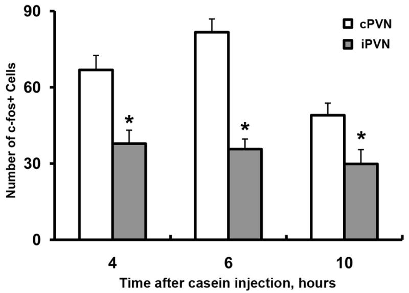
Number of c-fos positive cells in the ipsilateral (iPVN) vs. contralateral PVN (cPVN, relative to the injection site). * indicates statistical significance (p<0.05, iPVN vs. cPVN).
Figure 4.

Representative low-magnification microphotographs show labeling of c-fos (A–C) in coronal sections containing PVN. All animals received left sciatic nerve transection and were sacrificed 6 h after intramuscular injections. A: saline was injected into the right hindlimb. B: saline was injected into the left hindlimb. C: casein was injected into the right hindlimb. D: casein was injected into the left hindlimb. Arrow points to c-fos positive cells in the PVN; arrowheads point to c-fos positive cells in the SON.
We then compare c-fos response patterns in the PVN after casein was given to different body parts. Figure 5 shows representative microphotographs of c-fos labeled PVN sections. Animals were sacrificed 6 h post-injection. PVN response to casein injection to the right forelimb is shown in the left panels (Figs. 5A–F); PVN response to casein injection to the right hindlimb is shown in the right panels (Figs. 5a–f). Sections of the PVN are arranged to display evenly spaced coronal sections throughout the entire PVN in the anterior to posterior order (Figs. 5A–F are serial sections from one animal and Figs. 5a–f from another). These sections correspond to the coronal planes of Bregma −0.58 mm to Bregma −9.0 mm of the Mouse Brain Atlas by Franklin and Paxinos. Injection of casein into either the forelimb or the hindlimb on the right side induced left-dominant PVN c-fos expression throughout the entire PVN. Further, forelimb injection induced stronger c-fos expression in the anterior PVN (aPVN: from Bregma −0.58 mm to Bregma −0.70 mm) whereas hindlimb injection induced stronger c-fos expression in the posterior PVN (pPVN: from Bregma −0.70 mm to Bregma −0.90 mm). Figure 6 show quantitative analysis of the number of c-fos positive cells induced by injection of casein into forelimb or hindlimb in regards to the distribution of c-fos positive cells in the aPVN vs. pPVN. Average numbers of c-fos positive cells per section are graphed. Forelimb injection induced significantly more c-fos positive cells in the aPVN than in pPVN (F(1,10) = 39.22, p<0.05). In contrast, hindlimb injection induced significantly more c-fos positive cells in the pPVN than in the aPVN (F(1,10) =29.44, p<0.05).
Figure 5.

Representative high-magnification microphotographs show labeling of c-fos in coronal sections containing PVN after intramuscular casein injection into the right forelimb (A–F) or into the right hindlimb (a-f). Serial sections in the anterior to posterior order from two animals are shown. aPVN: anterior PVN; pPVN: posterior PVN.
Figure 6.
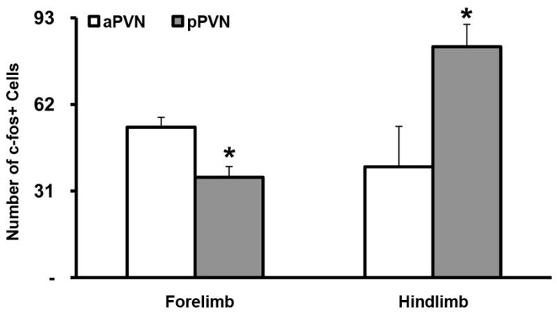
Quantitative analysis of the number of c-fos positive cells in the left PVN after casein injection into the right forelimb or the right hindlimb. * indicates statistical significance (p<0.05, aPVN vs. pPVN).
The spinal cord c-fos expression patterns in relation to the locations of casein injection were also studied. Figure 7 shows representative c-fos labeled section of the spinal cord. Casein injection into the left forelimb induced c-fos expression in the left superficial layer of the dorsal horn between the C5-Th1 spinal levels (Figure 7A, arrows point to c-fos positive cells); no c-fos positive cells were found on the contralateral side (Figure 7B). Similarly, casein injection into the left hindlimb induced c-fos expression in the left superficial layer of the dorsal horn between the spinal levels L2-S2 (Figure 7C); no c-fos positive cells were found on the contralateral side (Figure 7D). Further, forelimb casein injection did not induce c-fos expression in L2-S2 of the spinal cord and hindlimb casein injection did not induce c-fos expression in C5-Th1 of the spinal cord (data not shown).
Figure 7.

Representative high-magnification microphotographs show labeling of c-fos in transverse sections of the spinal cord after intramuscular casein injection into the left forelimb (A–B) or into the left hindlimb (C–D). Dye on the edge marks the side of casein injection. Arrows point to c-fos positive cells in the dorsal horn in the ipsilateral spinal cord (A and C). No c-fos induction was found in the contralateral spinal cord (B and D). A and B were taken from spinal level C5-TH1; C and D were taken from spinal level L2-S2.
Local administration of casein induced neuronal activation in other brain regions. Figure 8 shows representative c-fos labeled sections containing brain regions that are known to be activated during peripheral immune challenge. Similar to previous reports (Gaykema et al., 2007a), essentially no c-fos expression was induced after saline injection (data not shown). Six hours after casein injection into right hindlimb c-fos expression was induced bilaterally in the nucleus of the solitary tract (NTS), the parabrachial nucleus (PB), the bed nucleus of the stria terminalis (BST) and the anterior hypothalamus (AH); it did not induced c-fos expression in the ventrolateral medulla (VLM), central nucleus of amygdala (CeA), and locus coeruleus (LC). In the next study, the importance of inflammatory factors for casein induced c-fos expression in the PVN was determined. Figure 9 shows representative c-fos labeled PVN sections. Casein injection into the right hindlimb did not induce c-fos expression in the PVN in C3H/HeJ (Figure 9A), IL-1R1 KO (Figure 9B), IL-6 KO (Figure 9C), and TNFα KO (Figure 9D) animals. Localized live E. coli infection also induces location-specific PVN activation. Figure 10 shows representative microphotographs of c-fos and COX-2 labeled sections, comparing CNS responses to the injections of LPS and live E. coli into the right hindlimb. LPS induced bilateral PVN c-fos expression (Figure 10A), c-fos expression in the subfornical organ (SFO) (Figure 10C), the area postrema (AP) and the surrounding NTS (Figure 10E), and COX-2 expression in brain endothelial cells (Figure 10G). E. coli induced contralateral c-fos expression in the PVN (Figure 10B), no c-fos expression in the SFO (Figure 10D) and AP, bilateral c-fos expression in the NTS (Figure 10F), and COX-2 expression in the brain endothelial cells (Figure 10H).
Figure 8.

Representative microphotographs show c-fos labeled sections containing various supraspinal brain regions in both left (L) and right (R) sides of the brain. Animals were sacrificed 6 h after casein was injected into the right hindlimb muscles. The scale bar in C shows magnification level for pictures A-I. The magnification level in J is marked by its own scale bar. VLM: ventrolateral medulla; PB: parabrachial nucleus; AP: area postrema; NTS: nucleus of the solitary tract; LC: locus coeruleus; CeA: central nucleus of the amygdala; BST: bed nucleus of the stria terminalis; AH: anterior hypothalamus.
Figure 9.
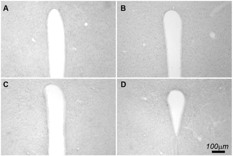
Representative high-magnification microphotographs show c-fos labeled coronal sections containing PVN after intramuscular casein injection into the right hindlimb of C3H/HeJ (A), IL-1R1 KO (B), IL-6 KO (C), and TNFα KO (D) animals.
Figure 10.

Representative microphotographs show c-fos labeled coronal sections containing PVN (A and B), SFO (C and D), and AP/NTS (E and F) after intramuscular injection into the right hindlimb of LPS (left panels), or live E. coli (right panels). COX-2 labeled sections are also shown (H and I).
Discussion
The induction of asymmetrical PVN activation after the injection of casein into the right hindlimb muscles was a surprising and serendipitous observation (Figure 1C). The induced c-fos expression, however, was not confined to the cells of the PVN. It should be noted that induction of c-fos in some brain regions, e.g., the LA (Figure 1B), occurred after both casein and saline injections. Thus, part of the c-fos expression pattern is probably induced by the injection stress, not by local inflammation induced by casein. Casein injection, however, is the cause of the induced c-fos expression in the SON and PVN (Figure 1C), which did not show c-fos positive cells after the saline injection. Whereas the bilateral SON activation pattern does not implicate SON in location specific signaling of the CNS, the left dominant PVN activation after casein injection into the right hindlimb suggests that signals originated in the right hindlimb are relayed to the left PVN via ascending nerve fibers that decussated to the left side of the brain. Consistent with this crossing-over neural pattern, when left hindlimb was injected with casein, right dominant PVN activation was observed, showing that the PVN activation patterns can clearly differentiate local inflammation originated from the left vs. the right side of the body. This PVN activation can be observed between 4–10 h after the casein injection, during which time casein induces local inflammation and febrile response (Zhang et al., 2008), indicating that local inflammation may continually send sensory information to the PVN for at least a 6-h time span. After left sciatic nerve transection, casein injection into the left hindlimb, innervated by the left sciatic, no longer induced c-fos expression in the PVN and SON. In contrast, casein injection into the right hindlimb induced c-fos expression in the PVN (mainly in the left PVN) and in the SON (bilaterally in both SONs), resembling the response observed in intact animals. These results demonstrate that PVN and SON c-fos expression induced by casein injection into the hindlimb is indeed relayed to the brain via peripheral nerves innervating the site of inflammation. Interestingly, this contralateral PVN activation pattern has been noticed in a previous study in which turpentine, another local inflammatory agent, was injected into the hindlimb (Turnbull et al., 2003). Thus, local inflammation, rather than a peculiar biochemical property of casein, is likely the cause of the observed PVN activation. In addition, the casein-induced activation of PVN was absent in animals deficient in IL-1R1, IL-6, TNFα, or TLR4, suggesting that the activated peripheral sensory nerves may be designed specifically to sense the presence of multiple inflammatory signaling molecules.
The pattern of c-fos expression in the PVN suggests that neural signaling of the CNS by peripheral inflammation is coded in the PVN in a location specific manner. This is further investigated by comparing PVN c-fos patterns induced by forelimb vs. hindlimb casein injections. Both right forelimb and right hindlimb casein injections induced left dominant PVN c-fos expression. Close examination of the serial coronal PVN sections in this experiment shows the left dominant PVN c-fos expression to be true for all the sections throughout the entire PVN. In addition, more c-fos expression was found in the anterior PVN after the forelimb casein injection, but in the posterior PVN after the hindlimb casein injection. Therefore, different PVN c-fos patterns can be discerned between forelimb- vs. hindlimb-inflammation initiated responses. It is also possible that the c-fos expressing cells in the same regions of the PVN induced by the forelimb casein injection and those induced by hindlimb casein injection are not the same cells. Our method is unable to determine this, but the pattern difference suggests that different, maybe partially overlapping, cell groups are activated by the hindlimb and forelimb inflammation. The activation of the contralateral PVN cells correlated with the c-fos expression in cells of the ipsilateral dorsal horn of the spinal cord. Whereas forelimb casein injection induced ipsilateral c-fos expressing cells in the dorsal horn between Th5 and C1 levels of the spinal cord, hindlimb casein injection induced c-fos expression between L2 and S2 levels. This is consistent with the neuroanatomical structure of the spinal cord which receives ipsilateral sensory input from the forelimb in the Th5-C1 and those from the hindlimb in the L2-S2. Thus, local inflammations in the forelimb and in the hindlimb are definitely projected to different neurons of the spinal cord. The observed PVN c-fos patterns suggest that the discrete spinal neuronal responses to local inflammation originated in different parts of the body are maintained at the PVN.
Many supraspinal neural structures are known to be activated by peripheral systemic inflammation (Gaykema et al., 2007a; Gaykema et al., 2007b; Gaykema et al., 2009). These regions include the ventral lateral medullar (VLM), the nucleus of the solitary tract (NTS), the parabrachial nucleus (PB), the PVN, the bed nucleus of the stria terminalis (BST), the central nucleus of the amygdala (CeA), and the circumventricular organs (CVOs). Surprisingly, casein injection did not induce c-fos expression in the VLM and the CeA. Further, casein injection induced bilateral c-fos expression in the NTS, the PB, and the BST. Ericsson et al. were the first to show that VLM and NTS are critical relays that project ipsilaterally to the PVN via the ascending medullary catecholaminergic tract when the PVN is activated by circulating interleukin-1 (Ericsson et al., 1994). Recent studies by Gaykema et al. showed that peripheral LPS induced PVN activation is mediated by immune-responsive inputs predominantly from the VLM, whereas NTS provides a greater contribution to the BST (Gaykema et al., 2007b). The present observation that asymmetrical activation of PVN was associated with symmetrical, not asymmetrical, activation of the NTS and no c-fos expression in the VLM suggests that PVN activation by local inflammation may not be relayed via these intermediates. The intriguing fact is while casein injection induced PVN activation in a location related manner, it also induced activation of neurons in the SON, NTS, BST, AH, and PB in a manner that does not appear to reflect location specificity.
This situation can be compared with the complex neural pathways that relay nociceptive signals to the brain. While the discriminative-sensory pathway of the spinothalamic tract (STT) sends its signal to the contralateral brain to encode location information, the affective-cognitive pathways send signals bilaterally to the supraspinal centers to trigger emotional response (Millan, 1999). Similar to the present study, a previous study showed that unilateral masseter inflammation induces bilateral activation of the NTS (Imbe et al., 1999). Therefore, local inflammation may signal the brain in a location non-specific manner at the level of NTS. In a study by Bester et al., increasing levels of pain of the hindpaw induced by noxious heat in rats first induced bilateral, then predominantly contralateral c-fos expression in the PB (Bester et al., 1997). Thus, the present result that bilateral, not contralateral, PB c-fos expression is induced by the casein injection suggest that local inflammation induced PB activation resembles that induced by low level, not high level, of pain. No contralateral PVN activation was reported in the Bester’s study; it is possible that discriminative thermal pain signaling without an inflammatory component is targeted to the PB, not PVN. Regarding PVN, it has been shown that pain signaling from the sciatic nerve causes bilateral, not unilateral, activation of the PVN (Bomholt et al., 2005; Liu et al., 1998). In addition, non-noxious stimulation of the hindlimb does not stimulate PVN neurons (Akaishi et al., 1988). Therefore, the PVN activation patterns induced by the casein injections in the present study appear to be separable from those induced by other stimuli.
It should be noted, however, heightened pain sensitivity at the site of inflammation is a cardinal feature of inflammation. It has been shown that nociceptors can specifically sense IL-1 (Binshtok et al., 2008) and that electrical stimulation of C-fibers can dramatically increase the release of IL-1 in the spinal cord (Whitehead et al., 2010). In addition, IL-6 can sensitize C-fibers to cause mechanical hypersensitivity (Brenn et al., 2007). Therefore, pain sensory fibers could well play an important role in relaying location specific information on local inflammation to the PVN in the present study, even though painful stimulation per se (without inflammation) is not likely the cause of the observed patterns of PVN activation.
The casein-induced pattern of PVN activation is also induced by the injection of live E. coli into the hindlimb, suggesting that localized infection by live bacteria could activate the CNS via the same neural routes revealed by the casein injections. In contrast to casein and live E. coli, LPS injection into the same hindlimb muscle induced bilateral symmetrical activation of PVN. In addition, LPS, not casein, induced c-fos expression in the circumventricular organs (CVOs) and COX-2 expression in brain endothelium. Previous studies showed that intramuscular (im) injection of LPS induces significant increases of pro-inflammatory cytokines in the blood (Jansky et al., 1995; Roth et al., 1997) and that intravenous injection of inflammatory cytokines induces the expression of c-fos in the CVOs (Brady et al., 1994) and COX-2 in brain vasculature (Kagiwada et al., 2004; Rummel et al., 2006). Therefore, activation of PVN after the im LPS injection might be mediated primarily by circulating cytokines which activate PVN via humoral routes; the PVN activation pattern induced by the neural routes under this condition may be masked by that induced by the humoral routes, which will not be able to carry location information.
The fact that injection of 3×10⁸ live E. coli into the hindlimb muscle induced the contralateral pattern of PVN activation suggests localized infection not only stimulates the CNS via neural routes, but such stimulation is also coded by PVN activation patterns that correspond to the site of infection. Previous studies showed that bacterial infection can stimulate CNS without the induction of cytokines in the blood (Campisi et al., 2003; Goehler et al., 2005) and that the release of free LPS, a component of bacteria cell wall, from invading E. coli is highly restricted by the immune response (Creasey et al., 1991). Therefore, immune signals originating from restricted local bacterial infection might be relayed to the brain in the location-specific manner by neural routes; should the local immunological control deteriorates to the point that systemic inflammation is threatened due to the release of free LPS, humoral pathways may be recruited to dominate immune-to-brain communication at the expense of the loss of location information regarding the site of inflammation. This would suggest that if an immunological location detector does exist, it may be detected only when signals of localized immunological events are not overwhelmed by a concurrent systemic inflammation. It should also be noted that injection of E. coli induced both the location specific PVN response and the COX-2 expression in brain endothelial cells. Thus, signals from both the neural and the humoral routes may be transmitted to brain during real life local inflammation.
Recent studies have suggested that signals of localized immune responses might be transmitted to the brain via primary sensory nerve tracts (Goehler et al., 2005; Roth and De Souza, 2001). The neural routes of the immune-to-brain afferent pathways, however, may not simply follow the conventional somatosensory afferents. Typically, the primary somatosensory afferents travel through gracile fasciculus or cuneate fasciculus and synapse with cells in the medulla which then sends secondary afferents to the thalamus where thalamic cells form the final projection to the somatosensory cortex. In the present study, although it is demonstrated that local inflammation induced signaling from the hindlimb traveled in the sciatic nerve, we did not find any c-fos expression in the sensory thalamus, e.g., in the lateral ventroposterior thalamic nucleus (VPL) and somatosensory cortex (data not shown). Liu et al. showed that pain signaling from the hindlimb via sciatic nerve induces c-fos expression in the contralateral VPL(Liu et al., 1998). Therefore, the afferents sensing inflammation may be destined to the PVN in the hypothalamus, not the cortex.
The results of the present study support the notion that immunological activities can be relayed to the brain in a location-specific manner. Whether such immune-to-brain communication results in location-specific modulation of immunological activities by the CNS remains to be determined. It is intriguing to note that somatosensory input may travel via the underappreciated spinal-hypothalamic tract to stimulate PVN neurons (Cliffer et al., 1991) and there are discrete efferent projections from individual cells of PVN to separate segments of the spinal cord (Naito et al., 1996). These pathways make it possible that local inflammation could signal the CNS at PVN which sends neural signals back to the site of inflammation, thereby modulating local immune responses by an integrated neuroimmune loop. Thus far, PVN has been recognized primarily as a center of autonomic control, generally involved in the regulation of body-wide homeostasis. Recent studies, however, showed that discrete PVN-spinal projections, not the conventional neuroendocrine mechanisms, allow PVN to specifically regulate functions of the heart and kidney via tissue specific sympathetic innervation (Coote, 2005). Therefore, PVN could modulate local immune responses in a similar manner. It should be noted that the contralateral patterns observed in the present study were not absolute–ipsilateral PVN was also activated to a lesser degree–and that the forelimb and the hindlimb casein injections appear to induce c-fos expression in many overlapping regions of the PVN. Although we do not know whether exactly the same cells in the PVN were induced to express c-fos by the casein injections into different body parts, the PVN clearly does not seem to have the precise mapping of body locations as the somatosensory cortex. The ability to recognize general areas, perhaps as body districts, of immune responses occurring from distant body parts, however, may be very useful. For example, if two wounds, one in the left leg and one in the right leg, occurred at different time such that one is undergoing the inflammatory phase while the other is undergoing the anti-inflammatory phase of the wound healing, then the coordination of these two different local immune responses by the CNS could optimize efficiency for both.
Acknowledgments
This study is supported by a grant from NIH (AI059089) to NQ.
Footnotes
Publisher's Disclaimer: This is a PDF file of an unedited manuscript that has been accepted for publication. As a service to our customers we are providing this early version of the manuscript. The manuscript will undergo copyediting, typesetting, and review of the resulting proof before it is published in its final citable form. Please note that during the production process errors may be discovered which could affect the content, and all legal disclaimers that apply to the journal pertain.
References
- Akaishi T, Robbins A, Sakuma Y, Sato Y. Neural inputs from the uterus to the paraventricular magnocellular neurons in the rat. Neurosci Lett. 1988;84:57–62. doi: 10.1016/0304-3940(88)90337-0. [DOI] [PubMed] [Google Scholar]
- Bester H, Matsumoto N, Besson JM, Bernard JF. Further evidence for the involvement of the spinoparabrachial pathway in nociceptive processes: a c-Fos study in the rat. J Comp Neurol. 1997;383:439–458. [PubMed] [Google Scholar]
- Binshtok AM, Wang H, Zimmermann K, Amaya F, Vardeh D, Shi L, Brenner GJ, Ji RR, Bean BP, Woolf CJ, Samad TA. Nociceptors are interleukin-1beta sensors. J Neurosci. 2008;28:14062–14073. doi: 10.1523/JNEUROSCI.3795-08.2008. [DOI] [PMC free article] [PubMed] [Google Scholar]
- Bomholt SF, Mikkelsen JD, Blackburn-Munro G. Normal hypothalamo-pituitary-adrenal axis function in a rat model of peripheral neuropathic pain. Brain Res. 2005;1044:216–226. doi: 10.1016/j.brainres.2005.03.005. [DOI] [PubMed] [Google Scholar]
- Brady LS, Lynn AB, Herkenham M, Gottesfeld Z. Systemic interleukin-1 induces early and late patterns of c-fos mRNA expression in brain. J Neurosci. 1994;14:4951–4964. doi: 10.1523/JNEUROSCI.14-08-04951.1994. [DOI] [PMC free article] [PubMed] [Google Scholar]
- Brenn D, Richter F, Schaible HG. Sensitization of unmyelinated sensory fibers of the joint nerve to mechanical stimuli by interleukin-6 in the rat: an inflammatory mechanism of joint pain. Arthritis Rheum. 2007;56:351–359. doi: 10.1002/art.22282. [DOI] [PubMed] [Google Scholar]
- Campisi J, Hansen MK, O’Connor KA, Biedenkapp JC, Watkins LR, Maier SF, Fleshner M. Circulating cytokines and endotoxin are not necessary for the activation of the sickness or corticosterone response produced by peripheral E. coli challenge. J Appl Physiol. 2003;95:1873–1882. doi: 10.1152/japplphysiol.00371.2003. [DOI] [PubMed] [Google Scholar]
- Cliffer KD, Burstein R, Giesler GJ., Jr Distributions of spinothalamic, spinohypothalamic, and spinotelencephalic fibers revealed by anterograde transport of PHA-L in rats. J Neurosci. 1991;11:852–868. doi: 10.1523/JNEUROSCI.11-03-00852.1991. [DOI] [PMC free article] [PubMed] [Google Scholar]
- Coote JH. A role for the paraventricular nucleus of the hypothalamus in the autonomic control of heart and kidney. Exp Physiol. 2005;90:169–173. doi: 10.1113/expphysiol.2004.029041. [DOI] [PubMed] [Google Scholar]
- Creasey AA, Stevens P, Kenney J, Allison AC, Warren K, Catlett R, Hinshaw L, Taylor FB., Jr Endotoxin and cytokine profile in plasma of baboons challenged with lethal and sublethal Escherichia coli. Circ Shock. 1991;33:84–91. [PubMed] [Google Scholar]
- Ericsson A, Kovacs KJ, Sawchenko PE. A functional anatomical analysis of central pathways subserving the effects of interleukin-1 on stress-related neuroendocrine neurons. J Neurosci. 1994;14:897–913. doi: 10.1523/JNEUROSCI.14-02-00897.1994. [DOI] [PMC free article] [PubMed] [Google Scholar]
- Gaykema RP, Balachandran MK, Godbout JP, Johnson RW, Goehler LE. Enhanced neuronal activation in central autonomic network nuclei in aged mice following acute peripheral immune challenge. Auton Neurosci. 2007a;131:137–142. doi: 10.1016/j.autneu.2006.07.005. [DOI] [PubMed] [Google Scholar]
- Gaykema RP, Chen CC, Goehler LE. Organization of immune-responsive medullary projections to the bed nucleus of the stria terminalis, central amygdala, and paraventricular nucleus of the hypothalamus: evidence for parallel viscerosensory pathways in the rat brain. Brain Res. 2007b;1130:130–145. doi: 10.1016/j.brainres.2006.10.084. [DOI] [PubMed] [Google Scholar]
- Gaykema RP, Daniels TE, Shapiro NJ, Thacker GC, Park SM, Goehler LE. Immune challenge and satiety-related activation of both distinct and overlapping neuronal populations in the brainstem indicate parallel pathways for viscerosensory signaling. Brain Res. 2009;1294:61–79. doi: 10.1016/j.brainres.2009.07.076. [DOI] [PMC free article] [PubMed] [Google Scholar]
- Goehler LE, Busch CR, Tartaglia N, Relton J, Sisk D, Maier SF, Watkins LR. Blockade of cytokine induced conditioned taste aversion by subdiaphragmatic vagotomy: further evidence for vagal mediation of immune-brain communication. Neurosci Lett. 1995;185:163–166. doi: 10.1016/0304-3940(95)11251-q. [DOI] [PubMed] [Google Scholar]
- Goehler LE, Gaykema RP, Opitz N, Reddaway R, Badr N, Lyte M. Activation in vagal afferents and central autonomic pathways: early responses to intestinal infection with Campylobacter jejuni. Brain Behav Immun. 2005;19:334–344. doi: 10.1016/j.bbi.2004.09.002. [DOI] [PubMed] [Google Scholar]
- Imbe H, Dubner R, Ren K. Masseteric inflammation-induced Fos protein expression in the trigeminal interpolaris/caudalis transition zone: contribution of somatosensory-vagal-adrenal integration. Brain Res. 1999;845:165–175. doi: 10.1016/s0006-8993(99)01913-7. [DOI] [PubMed] [Google Scholar]
- Jansky L, Vybiral S, Pospisilova D, Roth J, Dornand J, Zeisberger E, Kaminkova J. Production of systemic and hypothalamic cytokines during the early phase of endotoxin fever. Neuroendocrinology. 1995;62:55–61. doi: 10.1159/000126988. [DOI] [PubMed] [Google Scholar]
- Kagiwada K, Chida D, Sakatani T, Asano M, Nambu A, Kakuta S, Iwakura Y. Interleukin (IL)-6, but not IL-1, induction in the brain downstream of cyclooxygenase-2 is essential for the induction of febrile response against peripheral IL-1alpha. Endocrinology. 2004;145:5044–5048. doi: 10.1210/en.2004-0054. [DOI] [PubMed] [Google Scholar]
- Liu RJ, Qiang M, Qiao JT. Nociceptive c-fos expression in supraspinal areas in avoidance of descending suppression at the spinal relay station. Neuroscience. 1998;85:1073–1087. doi: 10.1016/s0306-4522(97)00662-3. [DOI] [PubMed] [Google Scholar]
- Millan MJ. The induction of pain: an integrative review. Prog Neurobiol. 1999;57:1–164. doi: 10.1016/s0301-0082(98)00048-3. [DOI] [PubMed] [Google Scholar]
- Naito J, Ishikawa H, Ohmori Y, Watanabe T. Cells of origin of spinal projections from the paraventricular nucleus (PVN) of the hypothalamus in the chicken. J Auton Nerv Syst. 1996;61:321–323. doi: 10.1016/s0165-1838(96)00077-x. [DOI] [PubMed] [Google Scholar]
- Oke SL, Tracey KJ. From CNI-1493 to the immunological homunculus: physiology of the inflammatory reflex. J Leukoc Biol. 2008;83:512–517. doi: 10.1189/jlb.0607363. [DOI] [PubMed] [Google Scholar]
- Quan N, Banks WA. Brain-immune communication pathways. Brain Behav Immun. 2007;21:727–735. doi: 10.1016/j.bbi.2007.05.005. [DOI] [PubMed] [Google Scholar]
- Roth J, Aslan T, Storr B, Zeisberger E. Lack of cross tolerance between LPS and muramyl dipeptide in induction of circulating TNF-alpha and IL-6 in guinea pigs. Am J Physiol. 1997;273:R1529–1533. doi: 10.1152/ajpregu.1997.273.4.R1529. [DOI] [PubMed] [Google Scholar]
- Roth J, De Souza GE. Fever induction pathways: evidence from responses to systemic or local cytokine formation. Braz J Med Biol Res. 2001;34:301–314. doi: 10.1590/s0100-879x2001000300003. [DOI] [PubMed] [Google Scholar]
- Rummel C, Sachot C, Poole S, Luheshi GN. Circulating interleukin-6 induces fever through a STAT3-linked activation of COX-2 in the brain. Am J Physiol Regul Integr Comp Physiol. 2006;291:R1316–1326. doi: 10.1152/ajpregu.00301.2006. [DOI] [PubMed] [Google Scholar]
- Travers SP, Travers JB. Taste-evoked Fos expression in nitrergic neurons in the nucleus of the solitary tract and reticular formation of the rat. J Comp Neurol. 2007;500:746–760. doi: 10.1002/cne.21213. [DOI] [PubMed] [Google Scholar]
- Turnbull AV, Prehar S, Kennedy AR, Little RA, Hopkins SJ. Interleukin-6 is an afferent signal to the hypothalamo-pituitary-adrenal axis during local inflammation in mice. Endocrinology. 2003;144:1894–1906. doi: 10.1210/en.2002-220964. [DOI] [PubMed] [Google Scholar]
- Whitehead KJ, Smith CG, Delaney SA, Curnow SJ, Salmon M, Hughes JP, Chessell IP. Dynamic regulation of spinal pro-inflammatory cytokine release in the rat in vivo following peripheral nerve injury. Brain Behav Immun. 2010;24:569–576. doi: 10.1016/j.bbi.2009.12.007. [DOI] [PubMed] [Google Scholar]
- Zhang H, Ching S, Chen Q, Li Q, An Y, Quan N. Localized inflammation in peripheral tissue signals the CNS for sickness response in the absence of interleukin-1 and cyclooxygenase-2 in the blood and brain. Neuroscience. 2008;157:895–907. doi: 10.1016/j.neuroscience.2008.09.038. [DOI] [PMC free article] [PubMed] [Google Scholar]