Abstract
Background & Aims
Acute pancreatitis is characterized by an activation cascade of digestive enzymes in the pancreas. The first of these, trypsinogen, can be converted to active trypsin by the peptidase cathepsin B (CTSB). We investigated whether cathepsin L (CTSL), the second most abundant lysosomal cysteine proteinase, can also process trypsinogen to active trypsin and has a role in pancreatitis.
Methods
In CTSL-deficient (Ctsl−/−) mice, pancreatitis was induced by injection of cerulein or infusion of taurocholate into the pancreatic duct. Human tissue, pancreatic juice, mouse pancreatitis specimens, and recombinant enzymes were studied by enzyme assay, immunoblot, N-terminal sequencing, immunocytochemistry, and electron microscopy analyses. Isolated acini from Ctsl−/− and Ctsb−/− mice were studied.
Results
CTSL was expressed in human and mouse pancreas, where it colocalized with trypsinogen in secretory vesicles and lysosomes and was secreted into pancreatic juice. Severity of pancreatitis was reduced in Ctsl−/− mice, compared with wild-type controls, whereas apoptosis and intrapancreatic trypsin activity were increased in Ctsl−/− mice. CTSL induced cleavage of trypsinogen occurred 3 amino acids toward the C terminus from the CTSB activation site and resulted in a truncated, inactive form of trypsin and an elongated propeptide (TAP). This elongated TAP was not detected by ELISA but was effectively converted to an immunoreactive form by CTSB. Levels of TAP thus generated by CTSB were not associated with disease severity, although this is what the TAP-ELISA is used to determine in the clinic.
Conclusions
CTSL inactivates trypsinogen and counteracts the ability of CTSB to form active trypsin. In mouse models of pancreatitis, absence of CTSL induces apoptosis and reduces disease severity.
Keywords: pancreatitis acute, lysosomal enzymes, cathepsins, proteases, trypsin
INTRODUCTION
Acute pancreatitis is thought to be caused by autodigestion of the pancreas by its own digestive enzymes. Physiologically, most digestive proteases are secreted as precursor zymogens and only acquire activity after cleavage and activation by proteolytic processing. The most notable example is trypsinogen, which reaches the small intestine as precursor zymogen, is activated by brush-border enterokinase, and then acts as the master enzyme in activating other digestive proteases. A pivotal role of trypsin in the initiating events of pancreatitis can be assumed because trypsin mutations are the most common autosomal dominant changes associated with pancreatitis1,2,3. In the absence of enterokinase in the pancreas, other mechanisms must be operative through which a premature and intrapancreatic activation of trypsinogen can be initiated. One of the best documented of these mechanisms is trypsinogen activation by cathepsin B (CTSB), a lysosomal protease. Biochemically CTSB has long been shown to be a trypsinogen activator4 and in experimental studies it was demonstrated that inhibition of CTSB or genetic deletion of the ctsb gene protects not only against premature trypsinogen activation but also against pancreatitis5. While the role of CTSB in trypsinogen activation and pancreatitis is now firmly established6,7,8 little is known about a potential role of other lysosomal enzymes.
Cathepsin L (CTSL) is another member of the papain family of cysteine proteinases with similar enzymatic properties. CTSL exhibits a much stronger endoproteolytic activity than CTSB9, and could therefore, if it were expressed in the pancreas, be an even more important regulator of protease activation in pancreatitis. Recently established functions of CTSL include distinct steps in major histocompatibility complex (MHC) processing, the maturation of enkephalin in neuroendocrine cells and the degradation and recycling of growth factor receptors in keratinocytes10,11,12. When released into the cytosol, CTSL has been suggested to regulate apoptosis and to process nuclear transcription factors13.
In the present study using human and mouse material, we found that CTSL is abundantly expressed in the exocrine pancreas, sorted into the lysosomal as well as the secretory pathway of acinar cells, and secreted into pancreatic juice. In mice, in which the ctsl gene was deleted, experimental pancreatitis was significantly less severe and involved a dramatic shift to cell injury via apoptosis. Surprisingly, and in complete contrast to CTSB, CTSL was found to very effectively inactivate trypsinogen and trypsin in vivo, in isolated acini, and in vitro.
MATERIALS AND METHODS
Due to the journal's space limitations please refer to supplemental files for more detailed descriptions of methods.
Induction of experimental pancreatitis in cathepsin L-deficient mice
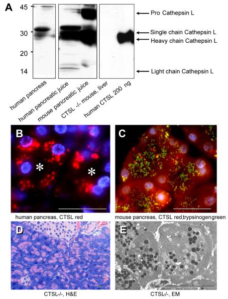
CTSL-deficient mice were generated by gene targeting in E14 mouse embryonic stem cells as described by Roth et al.14 The CTSL-deficient (Ctsl−/−) mice lack CTSL activity in all organs but do not show phenotypic alterations of the pancreas (fig.1). Cerulein-pancreatitis was induced by seven hourly injections of supramaximal (50μg/kg/bw/i.p) cerulein injections and taurocholate-induced pancreatitis by infusion of 50μl 2% taurocholate into the pancreatic duct as previously described5,15.
Figure 1. CTSL in the human and mouse pancreas and the Ctsl−/− pancreas.
In panel A, by Western blots with CTSL antibody pro-CTSL, single and heavy chain CTSL in human pancreas and pancreatic juice were detected. Panel B indicates expression and subcellular localization of CTSL in normal human pancreas (red fluorescence) and panel C colocalization of trypsinogen (green) with CTSL (red) in normal mouse pancreas. The pancreas of Ctsl−/− mice appears completely normal on H&E-stained sections (D, with an islet and duct at the top) and EM (E, with normal arrangement of zymogen granules and mitochondria). Bars indicate 50 μm and asterisks the acinar lumen.
Preparation of serum and tissue samples
Mice were sacrificed at intervals between 1 and 24 hours after the first injection of cerulein, and 6h after intraductal taurocholate application. Blood and tissue were harvested as previously reported5. Pancreatic acini were prepared using purified collagenase (Serva, Germany)5,15.
Biochemical assays
Trypsin, and trypsinogen after enterokinase activation, were measured at 37°C using 64 μM BOC-Gln-Ala-Arg-7-amido-4-methylcoumarin as a substrate5,15. Serum amylase and lipase activities were determined enzymatically by commercially available assays (Roche-Hitachi, Basel). TAP was assayed using an enzyme immunoassay (Biotrin, Ireland) and protein concentrations were determined according to Bradford. Serum cytokines were measured by FACS analysis using a CBA-mouse-assay (Becton-Dickenson). Caspase-3 activity was measured fluorometrically using Rodamin110-DEVD as substrate (Invitrogen, Eugene, OR) Lung myeloperoxidase was measured using O-dianisidine and H2O2 as previously reported5.
Human and animal material for morphology and morphometry
Human material from donor pancreas as well as pancreatic juice from controls and pancreatitis patients was obtained as previously described16 under an ethics committee approved protocol and with informed patient consent. For animal experiments, we collected tissue samples at selected time intervals of pancreatitis as previously reported5,8. For experiments involving the detection of apoptotic cells, free 3′OH-DNA termini were labeled using the terminal deoxynucleotidyl-transferase (TdT) method with fluorescein-labeled digoxigenin nucleotides5,8. Methods for electron microscopy and the quantification of areas of necrosis by morphometry are described in detail elsewhere8.
Proteolytic processing of trypsin and trypsinogen by CTSB and CTSL
The human cationic trypsin (PRSS1) expression plasmid was constructed as previously reported and expressed in E. coli Rosetta (DE3)17. Bovine trypsinogen, enterokinase and CTSB were from Sigma-Aldrich, and bovine trypsin and CTSL from Calbiochem. For CTSL detection, we used antibodies 33/2 and 3G10 (mouse monoclonal) kindly provided by H. Kirschke/E. Weber (Halle/Saale, Germany). For trypsin(ogen) detection, we used trypsin antibody from Chemicon (clone AB1832A). Trypsin activity, trypsinogen content and TAP (Biotrin, Ireland) were determined as previously reported5. Isolated pancreatic acini were prepared as described in detail elsewhere15 with Ile-Pro-Arg-rhodamin-110 as trypsin substrate, PhiPhiLux (Calbiochem) as caspase-3 substrate and propidium iodide exclusion (Roth) to quantitate necrosis.
Enzyme activity measurements and cleavage product detection
CTSL activity was determined using the fluorogenic substrate Z-Phe-Arg-AMC (64μM final concentration)8. CTSL-treated trypsin and trypsinogen samples or PARP cleavage during apoptosis was investigated by SDS-PAGE and cleavage products identified by Western blotting and N-terminal sequencing18. Mass-spectrometry was used as previously described18. CTLS in pancreatic juice was detected with monoclonal anti-cathepsin L antibody (3G10, 1:1000 dilution)8.
Data presentation and statistical analysis
Data in graphs are expressed as means±SEM from 5 or more experiments per group. Statistical comparison between the Ctsl−/− and the Ctsl+/+ group at various time intervals was done by STUDENT'S t-test for independent samples using SPSS for Windows. Differences were considered significant at a level of p<0.05. Data presentation was performed with Origin 7.0 for Windows.
RESULTS
Subcellular localization and sorting of CTSL into the secretory pathway
A prerequisite for a biological relevance of CTSL in pancreatitis would be its expression in the exocrine pancreas and its co-localization with trypsinogen. All of these have previously been established for CTSB5,8 and could also be operative for CTSL because of great similarities between the two lysosomal proteases. Blotting CTSL in the pancreatic juice of mice and humans resulted in a strong signal for pro-CTSL (31 kDa), single chain CTSL, as well as heavy chain CTSL (fig.1A). Detection of CTSL in pancreatic juice is direct evidence that some CTSL is sorted into the secretory pathway of the pancreas and actively secreted into the ducts. Immunofluorescence labeling of human pancreatic tissue from an organ donor localized CTSL to an intracellular vesicle compartment in acinar cells consistent with zymogen granules and lysosomes (fig.1B). Double-labeling with trypsinogen in untreated mice showed a distinct staining of CTSL predominately in the lysosmal compartment (Cy3 red) and of trypsinogen (FITC green) in zymogen granules (fig.1C) with some colocalization of both enzymes (yellow fluorescence). That subcellular redistribution increases rapidly during experimental pancreatitis (see below for fig.2). Untreated Ctsl−/− mice have a completely normal appearing exocrine pancreas on either light or electron-microscopy (fig.1D&E)
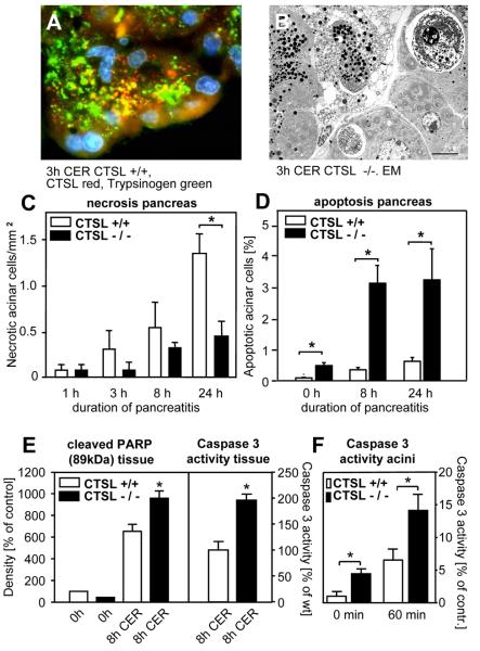
Figure 2. Cell injury and apoptisis in cerulein-induced pancreatitis.
Panel A: 3h of pancreatitis in a wild-type animal labeled with antibody against trypsinogen (green) and CTSL (red fluorescence). B: electronmicroscopy of 3h pancreatitis in the Ctsl−/− mouse. Note whirl-like arrangement of the ER in lower right corner, large autophagic vacuoles, of which one contains a nucleus, and a necrotic cell in the top centre. C: morphometry of acinar tissue necrosis in wild type and Ctsl−/− animals over 24h of pancreatitis. D: 3-OH-nick-end labeling (TUNEL) of apoptotic nuclei in Ctsl−/− animals over 24h of pancreatitis. This difference is already present in untreated animals. E: PARP cleavage and caspase-3 activity in pancreatic tissue after 8h of pancreatitis. F: Caspase-3 activity in living pancreatic acini after 60min of supramaximal cerulein stimulation. Bars indicate 1 μm. Values denote means ± SEM for 5 or more measurements.
Localization of Cathepsin L and pancreatic injury
Induction of pancreatitis lead to an increased colocalization of trypsin (green) and CTSL (red, fig.2A) at the apical portion of acinar cells and in cytoplasmic vesicles. An additional presence of both enzymes in the cytosol could also not be excluded (lower tissue margin in 2A). In subcellular fractionation studies, a shift of CTSL from a lysosome-enriched to a zymogen-granule-enriched fraction was found (not shown). Both observations indicate that the colocalization of CTSL and trypsin increases during the early course of pancreatitis. On EM (fig.2B) and light microscopy, wild-type and Ctsl−/− animals developed morphological signs of acute pancreatitis including acinar cell vacuolization, the formation of autophagic vesicles and overt necrosis. When quantitated by morphometry, CTSL-deleted animals had less extensive necrosis than wild-type animals (fig.2C). When the pancreas was stained for apoptotic cells by TUNEL assay (fig.2D) the result was reversed. CTSL-deleted animals developed a much greater extent of acinar cell apoptosis (fig.2F) than their wild-type littermates and this difference was already significant in untreated control animals. We investigated this further by quantitating the cleavage of PARP and the generation of caspase-3 activity in either pancreatic homogenates from pancreatitis animals or in isolated acini following supramaximal cerulein stimulation (fig.2E&F). In all experiments apoptosis dependent mechanisms were upregulated in the absence of CTSL. This indicates that CTSL is involved in the cell-death pathways of pancreatic acinar cells including, when CTSL is deleted, a prominent shift from necrosis-dominant acinar cell injury to apoptosis during pancreatitis.
Serum pancreatic enzymes and trypsinogen activation
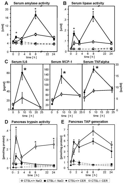
Induction of cerulein pancreatitis was followed by a rapid and biphasic increase in serum activities of amylase (fig.3A) and lipase (fig.3B) which is known to correspond to acinar cell injury5. As previously reported for Ctsb−/− mice5, the CTSL-deficient animals had a much milder disease course, i.e. 33% lower amylase and 25% lower lipase activities. This corresponded not only to the lower extent of necrosis in exocrine tissue (fig.2C&D) but also to a greatly reduced inflammatory response of the animals as indicated by the reduction of serum IL6, MCP-1 and TNFalpha (fig.2C,D,E). Under most clinical and experimental conditions the degree of disease severity in pancreatitis is paralleled by the extent of intrapancreatic trypsinogen activation. It therefore came as a complete surprise to find greatly increased free trypsin activities in the pancreas of Ctsl−/− mice during experimental pancreatitis (fig.3C). The different trypsin activities in the pancreas were not due to different expression levels of trypsinogen since Ctsb−/−, Ctsl−/− and wild-type mice had comparable trypsinogen levels in the pancreas under resting conditions (not shown). The observation was made even more puzzling by the fact that recovery of TAP, the trypsinogen activation peptide that is generated during activation of trypsinogen to trypsin, did not much differ between wild-type and Ctsl−/− animals (fig.3D). This suggests that trypsinogen activation is not altered in Ctsl−/− mice whereas the degradation of trypsinogen and trypsin is highly dependent on the presence of CTSL. It should be noted that the total tissue content of TAP in molar terms exceeded that of measurable trypsin activity by one order of magnitude. This excess of TAP over trypsin activity can indicate an inactivation of newly formed trypsin by CTSL and other proteases15 or an inhibition of trypsin activity by endogenous trypsin inhibitors. On the other hand, TAP may also be formed by sequential cleavage via CTSL and CTSB, a possibility we tested in the experiments below. These findings also indicate that CTSL deletion has completely opposite effects on the severity of pancreatitis and on the intrapancreatic activity of trypsin.
Figure 3. Disease severity and trypsinogen activation.
Deletion of CTSL greatly decreased pancreatitis-associated hyperamylasemia (A), hyperlipasemia (B), as well as serum levels of IL6, MCP-1, and TNFalpha (C) over 24h. E: trypsin activity and F: TAP generation in pancreatic tissue homogenates over 24h of pancreatitis. In strong contrast to serum pancreatic enzyme levels and the extent of tissue necrosis (fig.2), intrapancreatic trypsin activity is greatly increased in Ctsl−/− animals whereas TAP levels are similar in both groups. Values denote means ± SEM for 5 or more measurements.
Taurocholate-induced pancreatitis
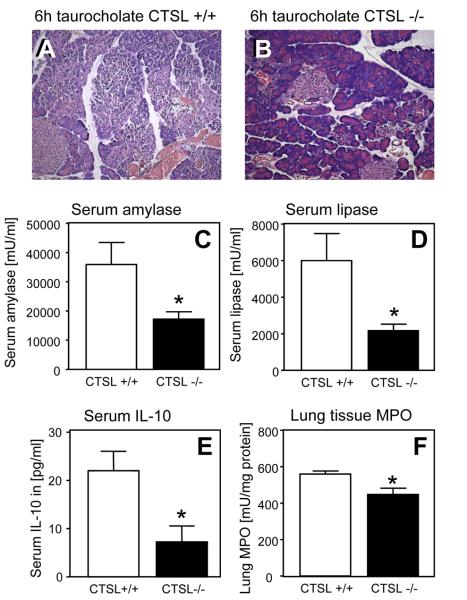
In order to test whether the absence of CTSL has a similar severity-reducing effect on other models of pancreatitis that are largely independent of trypsinogen activation, we induced taurocholate-induced pancreatitis in mice. Here again the morphological damage after 6h (fig.4A&B), or the increase in serum pancreatic enzymes (fig.4C&D), or the markers of inflammation in serum (fig.4E) and lung tissue (fig.4F) were found to be reduced in Ctsl−/− animals.
Figure 4. Taurocholate-induced pancreatitis.
Ctsl−/− and Ctsl+/+ animals were sacrificed after 6h of taurocholate-induced pancreatitis. On morphology (A, B), by serum pancreatic enzyme activities (C, D) and according to parameters of systemic inflammation (IL-10:E, Lung myeloperoxidase:F) the disease severity is reduced in the absence of CTSL. Values denote means ± SEM for 5 or more measurements.
Proteolytic cleavage of trypsinogen by CTSL
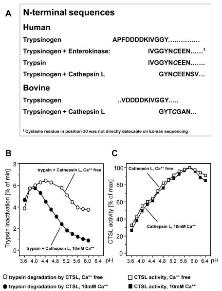
In order to characterize the effect of CTSL on trypsinogen and trypsin further we used purified or recombinant enzymes. The proteolytic processing of trypsinogen was followed by Western blotting, by measuring trypsin activity, and by quantification of the trypsinogen activation peptide (TAP). In fig.5A immunoblots of incubations with enterokinase under optimal catalytic conditions show a rapid and total conversion of bovine trypsinogen to trypsin. Control incubations at pH5.5 in the presence of 10mM Ca++ indicated that bovine trypsinogen did not autoactivate over 3 hours. The concentrations of CTSB for proteolytic activation of trypsinogen were chosen to reflect the conditions in experimental pancreatitis in which only 0.1 to 1% of total trypsinogen is known to be activated to trypsin. Under these conditions CTSB produced only a very weak trypsin band (fig.5A). At the same concentration CTSL produced a much more effective cleavage of trypsinogen resulting in a protein product in the molecular weight range of active trypsin (compared to enterokinase). 10-100-fold higher concentrations of CTSB were needed to produce similarly prominent trypsinogen cleavage (inset below CTSB). In contrast to the incubation with CTSB, in which the cleavage resulted in the generation of trypsin activity (fig.5B) and the formation of TAP (fig.5C), the trypsinogen processing by CTSL resulted neither in active trypsin nor in the generation of TAP. To determine its structure, the trypsin fragment generated by CTSL-cleavage from human and bovine trypsinogen was N-terminally sequenced (fig.6A). CTSL cleaved trypsinogen at the IVG-GYN site (G26-G27 in bovine and human cationic trypsinogen). Hereby, a truncated trypsin without the first three amino acids IVG is formed, and this protein completely lacks trypsin activity. On the other hand, the cleaved trypsinogen activation peptide that is elongated by three amino acids (IVG) is not immunoreactive against the TAP antibody. These experiments show that, while CTSB cleavage of trypsinogen results in active trypsin and immunoreactive TAP, the cleavage by CTSL three amino acids towards the C terminus from the CTSB-processing-site degrades trypsinogen to an inactive degradation product and a non-immunoreactive TAP-IVG peptide. This peptide generated by CTSL is rapidly processed further by CTSB as shown in figure 3 of the supplemental material.
Figure 5. Proteolytic processing of trypsinogen.
Cleavage of trypsinogen by CTSB and CTSL was studied in vitro. A: CTSL cleaves trypsinogen to a lighter protein that corresponds to active trypsin generated by enterokinase (EK) but is processed much more rapidly than by CTSB. Unlike CTSB-generated trypsin, however, this protein has no trypsin activity (B), nor does CTSL-cleavage generate immunoreactive TAP (C). CTSB and CTSL were used in equimolar amounts. To reach comparable cleavage efficiency a 100-fold CTSB concentration had to be used (inset below CTSB).
Figure 6. Trypsin cleavage by CTSL and effect of pH and Ca++.
Human cationic trypsinogen or active trypsin were incubated for 3h with CTSL or enterokinase (EK) and submitted to SDS-PAGE followed by N-terminal sequencing. Note that the inactive CTSL-generated protein is three amino acids shorter (IVG) than active trypsin generated by enterokinase (A). Inactivation of human trypsin was measured as residual trypsin activity after incubation with CTSL at the pH indicated (B). Proteolytic cleavage of trypsin by CTSL exhibited a completely different dependence on pH and Ca++ compared to CTSL-cleavage of the peptide substrate (C).
The role of pH and Ca++ on proteolytic cleavage
Previous investigations of the initial intracellular events during pancreatitis have shown that trypsinogen activation begins in vesicular organelles19 and that they represent an acidic compartment20. Furthermore, it has been demonstrated that trypsinogen activation after supramaximal cerulein stimulation depends on a sustained Ca++ rise in acinar cells21. As CTSL activity is pH-dependent and Ca++ stabilizes the trypsin(ogen) structure22, the processing of trypsin(ogen) by CTSL may depend on the ionic environment within that subcellular compartment. We therefore investigated the effect of pH on CTSL activity measured as degradation of human recombinant trypsin (reduction in trypsin activity) and cleavage of a CTSL peptide-substrate Z-Phe-Arg-AMC (fig.6B&C). The rate of trypsin inactivation was strongly pH-dependent in the range between 4.2 and 4.8 in the presence of Ca++, and in the range between 5.2 and 5.8 in the absence of Ca++ (fig.6B). Activity of CTSL increased from pH3.6 to 6.2 and was independent of the calcium concentration (fig.6C). The rate of trypsin inactivation by CTSL is thus approximately 10-times faster at pH4.0 than at pH5.5. It can therefore be concluded that the rate of trypsin cleavage by CTSL is mainly determined by pH-dependent changes in the surface charge of the trypsin molecule which results in a higher substrate affinity to CTSL. CTSL activity is, in itself, not calcium dependent. We then studied the inactivation and protein processing via CTSL for up to three hours (supplemental fig1.A). We found that EDTA-removal of calcium greatly increased trypsinogen and trypsin degradation by CTSL and, more prominently so, at an acidic pH. We could further identify an additional CTSL-induced cleavage site in active trypsin at position E82-G83. The detailed results of these experiments are found in the supplemental figures 1&2.
Processing of TAP by CTSB
When we synthetized the activation peptide cleavage product of CTSL (TAP-IVG or APFDDDDKIVG), that is not immunoreactive in the TAP-ELISA, and exposed it to CTSB it was rapidly converted to TAP (six fold faster compared to the TAP generation from trypsinogen by CTSB). This indicates that sequential cleavage of trypsinogen by first CTSL and subsequently CTSB generates very large amounts of TAP (50% of equimolar enterokinase). TAP generated under these conditions no longer reflects active trypsin or disease severity23. The details and data of this experiment are found in the supplemental materials.
Trypsin activity and TAP in isolated pancreatic acini
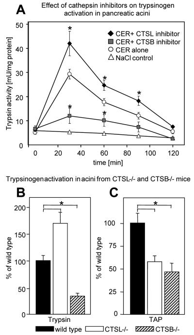
While CTSB is a trypsinogen-activating enzyme5, our data show that CTSL degrades trypsinogen and trypsin. To study whether this is a direct effect on acinar cells, we performed a series of in vitro experiments using freshly isolated acini. In these we inhibited CTSL or CTSB before measuring trypsinogen activation in response to supramaximal cerulein. Figure 7A shows the effect of a specific CTSL-inhibitor (1-Naphthalenylsulfonyl-Ile-Trp aldehyde) and the CTSB-inhibitor CA-074Me. CTSB-inhibition reduced trypsinogen activation by more than 70% whereas the inhibition of CTSL led to an increased level of active trypsin (+50%). To confirm this we also compared acinar cell preparations from Ctsl−/−, Ctsb−/− and wild-type mice (Ctsb/Ctsl+/+) in response to supramaximal cerulein. In the absence of CTSB trypsinogen activation was greatly decreased as indicated by significantly reduced levels of active trypsin and TAP (fig.7B&C). In the absence of CTSL, on the other hand, trypsin activity was increased to 170% compared to wild-type controls, but this was paralleled by a reduced rate of TAP formation. Lower TAP formation in the absence of CTSL may therefore indicate that a significant portion of TAP in wild-type animals is generated via the pathway identified above, in which CTSL rapidly generates TAP-IVG and this is subsequently converted to TAP by CTSB. The alternative explanation of an extended half life of trypsin (rather than higher activity) is less likely because under CTSL inhibition (fig.7A) in acini or in acini of Ctsl−/− animals (not shown) the time interval when trypsin activity returns to pre-stimulation levels is the same as in controls. It should be noted that the amount of TAP greatly exceeded the trypsin activities in molar terms in acini as well as in vivo. (fig.3F&G). CTSB is thus physiologically and critically involved in both pathways of TAP generation.
Figure 7. Inhibition or deletion of CTSB and CTSL in pancreatic acini.
In panel A, pancreatic acini were pre-incubated for 30 minutes with 10 nM CTSL inhibitor (1-Naphthalenylsulfonyl-Ile-Trp-aldehyde) or 1 μM CTSB inhibitor CA-074Me, then stimulated with cerulein. Trypsin activity was compared over 120min. Inhibition of CTSL increased and the inhibition of CTSB decreased intra-acinar cell trypsin activity. In B and C, acini were prepared from wild-type, CTSL-deleted and CTSB-deleted animals. Trypsin activity and TAP were determined after cerulein stimulation for 1h. The absence of CTSL increased whereas absence of CTSB decreased intra-acinar cell trypsin activity. The generation of TAP was reduced in both knock-out strains. Data represent means of 5 or more experiments ± SEM. Significant differences are indicated with asterisks when p was <0.05.
DISCUSSION
The underlying mechanism of acute pancreatitis has long been thought to involve autodigestion of the pancreas by its own digestive proteases. Under physiological conditions, the pancreas is protected by a variety of mechanisms that include storage and processing of digestive enzymes in membrane-confined vesicles, transport of proteases to the lumen as inactive precursor zymogens, the presence of protease inhibitors and the absence of the physiological activator enterokinase from the pancreas. Several studies have shown that these protective mechanisms are apparently overwhelmed in the early phase of pancreatitis and protease activation, specifically the premature and intrapancreatic activation of trypsinogen, is an inherent characteristic of human and several experimental models of pancreatitis15,20,23.
One well documented mechanism that permits the protease activation cascade to begin within acinar cells is the activation of trypsinogen by cathepsin B (CTSB). Several studies have shown with purified or recombinant enzymes4,8, with isolated preparations of pancreatic acini, or with animal models of pancreatitis5,15 that CTSB is a potent activator of trypsinogen and its deletion or inhibition greatly reduces intrapancreatic protease activation and the severity of pancreatitis. A prerequisite for the activation of trypsinogen by CTSB is supposed to be a redistribution of lysosomal enzymes into the zymogen-containing secretory compartment and a colocalization of CTSB with trypsinogen. Both conditions are met when pancreatitis begins in either the human or animal pancreas5. Whether other lysosomal proteases can have similar functions in the pancreas or in pancreatitis was previously unknown.
In the present study we found that cathepsin L (CTSL), the second most common lysosomal cysteine proteinase besides CTSB, is abundantly present in human and mouse pancreatic acinar cells. We further found that CTSL and CTSB are both localized in the lysosomal as well as the pancreatic secretory compartment and that their colocalization with zymogens further increases during pancreatitis and may even spread to the cytosol. Deletion of the ctsl gene, which does not affect the pancreas under physiological conditions, has two effects in experimental pancreatitis: 1) it greatly increases intrapancreatic trypsin activity because CTSL is a trypsin(ogen) inactivator and thus an antagonist of CTSB and 2) it greatly reduces the severity of pancreatitis possibly by shifting the cellular effects of pancreatitis towards apoptosis.
CTSB and CTSL are widely expressed members of the papain family of cysteine proteinases9. Recent experimental results suggest that they not only catalyze bulk proteolysis but also take part in proteolytic processing of protein substrates9. For CTSB, we and others have shown that it acts as a trypsinogen-activating enzyme in vivo and in vitro4,5 and that this process involves the cleavage of a Lys-Ile bond releasing active trypsin and the pro-peptide TAP. Due to its optimum at a pH <6, CTSB exerts its catalytic activity in an acidic intracellular compartment, where also zymogen activation has been shown to take place20. We found that CTSL, the second most abundant cysteine proteinase, shares the same intracellular compartment and confirmed24 that it possesses a much higher endoproteolytic activity than CTSB.
We further found that CTSL very effectively cleaves trypsinogen at position G26-G27 resulting in its inactivation. Human cationic trypsinogen was found to be cleaved at the exact same position. This cleavage site is located three amino acids towards the C terminus from the physiological (i.e. for enterokinase and CTSB) cleavage-site Lys-Ile and removes the N terminus of mature trypsin, thus impairing the catalytic center of trypsin25. Incubation of mature trypsin with CTSL, on the other hand, resulted in a negligible loss of the (Ile-Val-)-N terminus. This indicates a conformational change of trypsinogen upon physiological cleavage of TAP by either CTSB or autoactivation which prevents further processing of the N terminus by CTSL.
We also identified an additional cleavage site for CTSL at position E82-G83. This cleavage site is located in the calcium-binding loop and affects trypsinogen as well as trypsin. Under conditions at which trypsin(ogen) binds Ca++, e.g. pH 5.5, the cleavage by CTSL is strongly suppressed. On the other hand, EDTA-removal of Ca++ or decreasing pH to 4.0 strongly enhanced trypsin(ogen) degradation. Consistent with these findings the initial inactivation mechanisms for trypsinogen or trypsin by CTSL follow different pathways: i) trypsinogen is primarily cleaved in the N-terminal region in a Ca++-independent manner (G26-G27), ii) the primary cleavage of trypsin occurs predominantly at the Ca++-binding site (E82-G83). Which of these two prevails in vivo is impossible to predict but during experimental pancreatitis the absence of CTSL results in a manifold increase in intrapancreatic trypsin activity.
The proteolytic processing of trypsinogen by enterokinase or CTSB produces active trypsin and the cleaved pro-peptide in equal stoechiometric amounts. The amount of trypsinogen activation peptide (TAP) therefore reflects the extent of trypsinogen activation much more accurately than activity measurements, e.g. when trypsin is rapidly inactivated by autodegradation23, endogenous inhibitors, or proteolytic degradation. Due to its relative stability and the availability of specific antibodies, TAP is increasingly recognized as a standalone parameter for pancreatitis severity and has found its way into clinical practise23. Our results now indicate a second pathway in which the generation of trypsin activity and TAP do not develop in parallel. We found that the primary cleavage of trypsinogen by CTSL creates a TAP-IVG peptide that escapes detection by the TAP antibody. However, its subsequent conversion by CTSB produces immunoreactive TAP to a much greater extent than via direct CTSB activation of trypsinogen. Obviously, this pathway combines the highly effective endoproteolytic activity of CTSL with the exoproteolytic activity of CTSB. In the pathological situation of acute pancreatitis, these large amounts of TAP may no longer reflect trypsin activity.
Our experiments, using isolated pancreatic acini with either specific enzyme inhibitors or from CTSL and CTSB knockout animals, confirmed this fundamental difference between the two lysosomal hydrolases. CTSB was confirmed as an activator of trypsinogen and of the intracellular protease cascade, whereas CTSL was identified as its antagonist and a potent inactivator of trypsinogen and trypsin. Taken together, these data suggest that alterations in the structure or function of CTSL could represent an important mechanism in the pathogenesis of human pancreatitis.
The effect of CTSL on disease severity seems to be completely uncoupled from its effect on trypsinogen activation. It not only affects pancreatic injury directly but also translates into less systemic inflammation, even in a model such as taurocholate-induced pancreatitis in which trypsinogen activation might not play such a crucial role in determining disease severity. It is also paralleled by increased caspase-3 activation, PARP cleavage and apoptosis in the pancreas of Ctsl−/− animals. That such a shift to apoptosis-dominant cell injury improves the outcome of pancreatitis has previously been reported26. Cathepsins, on the other hand, have long been considered to be guardians of the cellular homeostasis and, when released into the cytoplasm, to influence apoptotic pathways through a number of different mechanisms27. A release of cathepsins into the cytoplasm has recently been shown to also occur in pancreatitis28. Crossbreeding of Ctsl−/− with Ctsb−/− mice results in a lethal phenotype around postnatal day 12 and is caused by significant neuronal cell apoptosis, whereas neither the CTSL nor the CTSB knock-out alone has such an effect. This suggests that some in vivo function of CTSB can be compensated for by CTSL and vice versa29. Other experimental observations would also be in line with a predominantly antiapoptotic role of CTSL30,31. In the setting of pancreatitis, however, this antiapoptotic function of CTSL appears to contribute to disease severity and the absence or inhibition of CTSL would have a beneficial therapeutic effect.
In conclusion, the present study establishes CTSL as a potent trypsin(ogen)-inactivating factor in vivo and in vitro and thus as an antagonist of CTSB in the digestive protease cascade that triggers pancreatitis. The antiapoptotic function of CTSL, on the other hand, affects disease severity in a manner that is unrelated to the initiating protease cascade. As far as the understanding of acute pancreatitis is concerned, these data indicate further that: i) greater intrapancreatic trypsin activity does not necessarily correlate with greater acinar cell injury, and ii) higher TAP levels do not always reflect higher levels of active trypsin or disease severity. The sequence in which lysosomal proteases find their substrates, the subcellular compartment in which they either constitutively or pathophysiologically colocalize with digestive enzymes, as well as the biophysical properties of that compartment ultimately determine whether their action is disease provoking or protective.
Supplementary Material
Acknowledgements
This study was supported by grants from the Deutsche Forschungsgemeinschaft DFG HA2080/7-1, LE 625/8-1, LE 625/9-1, MA4115/1-2, DFG GRK 840 E3 and E4, Mildred Scheel Stiftung 10-2031-Le I and 10-6977-Re, BMBF-NBL3 01 ZZ 0403 and Alfried-Krupp Foundation (Graduiertenkolleg Tumorbiologie) and NIH DK058088 to MST.
The authors wish to thank Dr. B. Schmidt, Zentrum für Biochemie und Molekulare Zellbiologie, Göttingen, for sequencing assistance and V. Krause, C. Jechorek, K. Mülling und N. Loth for technical and D. Schwenn for writing assistance.
Footnotes
Conflict of Interest Statement: All authors declare to have no conflict of interest
REFERENCES
- 1.Whitcomb DC, Gorry MC, Preston RA, et al. Hereditary pancreatitis is caused by a mutation in the cationic trypsinogen gene. Nat Genet. 1996;14:141–5. doi: 10.1038/ng1096-141. [DOI] [PubMed] [Google Scholar]
- 2.Simon P, Weiss FU, Sahin-Tóth M, et al. Hereditary pancreatitis caused by a novel PRSS1 mutation (Arg-122>Cys) that alters autoactivation and autodegradation of cationic trypsinogen. J Biol Chem. 2002;277:5404–10. doi: 10.1074/jbc.M108073200. [DOI] [PubMed] [Google Scholar]
- 3.Simon P, Weiss FU, Zimmer KP, et al. Spontaneous and sporadic trypsinogen mutations in idiopathic pancreatitis. Jama. 2002;288:2122. doi: 10.1001/jama.288.17.2122. [DOI] [PubMed] [Google Scholar]
- 4.Greenbaum LM, Hirshkowitz A, Shoichet I. The activation of trypsinogen by cathepsin B. J Biol Chem. 1959;234:2885–90. [PubMed] [Google Scholar]
- 5.Halangk W, Lerch MM, Brandt-Nedelev B, et al. Role of cathepsin B in intracellular trypsinogen activation and the onset of acute pancreatitis. J Clin Invest. 2000;106:773–81. doi: 10.1172/JCI9411. [DOI] [PMC free article] [PubMed] [Google Scholar]
- 6.Mahurkar S, Idris MM, Reddy DN, et al. Association of cathepsin B gene polymorphisms with tropical calcific pancreatitis. Gut. 2006;55:1270–5. doi: 10.1136/gut.2005.087403. [DOI] [PMC free article] [PubMed] [Google Scholar]
- 7.Weiss FU, Behn CO, Simon P, et al. Cathepsin B gene polymorphism Val26 is not associated with idiopathic chronic pancreatitis in European patients. Gut. 2007;56:1322–3. doi: 10.1136/gut.2007.122507. [DOI] [PMC free article] [PubMed] [Google Scholar]
- 8.Kukor Z, Mayerle J, Kruger B, et al. Presence of cathepsin B in the human pancreatic secretory pathway and its role in trypsinogen activation during hereditary pancreatitis. J Biol Chem. 2002;277:21389–96. doi: 10.1074/jbc.M200878200. [DOI] [PubMed] [Google Scholar]
- 9.Kirschke H, Barrett AJ. Cathepsin L--a lysosomal cysteine proteinase. Prog Clin Biol Res. 1985;180:61–9. [PubMed] [Google Scholar]
- 10.Yasothornsrikul S, Greenbaum D, Medzihradszky KF, et al. Cathepsin L in secretory vesicles functions as a prohormone-processing enzyme for production of the enkephalin peptide neurotransmitter. Proc Natl Acad Sci U S A. 2003;100:9590–5. doi: 10.1073/pnas.1531542100. [DOI] [PMC free article] [PubMed] [Google Scholar]
- 11.Nakagawa T, Roth W, Wong P, et al. Cathepsin L: critical role in Ii degradation and CD4 T cell selection in the thymus. Science. 1998;280:450–3. doi: 10.1126/science.280.5362.450. [DOI] [PubMed] [Google Scholar]
- 12.Reinheckel T, Hagemann S, Dollwet-Mack S, et al. The lysosomal cysteine protease cathepsin L regulates keratinocyte proliferation by control of growth factor recycling. J Cell Sci. 2005;118:3387–95. doi: 10.1242/jcs.02469. [DOI] [PubMed] [Google Scholar]
- 13.Cirman T, Oresic K, Mazovec GD, et al. Selective disruption of lysosomes in HeLa cells triggers apoptosis mediated by cleavage of Bid by multiple papain-like lysosomal cathepsins. J Biol Chem. 2004;279:3578–87. doi: 10.1074/jbc.M308347200. [DOI] [PubMed] [Google Scholar]
- 14.Roth W, Deussing J, Botchkarev VA, et al. Cathepsin L deficiency as molecular defect of furless: hyperproliferation of keratinocytes and pertubation of hair follicle cycling. Faseb J. 2000;14:2075–86. doi: 10.1096/fj.99-0970com. [DOI] [PubMed] [Google Scholar]
- 15.Halangk W, Kruger B, Ruthenburger M, et al. Trypsin activity is not involved in premature, intrapancreatic trypsinogen activation. Am J Physiol Gastrointest Liver Physiol. 2002;282:G367–74. doi: 10.1152/ajpgi.00315.2001. [DOI] [PubMed] [Google Scholar]
- 16.Kukor Z, Tóth M, Pál G, et al. Human cationic trypsinogen. Arg(117) is the reactive site of an inhibitory surface loop that controls spontaneous zymogen activation. J Biol Chem. 2002;277:6111–7. doi: 10.1074/jbc.M110959200. [DOI] [PubMed] [Google Scholar]
- 17.Sahin-Tóth M. Human cationic trypsinogen. Role of Asn-21 in zymogen activation and implications in hereditary pancreatitis. J Biol Chem. 2000;275:22750–5. doi: 10.1074/jbc.M002943200. [DOI] [PubMed] [Google Scholar]
- 18.Mayerle J, Schnekenburger J, Kruger B, et al. Extracellular cleavage of E-cadherin by leukocyte elastase during acute experimental pancreatitis in rats. Gastroenterology. 2005;129:1251–67. doi: 10.1053/j.gastro.2005.08.002. [DOI] [PubMed] [Google Scholar]
- 19.Lerch MM, Saluja AK, Runzi M, et al. Luminal endocytosis and intracellular targeting by acinar cells during early biliary pancreatitis in the opossum. J Clin Invest. 1995;95:2222–31. doi: 10.1172/JCI117912. [DOI] [PMC free article] [PubMed] [Google Scholar]
- 20.Lerch MM, Saluja AK, Dawra R, et al. The effect of chloroquine administration on two experimental models of acute pancreatitis. Gastroenterology. 1993;104:1768–79. doi: 10.1016/0016-5085(93)90658-y. [DOI] [PubMed] [Google Scholar]
- 21.Kruger B, Albrecht E, Lerch MM. The role of intracellular calcium signaling in premature protease activation and the onset of pancreatitis. Am J Pathol. 2000;157:43–50. doi: 10.1016/S0002-9440(10)64515-4. [DOI] [PMC free article] [PubMed] [Google Scholar]
- 22.Sahin-Tóth M, Tóth M. High-affinity Ca++ binding inhibits autoactivation of rat trypsinogen. Biochem Biophys Res Commun. 2000;275:668–71. doi: 10.1006/bbrc.2000.3355. [DOI] [PubMed] [Google Scholar]
- 23.Neoptolemos JP, Kemppainen EA, Mayer JM, et al. Early prediction of severity in acute pancreatitis by urinary trypsinogen activation peptide: a multicentre study. Lancet. 2000;355:1955–60. doi: 10.1016/s0140-6736(00)02327-8. [DOI] [PubMed] [Google Scholar]
- 24.Rocken C, Menard R, Buhling F, et al. Proteolysis of serum amyloid A and AA amyloid proteins by cysteine proteases: cathepsin B generates AA amyloid proteins and cathepsin L may prevent their formation. Ann Rheum Dis. 2005;64:808–15. doi: 10.1136/ard.2004.030429. [DOI] [PMC free article] [PubMed] [Google Scholar]
- 25.Bode W, Fehlhammer H, Huber R. Crystal structure of bovine trypsinogen at 1-8 A resolution. I. Data collection, application of patterson search techniques and preliminary structural interpretation. J Mol Biol. 1976;106:325–35. doi: 10.1016/0022-2836(76)90089-9. [DOI] [PubMed] [Google Scholar]
- 26.Mareninova OA, Sung KF, Hong P, et al. Cell death in pancreatitis: caspases protect from necrotizing pancreatitis. J Biol Chem. 2006;281:3370–81. doi: 10.1074/jbc.M511276200. [DOI] [PubMed] [Google Scholar]
- 27.Chwieralski CE, Welte T, Buhling F. Cathepsin-regulated apoptosis. Apoptosis. 2006;11:143–9. doi: 10.1007/s10495-006-3486-y. [DOI] [PubMed] [Google Scholar]
- 28.Dawra R, Talukdar R, Dudeja V, et al. Release of cathepsin B into cytosol induces apoptosis in pancreatic acinar cells through mitochondrial pathways during pancreatitis. Gastroenterology. 2009;136(S1):A372. [Google Scholar]
- 29.Felbor U, Kessler B, Mothes W, et al. Neuronal loss and brain atrophy in mice lacking cathepsins B and L. Proc Natl Acad Sci U S A. 2002;99:7883–8. doi: 10.1073/pnas.112632299. [DOI] [PMC free article] [PubMed] [Google Scholar]
- 30.Zheng X, Chu F, Mirkin BL, et al. Role of the proteolytic hierarchy between cathepsin L, cathepsin D and caspase-3 in regulation of cellular susceptibility to apoptosis and autophagy. Biochim Biophys Acta. 2008;1783:2294–300. doi: 10.1016/j.bbamcr.2008.07.027. [DOI] [PubMed] [Google Scholar]
- 31.Zheng X, Chu F, Chou PM, et al. Cathepsin L inhibition suppresses drug resistance in vitro and in vivo: a putative mechanism. Am J Physiol Cell Physiol. 2009;296:C65–74. doi: 10.1152/ajpcell.00082.2008. [DOI] [PMC free article] [PubMed] [Google Scholar]
Associated Data
This section collects any data citations, data availability statements, or supplementary materials included in this article.


