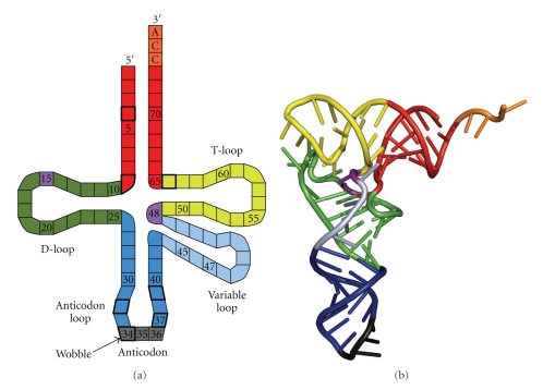
Figure 2.
tRNAs. (a) Schematic representation, showing the D-loop (green), anticodon loop (blue) that harbours the anticodon (grey), variable loop that is variable in length (light blue), the T-loop (yellow), the acceptor stem (red), and the CCA aminoacyl binding site (orange). The Levitt base pair is coloured purple. Nucleotides with thick boxes are often modified with variable modifications. (b) Tertiary structure of a yeast tRNAPhe, coloured similar to (a). Figure is rendered with PyMOL from data deposited in the Protein Data Bank (1 ehz).

