
Figure 2. Blocking Antibodies Against CD47 Enable Phagocytosis of NHL Cells by Macrophages and Synergize with Rituximab in Vitro.
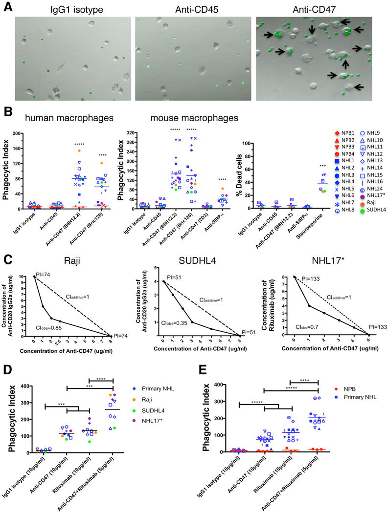
(A) CFSE-labeled NHL cells were incubated with human macrophages and the indicated antibodies and examined by immunofluorescence microscopy to detect phagocytosis (arrows). Photomicrographs from a representative NHL sample are shown. (B) Phagocytic indices of primary human NHL cells (blue), normal peripheral blood (NPB) cells (red), and NHL cell lines (purple, orange, and green) were determined using human (left) and mouse (middle) macrophages. Antibody-induced apoptosis (right panel) was tested by incubating NHL cells with the indicated antibodies or staurosporine without macrophages, and assessing the percentage of apoptotic and dead cells (% annexin V and/or PI positive). (C) Synergistic phagocytosis by anti-CD47 antibody (B6H12.2) and anti-CD20 mAbs was examined by isobologram analysis and determination of combination indices (CI). CIobs indicates observed results, and the dashed line indicates the expected results if antibodies were additive. (D,E) Synergy between anti-CD47 antibody and rituximab in the phagocytosis of NHL and NPB cells was assessed by determining the phagocytic index when incubated with a combination of both antibodies compared to either antibody alone at twice the dose, with either mouse (D) or human (E) macrophages. NHL17*: cell line derived from primary sample NHL17. ***p<0.001, ****p<0.0001, *****p<0.00001. Figure 2B p-values represent comparison against IgG1 isotype control antibody. See also Figure S2.